

Cytoplasm is a semi-fluid cellular material found inside the plasma membrane of a cell. It is present in all cells. In prokaryotic cells, the DNA, ribosomes and different cellular materials are present within this cytoplasmic region.
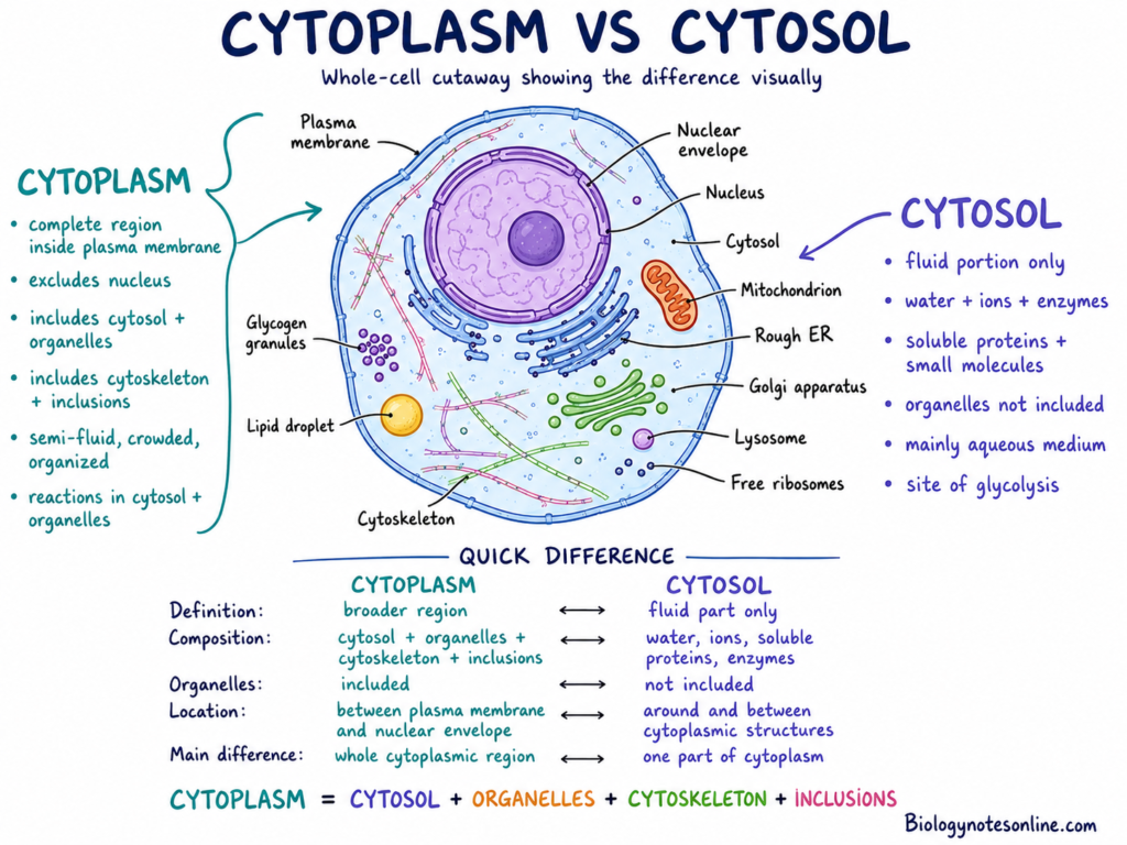
In eukaryotic cells, cytoplasm is the part present between the plasma membrane and the nuclear envelope. The nucleus is not included in cytoplasm. It remains separated by the nuclear envelope, while the different membrane-bound cell organelles are found in the surrounding cytoplasm.
Cytosol– It is the fluid portion of cytoplasm. Cytoplasm and cytosol are not the same. The cytoplasm consists of cytosol, cell organelles, ribosomes, cytoskeleton and different suspended materials. Thus, cytosol forms only one part of the cytoplasm.
The major functions of cytoplasm are cellular metabolism, protein synthesis and movement of different cellular materials. It holds the cell organelles and provides the region for transportation of molecules throughout the cell. The cytoskeleton present through the cytoplasm also has a role in maintaining the cell shape, cell movement and cell division.
What Is Cytoplasm?
Cytoplasm is the cellular material present inside the plasma membrane of a cell. In eukaryotic cells, it includes all the materials present outside the nucleus, but enclosed within the plasma membrane. It is made up of cytosol, cell organelles, cytoskeleton and different suspended cellular materials.
The simple cytoplasm definition is the material filling the inside of a cell. This is the region where the cell organelles are found and a large number of cellular reactions also takes place. Thus, the cytoplasm meaning is not only a fluid present within the cell.
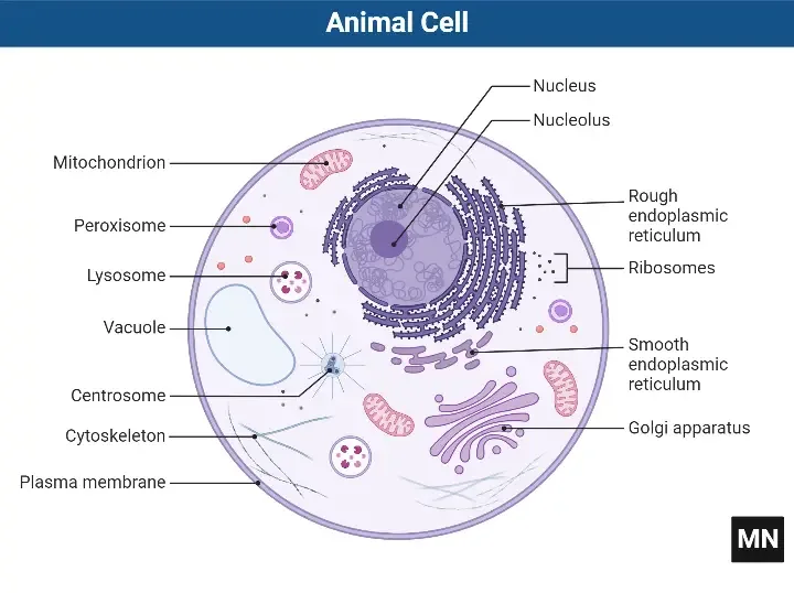
In eukaryotic cells, the cytoplasm lies between the plasma membrane and the nuclear envelope. The nucleus has its own membrane and it remains separated from the cytoplasm. Mitochondria, Golgi bodies, endoplasmic reticulum and other organelles are found within the cytoplasm.
Prokaryotic cells do not have a membrane-bound nucleus. The cytoplasm is present throughout the inner part of these cells. The genetic material is present in a nucleoid region, directly surrounded by cytoplasm, without having any nuclear envelope.
Cytoplasm is commonly described as the “cell fluid”. This description is not fully correct. The fluid portion of cytoplasm is known as cytosol. Cytoplasm contains this cytosol, along with organelles, ribosomes, cytoskeleton, stored materials and different large molecules.
The cytoplasm is an active cellular region. It is not a still fluid in which the organelles only remain floating. Different metabolic reactions are continuously carried out here. Molecules are moved from one region to another, organelles change their position and the cytoskeleton also rearranges according to the activities of cell.
Where Is It Located in a Cell?
The cytoplasm is located inside the plasma membrane in both prokaryotic and eukaryotic cells. The plasma membrane surrounds it and separates the internal materials of the cell from its external environment.
In eukaryotic cells, the cytoplasm is found outside the nuclear envelope. It fills the region between the nuclear envelope and the plasma membrane. The nucleus contains nucleoplasm, while the remaining cellular region outside the nucleus is the cytoplasm.
Cell organelles are suspended within the cytoplasm. Some organelles move through this region with the help of cytoskeletal filaments and motor proteins. Thus, the cytoplasm provides a surrounding region for the organelles and other materials of the cell.
In prokaryotic cells, cytoplasm spreads throughout the cell interior enclosed by the plasma membrane. There is no separate nuclear compartment. Ribosomes, the nucleoid, enzymes and different cellular molecules are present within the same cytoplasmic region.
Origin and Meaning of the Term
The term cytoplasm is formed from two words, “cyto” and “plasm”. “Cyto” refers to cell, while “plasm” refers to a formed material or substance. Therefore, cytoplasm refers to the material of the cell.
The term was historically associated with protoplasm, which was used for the living material present within a cell. Protoplasm was divided into cytoplasm and nucleoplasm. Cytoplasm is present outside the nucleus, while nucleoplasm is the material present inside the nucleus.
Structure and Main Components of Cytoplasm
The cytoplasm structure is not formed of a single fluid. It has cytosol, cell organelles, cytoskeleton and cytoplasmic inclusions. These form the main components of cytoplasm.
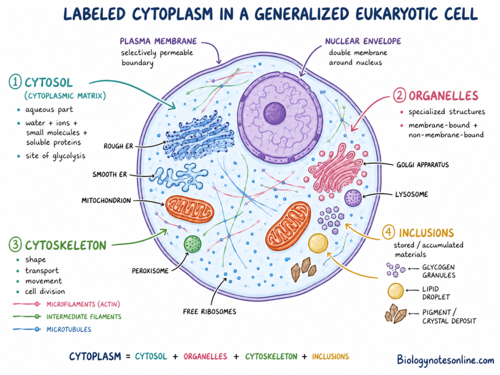
The main parts of cytoplasm are-
- Cytosol or cytoplasmic matrix
- Cytoplasmic organelles
- Cytoskeleton
- Cytoplasmic inclusions
Cytosol or Cytoplasmic Matrix
- Cytosol is the aqueous portion of cytoplasm. It is also called the cytoplasmic matrix.
- Water, inorganic ions and different small molecules are present in it. Soluble proteins and enzymes are also found.
- Cytosol is only one part of cytoplasm. Cell organelles are not included in cytosol.
- Glycolysis and many intermediary metabolic reactions takes place in this region.
Cytoplasmic Organelles
- Cytoplasmic organelles are specialized structures present within the cytoplasm.
- Membrane-bound organelles include mitochondria, endoplasmic reticulum, Golgi apparatus, lysosomes and peroxisomes.
- Mitochondria have a role in aerobic ATP production. Endoplasmic reticulum and Golgi apparatus are used in formation, modification and transportation of proteins and lipids.
- Lysosomes carry out intracellular digestion. Peroxisomes are used in different oxidative reactions.
- Ribosomes are non-membrane-bound structures. They are the site for protein synthesis.
- Chloroplasts and other plastids are present in plant cells. Chloroplasts carry out photosynthesis, while some other plastids store starch or pigments.
- The internal spaces of membrane-bound organelles form separate compartments. These spaces are not included in cytosol.
Cytoskeleton
- The cytoskeleton is a network of protein filaments extended through the cytoplasm.
- It is mainly formed of microfilaments, intermediate filaments and microtubules.
- Microfilaments are mainly formed of actin. They take part in cell movement, cell-shape changes and cytokinesis.
- Intermediate filaments provide mechanical strength to the cell.
- Microtubules help in organelle positioning, intracellular transport and chromosome movement during cell division.
- The cytoskeleton also maintains the internal arrangement of cell.
Cytoplasmic Inclusions
- Cytoplasmic inclusions are stored or accumulated materials present in the cytoplasm. Most of them are not permanent working organelles.
- Glycogen granules are present in many animal, fungal and bacterial cells. They store glucose units.
- Starch granules are found in plant cells. These granules are formed within chloroplasts or amyloplasts.
- Pigment granules, crystals and different cellular deposits may also be present. Their amount is different among different cell types.
- Lipid droplets store neutral lipids. They were earlier described only as inclusions, but are now considered active cellular organelles.
Cytoplasmic Inclusions
| Inclusion | Composition | Example cell | Purpose |
|---|---|---|---|
| Glycogen granules | Branched polymer of glucose | Liver and skeletal muscle cells | Storage of carbohydrate. It is broken down when glucose is required. |
| Lipid droplets | Triacylglycerols and steryl esters surrounded by a phospholipid monolayer | Adipocytes and liver cells | Storage of neutral lipids and supply of fatty acids. |
| Starch granules | Amylose and amylopectin | Plant storage cells and chloroplast-containing cells | Storage of carbohydrate in plants. |
| Melanin granules | Melanin pigment enclosed mainly within melanosomes | Melanocytes and pigmented epithelial cells | Provides pigmentation and helps in protection from ultraviolet radiation. |
| Lipofuscin granules | Oxidized lipids, proteins and other poorly degraded materials | Neurons and cardiac muscle cells | Accumulates with age. It is mainly a residual cellular material. |
| Secretory granules | Concentrated proteins, peptides or other secretory products | Pancreatic cells and endocrine cells | Temporary storage of materials before secretion. |
| Protein crystals | Crystallized proteins | Some glandular and liver cells | Storage or accumulation of specific proteins. |
| Calcium oxalate crystals | Calcium oxalate | Many plant cells | Storage of calcium and protection against herbivores. |
| Polyphosphate granules | Inorganic polyphosphate | Bacterial and fungal cells | Storage of phosphate and energy-related phosphate reserves. |
| Polyhydroxyalkanoate granules | Polyesters such as polyhydroxybutyrate | Many bacterial cells | Storage of carbon and energy. |
Components of Cytoplasm and Their Functions
| Component | Main Features | Functions |
|---|---|---|
| Cytosol | Aqueous portion of cytoplasm. Contains water, ions, enzymes and small molecules. | Site of glycolysis and many intermediary metabolic reactions. |
| Mitochondria | Double membrane-bound organelles. | Produce most of the ATP during aerobic respiration. |
| Endoplasmic reticulum (ER) | Membrane network present in cytoplasm. It may be rough or smooth. | Rough ER forms secretory and membrane proteins. Smooth ER is used in lipid synthesis and other reactions. |
| Golgi apparatus | Made up of flattened membrane sacs. | Modifies, sorts and packages proteins and lipids. |
| Lysosomes | Membrane-bound sacs containing hydrolytic enzymes. | Digest biological molecules, foreign materials and damaged cell parts. |
| Peroxisomes | Small membrane-bound organelles containing oxidative enzymes. | Carry out oxidation reactions. Catalase breaks down hydrogen peroxide. |
| Ribosomes | Non-membrane-bound structures. May be free or attached with rough ER. | Site for protein synthesis. |
| Chloroplasts | Plastids present in plants and algae. | Carry out photosynthesis. |
| Other plastids | Include chromoplasts and leucoplasts. | Store starch, pigments and other materials. |
| Cytoskeleton | Network of protein filaments present through cytoplasm. | Maintains cell shape, organelle arrangement, movement and cell division. |
| Microfilaments | Thin filaments mainly formed of actin. | Help in cell movement, change of cell shape and cytokinesis. |
| Intermediate filaments | Strong and stable protein filaments. | Provide mechanical strength and resistance against stretching. |
| Microtubules | Hollow filaments formed of tubulin. | Used in organelle transport, chromosome movement, cilia and flagella. |
| Cytoplasmic inclusions | Stored or accumulated materials. They are not active organelles in the usual sense. | Store nutrients, pigments and other cellular materials. |
| Glycogen granules | Carbohydrate granules found in animal, fungal and many bacterial cells. | Store glucose units for later use. |
| Starch granules | Found inside chloroplasts or amyloplasts of plant cells. | Store carbohydrate in plants. |
| Lipid droplets | Contain neutral lipids. These are now considered active cellular structures. | Store lipids and take part in lipid metabolism. |
| Pigment granules | Contain pigments such as melanin or lipofuscin. | Provide colour or store pigment materials. |
| Crystals and deposits | Accumulated protein, mineral or other materials. | May act as stored material or occur as cell-specific deposits. |
Chemical Composition and Physical Properties of Cytoplasm
The cytoplasm composition includes water, inorganic ions, proteins, carbohydrates, lipids, nucleic acids and different small molecules. Their amount is not the same in every cell. It changes according to cell type, age and metabolic condition.
Chemical Composition of Cytoplasm
1. Water
Water is the major component of cytoplasm. It acts as the medium for different cellular reactions. Ions, sugars, amino acids and other polar molecules remain dissolved in it.
The water content is generally about 70% or more of total cell mass. This amount is not fixed. Some cells contain more water, while some cells have lower amount.
2. Inorganic Ions
Different inorganic ions are present in cytoplasm. These include potassium (K⁺), sodium (Na⁺), magnesium (Mg²⁺), calcium (Ca²⁺), chloride (Cl⁻), phosphate and bicarbonate ions.
These ions are used in osmotic balance, electrical activity and enzyme reactions. Their concentration is controlled within the cell.
3. Proteins and Enzymes
Proteins form a major part of the non-water materials of cytoplasm. Some proteins remain dissolved in cytosol. Other proteins form cytoskeleton, ribosomes and different molecular complexes.
A large number of enzymes are also present in cytosol. These enzymes carry out glycolysis and many intermediary metabolic reactions.
4. Small Organic Molecules
Amino acids, simple sugars, nucleotides, vitamins and coenzymes are present in cytoplasm. Different intermediate products of metabolism are also found here.
Energy-carrying molecules such as ATP, ADP, NAD⁺ and NADH are continuously formed and used during cellular activities.
5. Carbohydrates
Carbohydrates are found as dissolved sugars and storage materials. Glucose and other simple sugars enter into different metabolic pathways.
Glycogen granules are present in many animal and fungal cells. In plant cells, starch is stored as starch granules inside plastids.
6. Lipids
Lipids are not freely soluble in the aqueous cytosol. Fatty acids and some other lipid molecules remain attached with proteins.
Neutral lipids are stored in lipid droplets. Membrane lipids are found in the plasma membrane and membranes of cell organelles.
7. Nucleic Acids
Different types of RNA are present in cytoplasm. These include messenger RNA (mRNA), transfer RNA (tRNA), ribosomal RNA (rRNA) and different regulatory RNAs.
In eukaryotic cells, most of the DNA is found inside nucleus. Mitochondria and chloroplasts also contain their own DNA. In prokaryotic cells, DNA is present in the nucleoid region within cytoplasm.
Physical Properties of Cytoplasm
1. Aqueous and Semi-fluid Nature
Cytoplasm has an aqueous nature because water is its major component. It allows movement of ions, small molecules and different cellular materials.
It is also described as a semi-fluid material. But the fluidity is not same throughout the cell.
2. Crowded Nature
Cytoplasm is highly crowded with proteins, RNA, ribosomes, organelles and molecular complexes. A large part of the internal space is occupied by these materials.
Due to this crowding, molecules do not move in the same way as they move in ordinary water. Large particles generally show slower movement.
3. Colloidal Nature
Cytoplasm is traditionally referred to as a colloidal system. Small molecules remain dissolved, while larger macromolecules remain dispersed in it.
It is not a simple colloidal solution. Organelles, cytoskeletal filaments and different molecular interactions are also present.
4. Viscosity
Cytoplasm is generally more viscous than water. Movement of large molecules and particles is affected by crowding and their interaction with other materials.
Its viscosity has no fixed value. It changes in different cell regions and under different cellular conditions.
5. Viscoelastic Property
Cytoplasm shows both viscous and elastic properties. This is referred to as viscoelasticity.
It may resist deformation for a short time. During longer period, it can flow like a viscous material.
6. Sol and Gel States
Some regions of cytoplasm may occur in a more fluid sol state. Other regions can form a more firm gel state.
These changes are related with the arrangement of cytoskeletal proteins. Sol-gel changes are commonly seen during amoeboid movement.
7. Heterogeneous Nature
Cytoplasm is not uniform throughout the cell. The region near nucleus may differ from the region near plasma membrane.
Organelles and cytoskeletal networks also produce local differences in chemical composition, crowding and movement.
8. Dynamic Nature
Cytoplasm is a dynamic cellular material. Molecules diffuse, organelles move and cytoskeletal filaments are continuously formed and broken down.
Different metabolic reactions also takes place continuously. It is not only a motionless jelly-like material present inside the cell.
9. pH and Osmotic Property
The cytosolic pH is generally maintained near the neutral range. The exact value is not the same in every cell.
Dissolved ions and molecules produce osmotic pressure. The plasma membrane and transport proteins regulate water and solute movement, which maintains the internal cytoplasmic condition.
Processes Occurring in Cytosol and Organelles
| Process | Cellular location | Main products | Important clarification |
|---|---|---|---|
| Glycolysis | Cytosol | Pyruvate, ATP and NADH | It occurs directly in cytosol. Mitochondria are not required for this stage. |
| Pentose phosphate pathway | Cytosol | NADPH and ribose-5-phosphate | It is a cytosolic pathway associated with nucleotide formation and reducing power. |
| Protein synthesis | Free ribosomes or ribosomes attached with rough ER | Polypeptide chains | Free ribosomes remain in cytosol. ER-bound ribosomes synthesize proteins entering the secretory pathway. |
| Citric acid cycle | Mitochondrial matrix | CO₂, NADH, FADH₂ and GTP or ATP | It does not occur freely in cytosol of eukaryotic cells. In prokaryotes, it occurs in cytoplasm. |
| Electron transport chain | Inner mitochondrial membrane | ATP and water | The enzymes are membrane-bound. It is not a reaction of the cytosol. |
| Oxidative phosphorylation | Inner mitochondrial membrane | ATP | A proton gradient is formed across the inner mitochondrial membrane. |
| Fatty acid β-oxidation | Mainly mitochondrial matrix | Acetyl-CoA, NADH and FADH₂ | Very-long-chain fatty acids are first shortened in peroxisomes. |
| Fatty acid synthesis | Mainly cytosol | Fatty acids | In animal cells, most fatty acid synthesis is carried out by cytosolic enzymes. |
| Protein folding and early modification | Rough ER lumen | Folded and partly modified proteins | Only proteins entering the ER pathway are processed here. |
| Protein sorting and modification | Golgi apparatus | Modified and sorted proteins or lipids | Golgi cisternae form a separate membrane-bound compartment. |
| Intracellular digestion | Lysosomes | Smaller breakdown products | Lysosomal enzymes function inside the acidic lysosomal lumen. |
| Hydrogen peroxide breakdown | Peroxisomes | Water and oxygen | Catalase acts inside peroxisomes. It prevents accumulation of hydrogen peroxide. |
| Light-dependent reactions | Thylakoid membranes of chloroplast | ATP, NADPH and oxygen | These reactions occur only in photosynthetic cells. |
| Calvin cycle | Chloroplast stroma | Carbohydrate precursors | It occurs in the stroma, not in the general cytosol. |
| DNA replication | Nucleus of eukaryotic cells | New DNA molecules | Mitochondrial and chloroplast DNA are replicated inside their own organelles. |
| Transcription | Mainly nucleus in eukaryotic cells | RNA | In prokaryotes, transcription occurs in cytoplasm because a nucleus is absent. |
| RNA translation in bacteria | Bacterial cytoplasm | Proteins | Transcription and translation may occur close together in prokaryotic cells. |
Mechanism of Cytoplasmic Streaming or Cyclosis
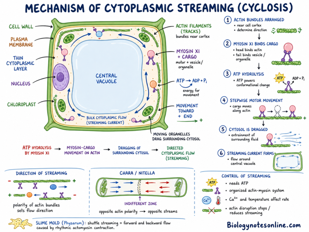
Cytoplasmic streaming, also called cyclosis, is the directed movement of cytoplasm within a cell. It is most clearly seen in large plant and algal cells. During this process, organelles, vesicles and different dissolved materials are moved through the cytoplasmic region.
The movement of cytoplasm in plant cells is mainly based on the interaction between actin filaments and myosin XI motor proteins. Chemical energy of ATP is changed into mechanical movement by the myosin proteins. The mechanism is as follows-
- Arrangement of actin filaments– Long actin filaments are arranged in bundles within the cytoplasm, commonly near the cell cortex. These filaments act as tracks for the movement of myosin and the materials attached with it. Their arrangement also determines the direction of cytoplasmic streaming.
- Attachment of myosin with cargo– Myosin XI is a plant-specific molecular motor involved in long-distance intracellular transport. The motor region of myosin binds with the actin filament. Its tail region binds with an organelle, vesicle or a cargo-associated membrane structure.
- Binding and hydrolysis of ATP– ATP binds with the motor region of myosin. The ATP is hydrolyzed into ADP and inorganic phosphate. Energy released during this process changes the shape of the myosin molecule, which allows it to move along the actin filament.
- Movement along actin filaments– Myosin XI now moves in a stepwise manner over the actin filament. The attached cargo is also carried with it. This movement is generally directed towards the plus end of actin filament.
- Movement of organelles and vesicles– The myosin-cargo complex moves rapidly within the cytoplasm. Endomembrane compartments, vesicles and several organelles show movement during streaming. Some organelles may be moved by direct motor attachment, while many other particles may be carried by the moving cytosol.
- Dragging of surrounding cytosol– The moving organelles and membrane compartments produce friction with the surrounding fluid. The cytosol is pulled or entrained with their movement. Large numbers of moving cargoes produce a continuous bulk flow, which is observed as cytoplasmic streaming.
- Formation of a streaming current– In large vacuolated plant cells, most cytoplasm occurs as a thin layer between the plasma membrane and the central vacuole. The cytoplasm moves through this region and forms a circulating current. Chloroplasts and other cytoplasmic materials may also move with this current.
- Direction of streaming– The direction of cyclosis depends mainly on polarity and arrangement of actin bundles. Myosin moves in a particular direction on each actin filament. Thus, parallel actin bundles produce an ordered flow of cytoplasm.
- Opposite streaming currents– In large cells of characean algae such as Chara and Nitella, actin bundles of opposite polarity are arranged in different regions. Myosin movement along these bundles produces two streams moving in opposite directions. The regions separating them are called indifferent zones.
- Maintenance of the streaming pattern– Actin filaments are not permanent motionless structures in all cells. Their arrangement can be changed and reorganized. Myosin XI also has a role in movement of cargo and remodelling of actin arrays, which affects the streaming pattern.
- Control of streaming– Cytoplasmic streaming requires ATP and a properly organized actin-myosin system. Changes in actin organization, motor activity, cellular calcium level and temperature can change its rate. Complete disruption of actin filaments greatly reduces or stops the streaming movement.
The major mechanism can be summarized as-
ATP hydrolysis by myosin XI → movement of myosin-cargo complex on actin → dragging of surrounding cytosol → directed cytoplasmic flow
In slime molds such as Physarum polycephalum, the mechanism is somewhat different. Rhythmic contraction and relaxation of the actomyosin network produces internal pressure differences. The cytoplasm flows forward and backward through the tubular regions. This repeated reversal is referred to as shuttle streaming.
Functions and Biological Importance of Cytoplasmic Streaming or Cyclosis
Cytoplasmic streaming or cyclosis is the movement of cytoplasm within a cell. It is important mainly in large plant, algal and some animal cells.
- Transport of organelles– It moves mitochondria, plastids, peroxisomes and other organelles within the cell.
- Movement of vesicles– Vesicles are carried from one region to another. It is important during secretion and cell growth.
- Distribution of nutrients– Sugars, ions and other materials are distributed through the cytoplasm.
- Mixing of cytoplasm– Cyclosis mixes cytosol and dissolved materials. It prevents their accumulation in one region.
- Faster internal movement– Diffusion is slow in large cells. Cytoplasmic streaming provides faster movement over longer distance.
- Positioning of organelles– It helps in changing the position of chloroplasts and other organelles according to cellular need.
- Cell growth– Materials required for cell expansion are moved through the cytoplasm. It is important in growing plant cells.
- Pollen tube growth– Vesicles and organelles are moved towards the growing pollen tube region.
- Root hair growth– Cytoplasmic movement supplies materials to the growing tip of root hairs.
- Response to light– Chloroplast movement is associated with changes in light condition. Cyclosis helps in their redistribution.
- Role in oocytes– In some animal oocytes, cytoplasmic flow helps in spindle positioning and unequal cell division.
- Embryonic development– It moves nuclei, granules and other materials during early development in some animals.
- Amoeboid movement– Flow of cytoplasm helps in pseudopodia formation and movement of amoeboid cells.
- Shuttle streaming– In slime molds such as Physarum polycephalum, cytoplasm moves forward and backward through the cell.
Examples of Cytoplasmic Streaming or Cyclosis
Cytoplasmic streaming or cyclosis is clearly observed in some large cells. The following are some common examples-
- Large plant cells– Large vacuolated plant cells show movement of cytoplasm around the central vacuole. Organelles and other particles are also moved with this stream.
- Characean algae– Chara and Nitella are the common examples. Their long internodal cells show rapid rotational streaming. Two cytoplasmic streams move in opposite directions.
- Amoebae– Amoeba proteus shows streaming during amoeboid movement. The inner endoplasm moves towards the growing pseudopodium. Cytoplasm is again rearranged in the posterior region.
- Slime moulds– The plasmodium of Physarum polycephalum shows forward and backward flow of cytoplasm. This type is called shuttle streaming.
- Pollen tubes– Pollen tubes show reverse-fountain streaming. Organelles and vesicles move towards the tip near the cell surface and return through the central region.
- Root hairs– Growing root hairs also show cytoplasmic streaming. Circulatory, reverse-fountain or helical movement may be found depending upon the cell and its growth stage.
Cytoplasm in Different Types of Cells
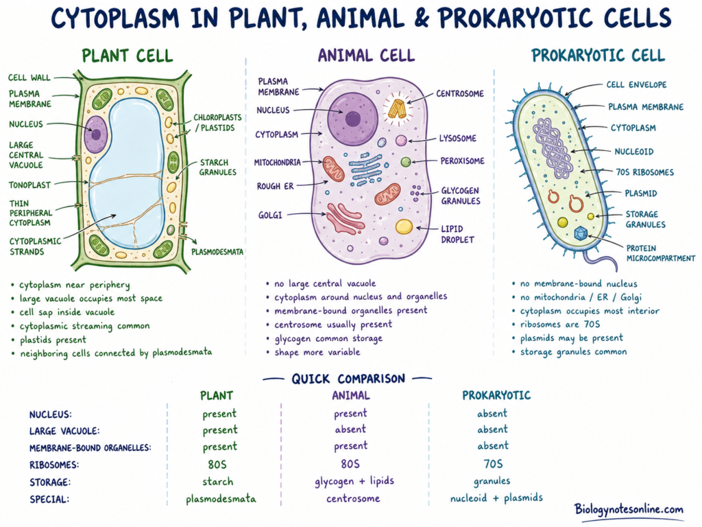
Cytoplasm is present in plant, animal and prokaryotic cells. Its arrangement and cell components are different in them.
In Plant Cells
- The cytoplasm in plant cell is present inside the plasma membrane and outside the nucleus.Mature plant cells generally contain a large central vacuole. It occupies most of the cell volume. Much of the cytoplasm remains as a thin layer near the cell periphery. Cytoplasmic strands may also pass across the vacuole.
- Chloroplasts are present in green plant cells. Other plastids include chromoplasts, leucoplasts and amyloplasts. Their presence depends upon the plant tissue.
- Starch granules are the common carbohydrate inclusions. They are formed within chloroplasts or storage plastids called amyloplasts.
- Plant cytoplasm commonly shows cytoplasmic streaming. The cytoplasmic materials move around the large vacuole.
- Plasmodesmata pass through the cell wall of neighbouring plant cells. They form a cytoplasmic connection between these cells.
- Cytoplasm and cell sap are different. Cell sap is the fluid present within the vacuole. It is enclosed by the tonoplast. Cytoplasm remains outside the vacuole.
In Animal Cells
- Animal cell cytoplasm is present inside the plasma membrane and around the nucleus.
- Animal cells do not contain a single large central vacuole like most mature plant cells. Small vesicles or vacuole-like compartments may be present.
- The cytoplasm contains mitochondria, endoplasmic reticulum, Golgi apparatus, lysosomes, peroxisomes and other organelles.
- The centrosome is generally present near the nucleus in animal cells. It acts as an important microtubule-organizing region.
- Glycogen granules are present in the cytoplasm of many animal cells. They are abundant in liver and muscle cells.
- Lipid droplets and pigment granules may also be found. Melanin and lipofuscin are examples of pigment materials. Their presence is different among cells.
- Animal cells show greater variation in shape. The amount and arrangement of cytoplasm also differs, such as in neurons, muscle cells, blood cells and epithelial cells.
In Prokaryotic Cells
- Bacterial cytoplasm occupies the inner region enclosed by the plasma membrane.
- A membrane-bound nucleus is absent. The main chromosome is present in an irregular DNA-containing region called the nucleoid.
- Small extra-chromosomal DNA molecules called plasmids may be present. They are not found in every bacterial cell.
- The cytoplasm contains a large number of 70S ribosomes. They occur freely, because rough endoplasmic reticulum is absent.
- Storage granules are found in many bacteria. Glycogen-like granules, polyphosphate and polyhydroxyalkanoate granules are some examples.
- Typical membrane-bound organelles such as mitochondria, Golgi apparatus and endoplasmic reticulum are absent.
- The bacterial cytoplasm is not completely unorganized. Nucleoid, ribosomes and proteins may remain concentrated in particular cell regions.
- Some bacteria also form protein-based microcompartments. These enclose particular metabolic enzymes without having a lipid membrane.
Comparison of Plant, Animal and Prokaryotic Cells
| Feature | Plant Cell | Animal Cell | Prokaryotic Cell |
|---|---|---|---|
| Cytoplasm location | Inside plasma membrane and outside nucleus | Inside plasma membrane and around nucleus | Throughout the region inside plasma membrane |
| Nucleus | Membrane-bound nucleus present | Membrane-bound nucleus present | Membrane-bound nucleus absent |
| Central vacuole | Large central vacuole commonly present | Large central vacuole absent | Absent |
| Cytoplasm distribution | Much of cytoplasm remains near cell periphery | Distributed around nucleus and organelles | Occupies most of cell interior |
| Membrane-bound organelles | Present | Present | Typical membrane-bound organelles absent |
| Plastids | Chloroplasts and other plastids present in many cells | Absent | Absent |
| Ribosomes | 80S cytoplasmic ribosomes | 80S cytoplasmic ribosomes | 70S ribosomes |
| Genetic material | Mainly inside nucleus | Mainly inside nucleus | Present in nucleoid region |
| Extra DNA | Present in mitochondria and plastids | Present in mitochondria | Plasmids may be present |
| Storage materials | Starch granules, oils and other materials | Glycogen granules, lipid droplets and pigments | Glycogen, polyphosphate and other granules |
| Cytoplasmic streaming | Common in many large cells | Present in some specialized cells | Not generally described as typical cyclosis |
| Cell shape | Generally fixed due to cell wall | More variable | Usually fixed by cell wall or cell envelope |
| Special feature | Plasmodesmata connect neighbouring cells | Centrosome commonly present | Biochemical regions may form without a nucleus |
Major Functions of the Cytoplasmic Region
The cytoplasm function is associated with metabolism, transport, storage and internal organization of cell. It is not only the material filling the space of cell. The following are some of the major functions of cytoplasm–
- Site for metabolic reactions– The cytosol is the site for most of the intermediary metabolism. Glycolysis also takes place in it. Small molecules are broken down and other molecules are synthesized, which are used as building materials for the cell.
- Protein synthesis and degradation– Protein synthesis begins on ribosomes present in cytosol. Free ribosomes produce the proteins which are released into cytosol, while ribosomes directed to rough endoplasmic reticulum produce proteins entering the ER. Cytosol also contains systems for the breakdown of damaged or unnecessary proteins.
- Holding of cell organelles– Different cytoplasmic organelles are present within the cytoplasm. The organelles do not remain placed randomly. Cytoskeleton and motor proteins are involved in their arrangement and positioning at different regions of cell.
- Intracellular transportation– Vesicles, organelles and other cell materials are moved through the cytoplasm. Microtubules and actin filaments act as tracks, while kinesin, dynein and myosin act as motor proteins. This movement transports materials from one region to another.
- Cytoplasmic streaming– In many large plant and algal cells, the cytoplasm shows a continuous directed movement called cytoplasmic streaming or cyclosis. It helps in movement and mixing of organelles, vesicles and other materials over longer cellular distances.
- Maintenance of cell shape– The cytoskeleton extends throughout the cytoplasm and forms the structural framework of cell. It provides shape and general organization to the cytoplasmic region. It also supports the plasma membrane and different internal structures.
- Cell movement– Cytoplasmic microfilaments, microtubules and their motor proteins take part in cell movement. These structures are used during amoeboid movement, contraction and different changes in cell shape. Microtubules also have a role in the movement of cilia and flagella.
- Role in cell division– The cytoskeletal structures of cytoplasm are rearranged during cell division. Microtubules form the mitotic spindle and separate the chromosomes. In animal cells, actin and myosin form a contractile ring, which divides the cytoplasm during cytokinesis.
- Storage of cellular materials– The cytoplasmic region contains stored carbohydrates, lipids and other materials. Glycogen granules store glucose units in many animal cells. Lipid droplets mainly store neutral lipids, but they are also active structures involved in lipid metabolism.
- Medium for molecular interactions– Cytoplasm provides the internal environment where proteins, RNA and other molecules interact. This region is crowded and active, not a simple dilute fluid. Molecular crowding and metabolic activity affect movement, assembly and reactions of different cell molecules.
The importance of cytoplasm is that most cellular activities are directly or indirectly associated with this region. Metabolism, protein formation, internal movement and division cannot be maintained without its organized condition.
Role of Cytoplasm During Cell Division
The cytoplasm has a role in movement of chromosomes, distribution of organelles and the final separation of a parent cell. During mitosis, the cytoskeleton present in cytoplasm is reorganized. Microtubules form the mitotic spindle. Other cytoplasmic components also change their position.
The division of cytoplasm is referred to as cytokinesis. Mitosis divides the nuclear material, while cytokinesis separates the cytoplasm and plasma membrane. Two daughter cells are formed by the end of this process. The contents of parent cell are also distributed between them.
Cytokinesis in Animal Cells
In animal cells, cytokinesis takes place by formation of a cleavage furrow. It appears at the cell surface, generally around the equatorial region. The furrow now moves inward and the cell becomes constricted in the middle.
An actin-myosin contractile ring is formed below the plasma membrane. Actin filaments and myosin II are the major components. Contraction of this ring pulls the plasma membrane inward, making the cleavage furrow deeper.
The furrow continues to grow until only a narrow cytoplasmic bridge remains between the daughter cells. This bridge is finally separated during abscission. Two individual daughter cells are now formed.
Mitochondria, endoplasmic reticulum, Golgi membranes and other cytoplasmic materials are distributed during the division. Some materials are moved with the help of cytoskeleton. Others are separated according to their number and position in the parent cell. The distribution is not always exactly equal, especially during an unequal cell division.
Cytokinesis in Plant Cells
Plant cells do not generally form a cleavage furrow. The rigid cell wall prevents the plasma membrane from being pinched inward like an animal cell. A new partition is formed within the central region of the dividing cell.
During this process, a cytoskeletal structure called the phragmoplast is formed between the two daughter nuclei. It is mainly made up of microtubules with actin filaments and other associated materials. The phragmoplast guides membrane vesicles towards the division region.
Vesicles mainly produced from the Golgi apparatus and trans-Golgi network are carried towards the centre. These vesicles accumulate and fuse with each other. A small membrane structure is formed, which is known as the cell plate.
The cell plate grows from the centre towards the outer region. New vesicles are continuously added along its growing margins. Finally, it reaches and joins with the parental plasma membrane.
The membranes of the cell plate form the two new plasma membranes. The materials present between these membranes develop into the middle lamella and new cell wall. The parent cell is separated into two daughter cells by this newly formed wall.
Animal-cell cytokinesis occurs by inward cleavage. Plant-cell cytokinesis takes place by outward growth of the cell plate.
Cytoplasm Under the Microscope
The cytoplasm has very little natural contrast. Its appearance is different in living and stained cells.
Appearance in Unstained and Stained Cells
- Living unstained cells– The cytoplasm is usually transparent or lightly coloured. Most of its water and dissolved molecules do not absorb much visible light.
- Granular appearance– Small granules may be seen within it. These are due to organelles, vesicles, stored materials and other particles having different refractive index.
- Ordinary light microscope– Cytoplasmic details are not clearly seen under normal brightfield microscopy. Many small organelles and molecular structures remain below its resolving power.
- Stained cells– Stains increase contrast between cytoplasm, nucleus and other cell parts. The cytoplasm may appear pink, blue or another colour depending upon the stain used.
- Fixation– Cells are generally fixed before permanent staining. Fixation preserves the general cell structure and kills the cell.
- Effect of preparation– Fixation, dehydration and staining may change some natural cellular features. Shrinkage, extraction of molecules or artificial deposits may occur. Thus, a stained image is not exactly similar to living cytoplasm.
Methods Used to Study It
- Phase-contrast microscopy– It converts small phase changes of light into visible contrast. Living and unstained cytoplasm can be viewed by this method. Organelles and cytoplasmic movement are seen more clearly.
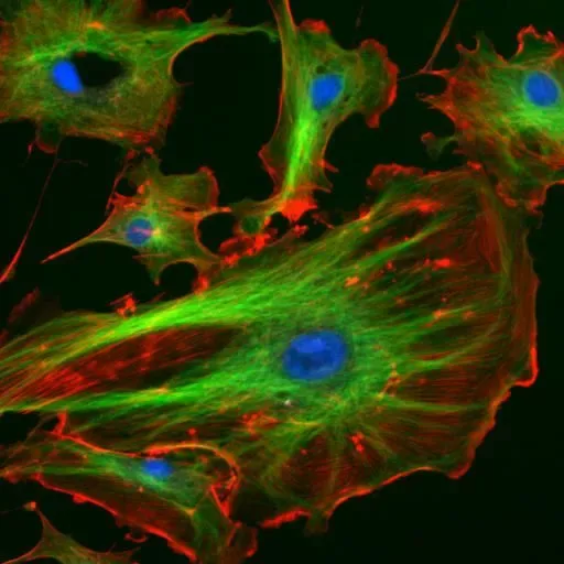
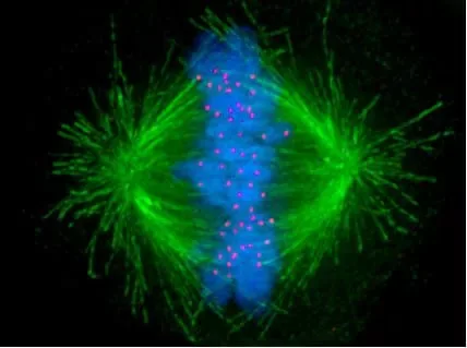
- Fluorescence microscopy– Fluorescent dyes, antibodies or fluorescent proteins are used to mark selected cytoplasmic components. A particular protein, organelle or filament can be detected against a dark background.
- Live-cell imaging– Living cells are recorded over a period of time. It is used to study cytoplasmic streaming, vesicle transport, organelle movement and changes during cell division.
- Cell fractionation– Cells are broken carefully to form a homogenate. Cytosol and different organelles are then separated, generally by differential or density-gradient centrifugation. Their chemical components and activities can now be studied separately.
- Particle tracking– Small particles, natural granules or fluorescent probes are followed through the cytoplasm. Their direction, speed and type of movement are measured.
- Microrheology– Movement of microscopic particles is used to study local viscosity, elasticity and mechanical differences of cytoplasm. It shows that cytoplasm is not physically same throughout the cell.
Cytoplasm vs Cytosol
| Feature | Cytoplasm | Cytosol |
|---|---|---|
| Definition | Complete material present inside the plasma membrane, excluding the nucleus in eukaryotic cells | Fluid portion of the cytoplasm |
| Composition | Cytosol, organelles, cytoskeleton and cytoplasmic inclusions | Water, ions, soluble proteins, enzymes and small molecules |
| Cell organelles | Included | Not included |
| Cytoskeleton | Included as a cytoplasmic component | Not considered as the complete cytoskeleton |
| Physical nature | Semi-fluid, crowded and structurally organized region | Mainly aqueous and soluble cellular medium |
| Location in eukaryotic cells | Between the plasma membrane and nuclear envelope | Present around and between the cytoplasmic structures |
| Major reactions | Includes reactions taking place in cytosol and cytoplasmic organelles | Glycolysis and many intermediary metabolic reactions take place here |
| Main difference | It is the broader cellular region | It is only one part of cytoplasm |
Cytoplasm vs Protoplasm
| Feature | Cytoplasm | Protoplasm |
|---|---|---|
| Definition | Cellular material present inside the plasma membrane, excluding the nucleus in eukaryotic cells | Complete living material present within the plasma membrane |
| Parts included | Cytosol, organelles, cytoskeleton and inclusions | Cytoplasm and nucleoplasm |
| Nucleus | Nucleus is not included | Nuclear material is included |
| Location | Present outside the nuclear envelope | Present throughout the living internal part of cell |
| Use of term | Commonly used in modern cell biology | Mainly an older and broader biological term |
| Nature | It is a defined cellular region | It refers to the total living substance of cell |
| Main difference | It is only one part of the protoplasm | It includes both cytoplasm and nuclear contents |
Cytoplasm vs Nucleoplasm
| Feature | Cytoplasm | Nucleoplasm |
|---|---|---|
| Definition | Cellular material present outside the nucleus and inside the plasma membrane | Material present inside the nuclear envelope |
| Location | Between plasma membrane and nuclear envelope | Within the nucleus |
| Main components | Cytosol, organelles, cytoskeleton and inclusions | Water, ions, proteins, nucleotides and nuclear enzymes |
| Organelles | Cytoplasmic organelles are present | Cytoplasmic organelles are absent |
| Genetic material | Not generally present as nuclear chromosomes | Surrounds chromatin and chromosomes |
| Major activities | Metabolism, protein synthesis and intracellular transport | DNA replication, transcription and RNA processing |
| Boundary | Enclosed by plasma membrane | Enclosed by nuclear envelope |
| Presence | Present in prokaryotic and eukaryotic cells | Present only in cells having a membrane-bound nucleus |
| Main difference | Forms the region outside the nucleus | Forms the internal matrix of nucleus |
Cytoplasm vs Cytoskeleton
| Feature | Cytoplasm | Cytoskeleton |
|---|---|---|
| Definition | Cellular material present inside plasma membrane, excluding nucleus in eukaryotic cells | Network of protein filaments present through the cytoplasm |
| Main components | Cytosol, organelles, cytoskeleton and inclusions | Microfilaments, intermediate filaments and microtubules |
| Nature | Semi-fluid and crowded cellular region | Fibrous and organized protein framework |
| Location | Between plasma membrane and nuclear envelope | Extended throughout the cytoplasm |
| Major role | Site for metabolism, transport and different cellular activities | Maintains cell shape and internal organization |
| Organelle movement | Provides the region where organelles remain and move | Helps in positioning and movement of organelles |
| Cell movement | Cytoplasmic materials take part in movement | Actin filaments and microtubules produce or support movement |
| Cell division | Cytoplasm is divided during cytokinesis | Forms spindle and contractile structures during division |
| Main difference | It is the complete cytoplasmic region | It is only one structural component of cytoplasm |
References
Books and Book Chapters
- Alberts, B., Johnson, A., Lewis, J., Raff, M., Roberts, P., & Walter, P. (2002). Molecular biology of the cell (4th ed.). Garland Science. https://www.ncbi.nlm.nih.gov/books/NBK21054/
- Baron, S. (Ed.). (1996). Medical microbiology (4th ed.). University of Texas Medical Branch at Galveston. https://www.ncbi.nlm.nih.gov/books/NBK7627/
- Chaudhry, R., & Varacallo, M. A. (2023). Biochemistry, glycolysis. In StatPearls. StatPearls Publishing. https://www.ncbi.nlm.nih.gov/books/NBK482303/
- Cooper, G. M. (2000). The cell: A molecular approach (2nd ed.). Sinauer Associates. https://www.ncbi.nlm.nih.gov/books/NBK9839/
- Daghlas, S. A., & Rahimi, N. (2026). Biochemistry, glycogen. In StatPearls. StatPearls Publishing. https://www.ncbi.nlm.nih.gov/books/NBK539802/
- Ellis, R. J. (2001). Protein misassembly: Macromolecular crowding and molecular chaperones. In Madame Curie Bioscience Database. Landes Bioscience. https://www.ncbi.nlm.nih.gov/books/NBK6375/
- Iotti, S., & Malucelli, E. (2011). Free magnesium concentration in the human brain. In R. Vink and M. Nechifor (Eds.), Magnesium in the central nervous system. University of Adelaide Press. https://www.ncbi.nlm.nih.gov/books/NBK507267/
- Khan, Y. S., & Farhana, A. (2025). Histology, cell. In StatPearls. StatPearls Publishing. https://www.ncbi.nlm.nih.gov/books/NBK554382/
- National Research Council. (1989). Opportunities in biology. National Academies Press. https://doi.org/10.17226/742
- O’Neill, M. A., Darvill, A. G., Etzler, M. E., Mohnen, D., Perez, S., Mortimer, J. C., & Pauly, M. (2022). Viridiplantae and algae. In A. Varki, R. D. Cummings, J. D. Esko, P. Stanley, G. W. Hart, M. Aebi, D. Mohnen, T. Kinoshita, N. H. Packer, J. H. Prestegard, R. L. Schnaar, and P. H. Seeberger (Eds.), Essentials of glycobiology (4th ed., Chapter 24). Cold Spring Harbor Laboratory Press. https://doi.org/10.1101/glycobiology.4e.24
- Rose, L. S., & Kemphues, K. J. (1997). Establishment of polarity in the one-cell embryo. In D. L. Riddle, T. Blumenthal, B. J. Meyer, and J. R. Priess (Eds.), C. elegans II (2nd ed.). Cold Spring Harbor Laboratory Press. https://www.ncbi.nlm.nih.gov/books/NBK20089/
- Siegel, G. J., Agranoff, B. W., Albers, R. W., Fisher, S. K., & Uhler, M. D. (Eds.). (1999). Basic neurochemistry: Molecular, cellular and medical aspects (6th ed.). Lippincott-Raven. https://www.ncbi.nlm.nih.gov/books/NBK28100/
- Snyder, A. N., Srikakolapu, S., Gaddy, T., & Crane, J. S. (2023). Histology, lipofuscin. In StatPearls. StatPearls Publishing. https://www.ncbi.nlm.nih.gov/books/NBK537358/
Journal Articles
- Amari, T., Nagata, N., Tominaga, M., & Takatsuka, H. (2025). Cytoskeleton as a generator of characteristic physical properties of plant cells: “Cell wall,” “large vacuole,” and “cytoplasmic streaming.” Biophysics and Physicobiology, 22(3), e220013. https://doi.org/10.2142/biophysico.bppb-v22.0013
- Bonucci, M., Shu, T., & Holt, L. J. (2023). How it feels in a cell. Trends in Cell Biology, 33(11), 924–938. https://doi.org/10.1016/j.tcb.2023.05.002
- Dembo, M. (1989). Mechanics and control of the cytoskeleton in Amoeba proteus. Biophysical Journal, 55(6), 1053–1080. https://doi.org/10.1016/S0006-3495(89)82904-2
- Feig, M., Yu, I., Wang, P.-H., Nawrocki, G., & Sugita, Y. (2017). Crowding in cellular environments at an atomistic level from computer simulations. The Journal of Physical Chemistry B, 121(34), 8009–8025. https://doi.org/10.1021/acs.jpcb.7b03570
- Gao, Q., & Goodman, J. M. (2015). The lipid droplet—A well-connected organelle. Frontiers in Cell and Developmental Biology, 3, Article 49. https://doi.org/10.3389/fcell.2015.00049
- Goldstein, R. E., & van de Meent, J.-W. (2015). A physical perspective on cytoplasmic streaming. Interface Focus, 5(4), Article 20150030. https://doi.org/10.1098/rsfs.2015.0030
- Gu, X., & Verma, D. P. S. (1997). Dynamics of phragmoplastin in living cells during cell plate formation and uncoupling of cell elongation from the plane of cell division. The Plant Cell, 9(2), 157–169. https://doi.org/10.1105/tpc.9.2.157
- Hashemi, H. F., & Goodman, J. M. (2015). The life cycle of lipid droplets. Current Opinion in Cell Biology, 33, 119–124. https://doi.org/10.1016/j.ceb.2015.02.002
- Htet, P. H., & Lauga, E. (2025). Analytical methods for cytoplasmic streaming in elongated cells. PNAS Nexus, 4(3), Article pgaf057. https://doi.org/10.1093/pnasnexus/pgaf057
- Kikuchi, K., & Mochizuki, O. (2015). Diffusive promotion by velocity gradient of cytoplasmic streaming in Nitella internodal cells. PLOS ONE, 10(12), Article e0144938. https://doi.org/10.1371/journal.pone.0144938
- Koenig, A. M., Liu, B., & Hu, J. (2023). Visualizing the dynamics of plant energy organelles. Biochemical Society Transactions, 51(6), 2029–2040. https://doi.org/10.1042/BST20221093
- Lebecq, A., Goldy, C., Fangain, A., Gascon, E., Belcram, K., Pastuglia, M., Bouchez, D., & Caillaud, M.-C. (2023). The phosphoinositide signature guides the final step of plant cytokinesis. Science Advances, 9(29), Article eadf7532. https://doi.org/10.1126/sciadv.adf7532
- Liu, D. (2026). Why is this journal called Protoplasma? A history of protoplasm theory and the divisions in cell biology before 1926. Protoplasma. Advance online publication. https://doi.org/10.1007/s00709-026-02158-1
- Lu, W., & Gelfand, V. I. (2023). Go with the flow—Bulk transport by molecular motors. Journal of Cell Science, 136(5), Article jcs260300. https://doi.org/10.1242/jcs.260300
- Matsumoto, K., Takagi, S., & Nakagaki, T. (2008). Locomotive mechanism of Physarum plasmodia based on spatiotemporal analysis of protoplasmic streaming. Biophysical Journal, 94(7), 2492–2504. https://doi.org/10.1529/biophysj.107.113050
- Najafi, J., Dmitrieff, S., & Minc, N. (2023). Size- and position-dependent cytoplasm viscoelasticity through hydrodynamic interactions with the cell surface. Proceedings of the National Academy of Sciences of the United States of America, 120(9), Article e2216839120. https://doi.org/10.1073/pnas.2216839120
- Obara, J., & Tominaga, M. (2025). Dynamics of full-length Arabidopsis myosin XI and its involvement in actin remodeling. Plant and Cell Physiology, 66(10), 1454–1467. https://doi.org/10.1093/pcp/pcaf097
- Onelli, E., Scali, M., Caccianiga, M., Stroppa, N., Morandini, P., Pavesi, G., & Moscatelli, A. (2018). Microtubules play a role in trafficking prevacuolar compartments to vacuoles in tobacco pollen tubes. Open Biology, 8(10), Article 180078. https://doi.org/10.1098/rsob.180078
- Paterlini, A. (2023). A year at the forefront of plasmodesmal biology. Biology Open, 12(10), Article bio060123. https://doi.org/10.1242/bio.060123
- Peremyslov, V. V., Cole, R. A., Fowler, J. E., & Dolja, V. V. (2015). Myosin-powered membrane compartment drives cytoplasmic streaming, cell expansion and plant development. PLOS ONE, 10(10), Article e0139331. https://doi.org/10.1371/journal.pone.0139331
- Qu, X., Jiang, Y., Chang, M., Liu, X., Zhang, R., & Huang, S. (2015). Organization and regulation of the actin cytoskeleton in the pollen tube. Frontiers in Plant Science, 5, Article 786. https://doi.org/10.3389/fpls.2014.00786
- Rieu, J.-P., Delanoë-Ayari, H., Takagi, S., Tanaka, Y., & Nakagaki, T. (2015). Periodic traction in migrating large amoeba of Physarum polycephalum. Journal of the Royal Society Interface, 12(106), Article 20150099. https://doi.org/10.1098/rsif.2015.0099
- Schiel, J. A., Childs, C., & Prekeris, R. (2013). Endocytic transport and cytokinesis: From regulation of the cytoskeleton to midbody inheritance. Trends in Cell Biology, 23(7), 319–327. https://doi.org/10.1016/j.tcb.2013.02.003
- Stewart, A. M., Stewart, K. L., Yeates, T. O., & Bobik, T. A. (2021). Advances in the world of bacterial microcompartments. Trends in Biochemical Sciences, 46(5), 406–416. https://doi.org/10.1016/j.tibs.2020.12.002
- Stricker, J., Falzone, T., & Gardel, M. L. (2010). Mechanics of the F-actin cytoskeleton. Journal of Biomechanics, 43(1), 9–14. https://doi.org/10.1016/j.jbiomech.2009.09.003
- Surovtsev, I. V., & Jacobs-Wagner, C. (2018). Subcellular organization: A critical feature of bacterial cell replication. Cell, 172(6), 1271–1293. https://doi.org/10.1016/j.cell.2018.01.014
- Takatsuka, H., Higaki, T., & Ito, M. (2023). At the nexus between cytoskeleton and vacuole: How plant cytoskeletons govern the dynamics of large vacuoles. International Journal of Molecular Sciences, 24(4), Article 4143. https://doi.org/10.3390/ijms24044143
- Tan, X., Li, K., Wang, Z., Zhu, K., Tan, X., & Cao, J. (2019). A review of plant vacuoles: Formation, located proteins, and functions. Plants, 8(9), Article 327. https://doi.org/10.3390/plants8090327
- Tominaga, M., Kimura, A., Yokota, E., Haraguchi, T., Shimmen, T., Yamamoto, K., Nakano, A., & Ito, K. (2013). Cytoplasmic streaming velocity as a plant size determinant. Developmental Cell, 27(3), 345–352. https://doi.org/10.1016/j.devcel.2013.10.005
- Weihs, D., Mason, T. G., & Teitell, M. A. (2006). Bio-microrheology: A frontier in microrheology. Biophysical Journal, 91(11), 4296–4305. https://doi.org/10.1529/biophysj.106.081109
- Woodhouse, F. G., & Goldstein, R. E. (2013). Cytoplasmic streaming in plant cells emerges naturally by microfilament self-organization. Proceedings of the National Academy of Sciences of the United States of America, 110(35), 14132–14137. https://doi.org/10.1073/pnas.1302736110
- Xu, X., Liu, Y., Wang, R., Xu, W., Shi, H., Huang, N., Teng, J., Meng, J., Zheng, P., & Chen, J. (2026). Pericentrosomal redistribution of the endoplasmic reticulum ensures organelle symmetric inheritance and mitotic progression. Advanced Science. Advance online publication. https://doi.org/10.1002/advs.76193
- Yi, K., Unruh, J. R., Deng, M., Slaughter, B. D., Rubinstein, B., & Li, R. (2011). Dynamic maintenance of asymmetric meiotic spindle position through Arp2/3-complex-driven cytoplasmic streaming in mouse oocytes. Nature Cell Biology, 13(10), 1252–1258. https://doi.org/10.1038/ncb2320
- Zhang, S., Hu, X., Zhang, B., Liu, J., Feng, H., Liu, C., Hui, Y., Fang, Y., Liu, L., & Zhang, X. (2025). Mapping cellular cytosolic pH in vivo under physiological and pathophysiological conditions. Journal of Biological Chemistry, 301(10), Article 110637. https://doi.org/10.1016/j.jbc.2025.110637