

Metaphase is a stage of the cell division that occurs before anaphase. It is found in mitosis and also in both divisions of meiosis. During this stage, chromosomes become arranged near the equatorial region of spindle, forming a metaphase plate.
The arrangement of chromosomes at the spindle equator is the main feature of metaphase. Spindle fibres coming from the opposite poles become attached with chromosomes through their kinetochores. Due to this, the chromosomes are held in the middle region of the cell.
In mitotic metaphase, each chromosome is formed of two sister chromatids. They remain joined with each other during this stage, mainly at the centromeric region. Their separation does not take place in metaphase. It starts during anaphase.
Metaphase is also present in meiosis I and meiosis II. In metaphase I, homologous chromosome pairs are arranged at the spindle equator. During metaphase II, the chromosomes again arrange at the equator, where sister chromatids are prepared for their separation.
Before starting the anaphase, attachment of every chromosome with spindle fibres is checked. This checking is carried out by the spindle assembly checkpoint (SAC). If any kinetochore is not properly attached, the beginning of anaphase is delayed.
What Is Metaphase?
Metaphase is a stage of nuclear division. It is present in mitosis and also in the two divisions of meiosis. During this stage, chromosomes are positioned around the middle region of the spindle.
In the five-stage model of mitosis, metaphase is the third stage. It occurs after prometaphase and before anaphase. The five stages are prophase, prometaphase, metaphase, anaphase, and telophase.
Mitosis is a continuous process. These stages are named for describing and identifying the major events. The process does not completely stop at one stage and then again starts in another stage. Most of the events pass gradually from one stage into the next.
Metaphase and M phase are not same. Metaphase is only one stage of mitosis. M phase is a larger part of cell cycle which includes nuclear division (mitosis) and division of cytoplasm (cytokinesis).
The term metaphase came into scientific use in 1887.
Position of Metaphase in the Cell Cycle
Interphase occurs before the M phase. In a usual cell cycle, it includes G₁, S, and G₂ phases. The cell grows and makes the required preparation during these phases.
The replication of DNA takes place during the S phase of interphase. It does not take place in metaphase. Therefore, the chromosomes entering into metaphase have already been replicated.
After interphase, mitosis starts with prophase. During prophase and prometaphase, chromosomes become condensed and the mitotic spindle is formed. The nuclear envelope is also broken down and spindle microtubules get access to the chromosome kinetochores. These stages prepare chromosomes and spindle for metaphase.
Metaphase occurs after these early events. The chromosomes remain attached with the spindle and become arranged around the spindle equator. After this arrangement and required checking, the cell enters into anaphase.
In mitosis, anaphase begins when sister chromatids start separating from one another. The same type of separation occurs during anaphase II. However, during anaphase I of meiosis, the homologous chromosomes are separated while sister chromatids remain joined.
Cytokinesis is the process of division of cytoplasm. It is different from the chromosome alignment of metaphase. It usually starts during anaphase and continues through telophase, but it is considered as a separate process from nuclear division.
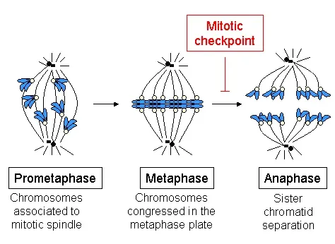

Main Characteristics of a Cell in Metaphase
The following are the main characteristics of a cell during metaphase–
- Highly condensed chromosomes – The chromosomes are highly condensed during this stage. They are visible as separate and distinct chromosome structures inside the dividing cell.
- Presence of sister chromatids – Each mitotic chromosome consists of two sister chromatids. The two chromatids are the replicated copies of a chromosome and remain together in metaphase.
- Chromosomes at equatorial region – The chromosomes occupy the equatorial region of the spindle. This arrangement at the middle region is referred to as the metaphase plate.
- Opposite attachment of sister kinetochores – The sister kinetochores become connected with spindle microtubules coming from the opposite poles. Thus, one sister kinetochore is oriented towards one pole and the other towards opposite spindle pole.
- Bipolar spindle – A complete bipolar mitotic spindle is present in the cell. It contains two spindle poles and microtubules are extended from both the poles towards chromosomes.
- Nuclear envelope is absent – In cells undergoing open mitosis, the nuclear envelope is normally not present during metaphase. It has already disassembled during prometaphase, allowing spindle microtubules to reach the kinetochores. This is different in organisms having closed mitosis.
- Cohesion between sister chromatids – The two sister chromatids remain connected by the cohesin complex. Cohesin holds them together, particularly at the centromeric region and acts against the pulling forces produced by spindle fibres.
- Chromosome separation is not started – Sister chromatids are not separated in metaphase. Their connection is removed at the metaphase to anaphase transition, and separation then begins during anaphase.
- Checking of spindle attachments – The cell maintains proper attachment between kinetochores and spindle microtubules. The spindle assembly checkpoint (SAC) delays anaphase when unattached kinetochores are present, while incorrect attachments are corrected.
What Happens During Metaphase?
During metaphase, the replicated chromosomes are brought near the middle region of the spindle. The spindle attachments are maintained and checked before chromosome separation. The following events take place during this stage-
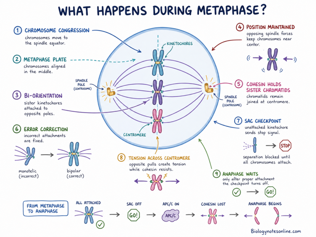
- Chromosomes reach the spindle equator – Chromosomes move towards the equatorial region after their attachment with spindle microtubules. This movement and arrangement of chromosomes is called chromosome congression. The chromosomes occupy an equatorial zone, referred to as the metaphase plate.
- Sister kinetochores become attached with opposite poles – The two sister kinetochores of a mitotic chromosome are connected with microtubules coming from opposite spindle poles. This type of attachment is called bi-orientation or amphitelic attachment. It is required for proper separation of sister chromatids.
- Chromosome position is maintained – The spindle microtubules produce forces from the two opposite directions. These forces maintain the chromosomes around the middle region. Chromosomes are not completely motionless here, and they may show small movements around the metaphase plate.
- Sister chromatids remain joined – Each mitotic chromosome still contains two sister chromatids. They are held together by the cohesin complex, mainly around the centromeric region. Their separation has not started in this stage.
- Incorrect spindle attachments are corrected – Some kinetochores may initially attach with microtubules in an unsuitable manner. Such attachments are made unstable and another attachment can be formed. During this process, correct bipolar attachment of chromosomes is established.
- Spindle attachment is checked – Unattached kinetochores produce a signal through the spindle assembly checkpoint (SAC). This checkpoint prevents early chromosome separation. It remains active until the required kinetochore-microtubule attachments are formed.
- Tension is developed across sister kinetochores – Microtubules attached from opposite poles pull the sister kinetochores in opposite directions. Cohesin acts against this pulling force. Due to this, tension is developed across the centromeric region and proper attachments become more stable.
- Anaphase is delayed until attachment is completed – A chromosome with an unattached kinetochore can maintain the checkpoint signal. Therefore, sister chromatids are not separated while attachment problems are present. The cell remains around metaphase until the inhibitory signal is removed.
- Metaphase ends and anaphase begins – After proper attachment of chromosomes, the checkpoint becomes inactive. The anaphase-promoting complex/cyclosome (APC/C) is then allowed to promote loss of sister chromatid cohesion. Sister chromatids start separating and the cell now enters into anaphase.

Metaphase in Mitosis
Metaphase in mitosis is the stage in which duplicated chromosomes become arranged individually at the equatorial region of the spindle. Homologous chromosomes do not form pairs here. Each chromosome is still composed of two sister chromatids.
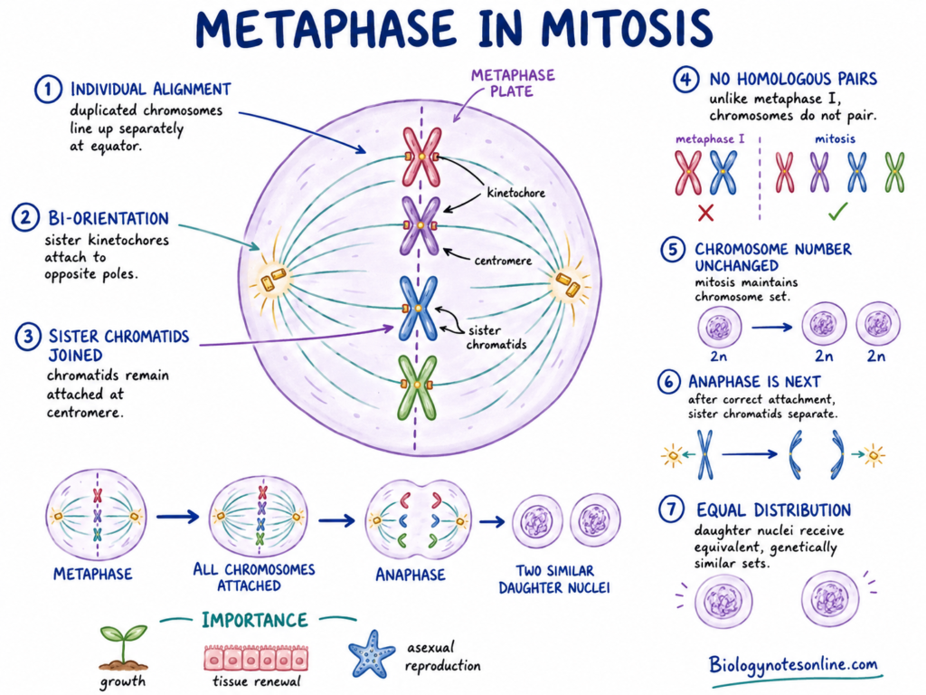
The following events are found during mitotic metaphase-
- Individual chromosome alignment – Each duplicated chromosome becomes arranged separately near the spindle equator. This is different from metaphase I, where homologous chromosomes are present as pairs.
- Orientation of sister chromatids – The kinetochore of one sister chromatid is connected towards one spindle pole. Kinetochore of the other chromatid is oriented towards the opposite pole. This opposite arrangement is called bi-orientation.
- Chromosome number remains unchanged – Mitosis does not reduce the chromosome number. After completion of division, each daughter nucleus normally receives the same number of chromosome sets as the original nucleus.
- Chromatids are not separated – Sister chromatids remain joined during metaphase. Their actual movement towards opposite poles has not started.
- Anaphase is the next stage – After correct spindle attachment, mitosis enters into anaphase. In this stage, sister chromatids separate and move towards two opposite poles.
- Equivalent chromosome sets are formed – The separated chromatids later become enclosed within two daughter nuclei. Normally, both nuclei receive equivalent and genetically similar chromosome sets. Errors in chromosome attachment or separation can change this normal distribution.
Mitotic metaphase is therefore a necessary part of equal chromosome distribution. This distribution is required during growth of the organism, renewal of old or damaged tissues and in asexual reproduction of many eukaryotic organisms.
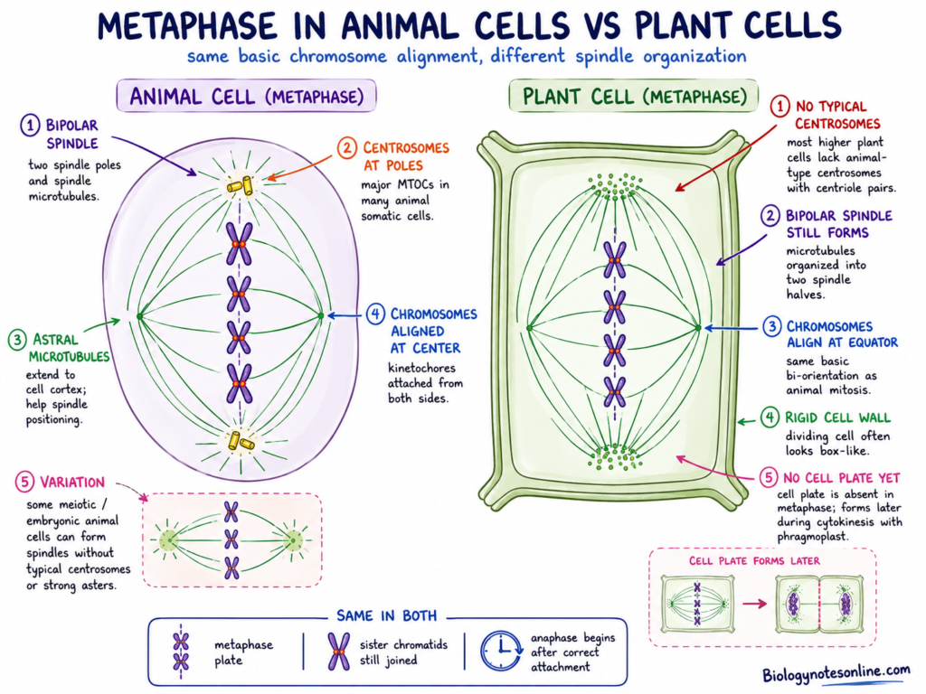
Metaphase in Animal Cells
The following are the main events of metaphase in animal cells-
- Bipolar spindle – A bipolar mitotic spindle is present. It has two opposite spindle poles with different groups of microtubules arranged between them.
- Centrosomes at spindle poles – In many animal somatic cells, duplicated centrosomes act as the major microtubule-organizing centres. They commonly form the two spindle poles.
- Astral microtubules – Astral microtubules may extend outwards from the spindle poles. Their outer ends interact with the cell cortex and help in positioning or orientation of the spindle.
- Central chromosome arrangement – Chromosomes become aligned near the central region of the cell. Their kinetochores are connected with microtubules coming from the two spindle halves.
- Variation among animal cells – Spindle organization is not exactly same in every animal cell. Some animal meiotic cells and certain embryonic cells form spindles without typical centrosomes or prominent asters.
Metaphase in Plant Cells
Metaphase in plant cells follows the same basic process of chromosome bi-orientation and alignment. But organization of the spindle has some differences.
- Absence of typical centrosomes – Most higher plant cells do not contain centrosomes having a pair of centrioles. Therefore, their spindle poles are not formed around typical animal-type centrosomes.
- Formation of bipolar spindle – A bipolar spindle is still formed from organized microtubules. Microtubule nucleation and their arrangement take place at several sites and the microtubules are organized into two spindle halves.
- Chromosome alignment – Duplicated chromosomes become arranged near the spindle equator. Sister kinetochores are connected towards opposite spindle poles, following the same fundamental principle present in animal mitosis.
- Appearance of dividing cell – Plant cells have a rigid cell wall. Due to this wall, many dividing cells may look rectangular or box-like in microscope preparations. The spindle and chromosomes are present within this fixed cell boundary.
- Cell plate is not present in metaphase – Formation of the cell plate does not take place during metaphase. It develops later during plant cytokinesis, with the help of the phragmoplast.
Metaphase I of Meiosis
Metaphase I is a stage of the first meiotic division. During this stage, the paired homologous chromosomes become arranged at the equatorial region of spindle. It occurs after prophase I or prometaphase I and before anaphase I.
The following are the main events taking place during metaphase I-
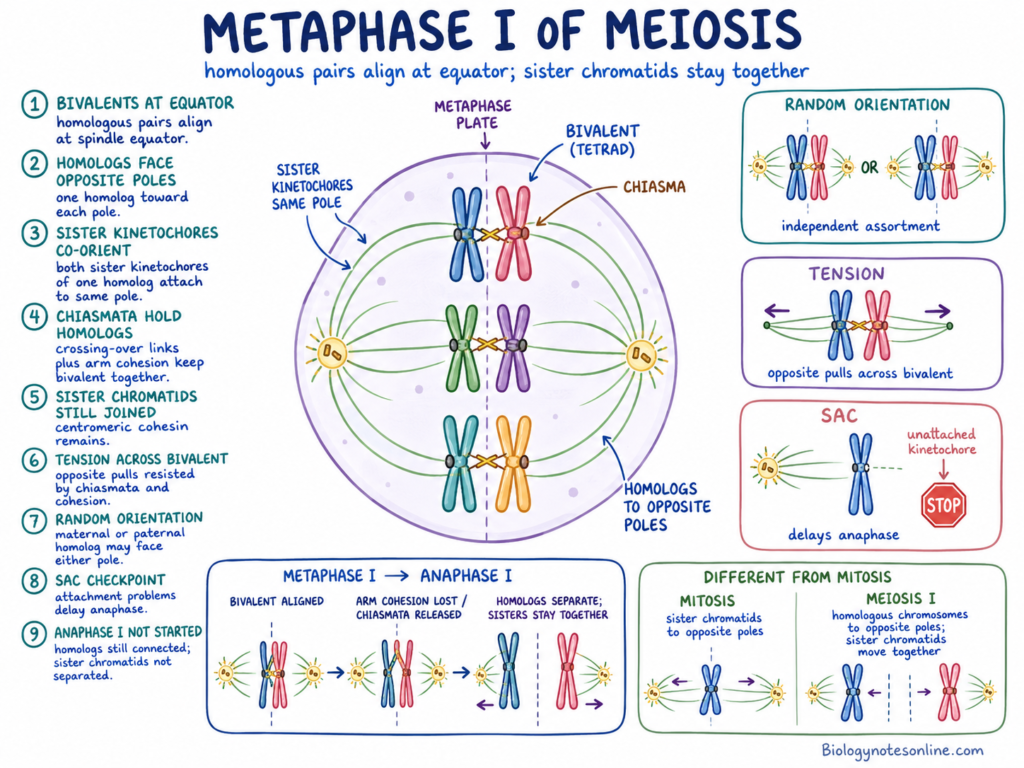
- Arrangement of bivalents – The homologous chromosome pairs are arranged at the spindle equator. Each pair is called a bivalent. Since one bivalent contains four chromatids, it is also referred to as a tetrad.
- Homologous chromosomes face opposite poles – One homologous chromosome becomes oriented towards one spindle pole. The second homolog is connected towards the opposite pole. Thus, the homologous chromosomes show bipolar orientation on the spindle.
- Sister kinetochores face same pole – The two sister kinetochores of one homolog are placed close with each other and normally act in same direction. Microtubules from the same spindle pole become attached with them. This is known as co-orientation or mono-orientation of sister kinetochores.
- Chiasmata hold the homologues – Homologous chromosomes remain joined at one or more chiasmata, which were formed due to crossing over in prophase I. Chiasmata together with cohesion along chromosome arms maintain the bivalent structure.
- Sister chromatids remain connected – Each homolog still consists of two sister chromatids. They are held together by the cohesin complex, particularly around centromeric region. Separation of sister chromatids does not take place in meiosis I.
- Tension is formed across homologues – Spindle microtubules pull the two homologous chromosomes towards opposite poles. The chiasmata and arm cohesion resist this force. Due to this, tension is produced across the bivalent and correct attachment becomes maintained.
- Orientation is random – The maternal or paternal homolog may face either spindle pole. Orientation of one bivalent is generally independent from the orientation of other bivalents. This random arrangement later contributes in different chromosome combinations in meiotic products.
- Spindle attachment is checked – The spindle assembly checkpoint (SAC) monitors attachment between kinetochores and spindle microtubules. When an attachment problem is present, beginning of anaphase can be delayed.
- Chromosome separation is not started – The homologous chromosomes are still connected during metaphase I. At the beginning of anaphase I, cohesion along chromosome arms is removed and chiasmata are released. The homologues then move towards opposite poles, but sister chromatids remain joined at their centromeres.
Metaphase I is different from mitotic metaphase. In mitosis, sister chromatids are oriented towards opposite poles. In metaphase I, homologous chromosomes are oriented towards opposite poles and the sister chromatids of each homolog move as one unit. Therefore, meiosis I is called a reductional division.
Metaphase II of Meiosis
Metaphase II is a stage of the second meiotic division. It takes place in the haploid cells formed after meiosis I. During this stage, duplicated chromosomes become arranged near the equatorial region of spindle.
The following are the main events of metaphase II-
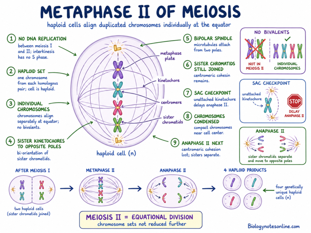
- No DNA replication – DNA is not replicated between meiosis I and meiosis II. In some organisms, a short stage called interkinesis is present, but it does not contain an S phase.
- Haploid chromosome set – Each cell has only one chromosome from every homologous pair. Therefore, the cell is haploid. However, every chromosome is still formed of two sister chromatids.
- Individual chromosome arrangement – Chromosomes become arranged individually at the middle region of spindle. Homologous chromosome pairs or bivalents are not formed here. This arrangement is more similar to mitotic metaphase.
- Opposite orientation of sister kinetochores – Each sister chromatid has its own kinetochore. The two sister kinetochores become attached with spindle microtubules coming from the opposite poles. Thus, sister chromatids are bi-oriented during metaphase II.
- Bipolar spindle is present – A spindle with two opposite poles is formed. Spindle microtubules extend towards the chromosomes and become attached with their kinetochores.
- Sister chromatids remain joined – Sister chromatids do not separate during metaphase II. They remain connected at the centromeric region by the remaining cohesin. This centromeric cohesion was protected during meiosis I.
- Spindle attachments are maintained – Correct attachment of sister kinetochores with opposite spindle poles is required. Improper or incomplete attachment can delay the beginning of anaphase II through the spindle assembly checkpoint (SAC).
- Chromosomes remain highly condensed – The chromosomes are highly condensed and positioned near the centre of the cell. Their separation has not started at this stage.
- Anaphase II follows metaphase II – During anaphase II, centromeric cohesion is removed and sister chromatids are pulled towards opposite spindle poles. The separated chromatids now become individual chromosomes.
Meiosis II is called an equational division. It does not further reduce the number of chromosome sets. At the end, the sister chromatids are distributed into haploid nuclei, usually producing four haploid meiotic products from the original diploid cell.
Mitosis, Metaphase I and Metaphase II Compared
The differences between mitotic metaphase, metaphase I, and metaphase II are given below-
| Feature | Mitotic metaphase | Metaphase I | Metaphase II |
|---|---|---|---|
| Type of cell division | It is a stage of mitosis. The division is equational. | It is present in the first meiotic division. This division is reductional. | It occurs in the second meiotic division. It is an equational division. |
| Number of chromosome sets | Chromosome-set number is same as the parent cell. In a diploid cell, it is 2n. | The cell is still 2n at metaphase I. DNA has already been replicated. | The cell has one chromosome set (n). Each chromosome, however, still has two chromatids. |
| Structure aligned | Duplicated chromosomes become aligned individually. | Homologous chromosome pairs or bivalents are aligned. | Duplicated chromosomes again arrange individually. Homologous pairs are not present. |
| Position of homologous chromosomes | Homologous chromosomes do not remain paired. Each chromosome behaves separately. | The two homologues are present together as a bivalent and face opposite spindle poles. | Only one chromosome from each original homologous pair is present in a cell. Therefore, homologues cannot form a pair. |
| Sister-kinetochore orientation | Sister kinetochores are oriented towards opposite spindle poles. This is bi-orientation. | Sister kinetochores of one homologue generally face the same pole. It is referred to as co-orientation. | Sister kinetochores again become oriented towards opposite poles, similar to mitosis. |
| Crossing-over status | Crossing over is not a normal event of mitotic metaphase. | Crossing over has already occurred during prophase I. The homologues may remain connected at chiasmata. | Crossing over does not newly occur here. Chromosomes may already contain recombinant chromatids formed during prophase I. |
| Source of genetic variation | Metaphase of normal mitosis does not itself produce a major new chromosome combination. Daughter cells are generally maintained similar. | Random orientation of bivalents produces independent assortment. Crossing over completed earlier also gives new allele combinations. | Different recombinant sister chromatids become distributed. No new crossing over takes place during this stage. |
| Structure separated in following anaphase | Sister chromatids separate during anaphase. | Homologous chromosomes separate during anaphase I. Sister chromatids remain joined. | Sister chromatids separate during anaphase II. |
| Effect on chromosome number | Chromosome number is maintained after division. | Chromosome-set number is reduced from diploid to haploid after meiosis I. | The haploid chromosome-set number is maintained. Only sister chromatids are divided. |
| Expected daughter-cell relationship | The two daughter nuclei normally receive equivalent chromosome sets. They are usually genetically very similar to each other and the parent nucleus. | Two haploid nuclei or cells are produced. Their chromosomes are still duplicated and the chromosome combinations may differ. | Four haploid meiotic products are normally obtained from the original cell. They are usually genetically different due to crossing over and chromosome assortment. |
Metaphase Compared with Other Mitotic Stages
Prometaphase vs Metaphase
The differences between prometaphase and metaphase are given below-
| Feature | Prometaphase | Metaphase |
|---|---|---|
| Nuclear envelope | Nuclear envelope breaks down in cells having open mitosis. | Nuclear envelope is normally already disassembled. |
| Kinetochore attachment | Initial capture of kinetochores by spindle microtubules takes place. Some attachments may remain incomplete. | Kinetochores generally have attachment with spindle microtubules from the opposite poles. |
| Chromosome congression | Chromosome movement towards the middle region is going on. | Chromosome congression is mostly completed. |
| Position of chromosomes | Chromosomes may remain scattered at different regions of spindle. | Chromosomes are arranged near the spindle equator. |
| Metaphase plate | A complete metaphase plate is not present. | Chromosomes form the metaphase plate. |
| Spindle attachment | Incorrect or single-pole attachments can be present. | Correct bipolar attachments are maintained and checked. |
| Boundary of stage | It gradually changes into metaphase. | It starts when chromosome alignment is mostly completed. The boundary is not perfectly abrupt. |
Metaphase vs Anaphase
The major differences between metaphase and anaphase are as follows-
| Feature | Metaphase | Anaphase |
| Chromosome condition | Duplicated chromosomes remain aligned at the spindle equator. | Sister chromatids separate and move towards the poles. |
| Sister chromatids | Sister chromatids are still joined. | Sister chromatids are no longer joined and become daughter chromosomes. |
| Cohesin | Centromeric cohesin remains intact. | Cohesin is cleaved at the beginning of anaphase. |
| Centromere condition | Sister centromeric regions have not separated. | Sister centromeres move apart. |
| Chromosome position | One chromosome group is present around the metaphase plate. | Two chromosome groups move in opposite poleward directions. |
| Metaphase plate | Metaphase plate is clearly present. | The metaphase plate disappears as chromosomes move apart. |
| Spindle checkpoint | Spindle assembly checkpoint (SAC) is active or being satisfied. | Checkpoint inhibition is removed and anaphase machinery becomes active. |
| Main event | Chromosome alignment and attachment checking. | Chromosome separation and poleward movement. |
Metaphase Plate vs Cell Plate
Metaphase plate and cell plate are different structures. Their differences are given below-
| Feature | Metaphase Plate | Cell Plate |
| Nature | It is an imaginary equatorial plane. | It is a physical membranous structure. |
| Stage | It is associated with metaphase. | It develops during plant cytokinesis. |
| Occurrence | It is used for describing chromosome position in dividing cells. | It is formed in dividing plant cells. |
| Formation | It is not actually formed as a solid structure. | It is formed by fusion of vesicles at the middle region of cell. |
| Function | It indicates the region where chromosomes become arranged. | It separates the cytoplasm and develops into the new partition between daughter cells. |
| Relation with chromosomes | Chromosomes are aligned around this plane. | It does not take part in metaphase chromosome alignment. |
| Time of appearance | Present before chromosome separation. | Develops later, mainly during cytokinesis. |
| Meaning | It is only a positional term. | It is an actual developing cellular structure. |
Therefore, metaphase plate and cell plate should not be considered as synonyms.
Why Is Metaphase Important?
Metaphase has an important role in proper distribution of chromosomes. During this stage, chromosomes remain arranged and attached with spindle before their separation. The following are some of the importance of metaphase-
- Orderly arrangement of chromosomes – Chromosomes become arranged around the equatorial region of spindle. Due to this arrangement, they are kept in a common region before their movement towards the two opposite poles.
- Formation of correct bi-orientation – Sister kinetochores of a mitotic chromosome are connected with microtubules coming from opposite spindle poles. This opposite attachment is required for proper separation of the sister chromatids.
- Correction of spindle attachment – Initial kinetochore-microtubule attachment can be incorrect. Unsuitable attachments are made unstable and another attachment can be formed. This reduces the chances of wrong chromosome movement during anaphase.
- Prevention of early chromosome separation – The spindle assembly checkpoint (SAC) delays the beginning of anaphase when unattached kinetochores are present. Therefore, sister chromatids are not separated before the required spindle attachment is obtained.
- Accurate distribution of genetic material – Proper alignment and bipolar attachment help the sister chromatids to move into opposite daughter cells. Thus, both daughter nuclei normally receive equivalent chromosome sets.
- Maintenance of genomic stability – Accurate chromosome segregation reduces the formation of cells having missing or additional chromosomes. It therefore supports genomic stability and decreases the chance of aneuploidy.
- Independent assortment during meiosis I – During metaphase I, maternal and paternal homologous chromosome pairs can become oriented in different combinations. This random orientation contributes to independent assortment and formation of genetically different meiotic products.
- Useful for cytogenetic observation – Chromosomes are highly condensed and clearly distinguishable during metaphase. For this reason, metaphase chromosome preparations are used in karyotyping, chromosome counting and detection of many numerical or large structural chromosome abnormalities.
Metaphase does not guarantee completely error-free chromosome separation. Some unsuitable attachments, especially merotelic attachments, may escape the spindle checkpoint and remain until anaphase. Such errors can result in chromosome mis-segregation.
Errors During Metaphase and Their Consequences
Errors during metaphase occur when chromosomes do not obtain the required spindle attachment or proper arrangement. Some of these errors are corrected before anaphase. If remain without correction, unequal chromosome separation can take place.
The following are the major metaphase errors and their consequences-
- Monotelic attachment – In this attachment, one sister kinetochore is connected with a spindle pole while the other kinetochore remains unattached. The unattached kinetochore activates the spindle assembly checkpoint (SAC). Due to this, anaphase is generally delayed until another attachment is formed.
- Syntelic attachment – Both sister kinetochores become attached with microtubules coming from the same spindle pole. Proper tension is not formed across the sister kinetochores. If this error remains, both chromatids may pass towards the same daughter cell.
- Merotelic attachment – A single kinetochore becomes attached with microtubules from both spindle poles. The chromosome may still reach the metaphase plate, therefore this attachment is not always detected by the spindle checkpoint. During anaphase, it can produce a lagging chromosome and chromosome mis-segregation.
- Failure of chromosome congression – One or more chromosomes fail to arrange properly near the spindle equator. They remain outside the normal metaphase plate or move irregularly in spindle. This can maintain metaphase delay, and also increases the chance of lagging chromosome during the next stage.
- Abnormal spindle formation – The spindle may have unsuitable pole separation or more than two temporary poles. Such spindle geometry promotes wrong kinetochore-microtubule attachments. Multipolar division can distribute chromosomes into three or more unequal groups, while transient multipolarity can still produce errors after formation of a bipolar spindle.
- Defect in sister chromatid cohesion – Weak or early loss of cohesin allows sister chromatids to separate before normal anaphase. A long metaphase delay can also produce gradual and irregular chromatid separation, which is called cohesion fatigue. These chromatids can form new wrong attachments and then become mis-segregated.
- Failure of spindle assembly checkpoint – A defective checkpoint may allow the cell to start anaphase while some kinetochores are still unattached. Incorrect attachments are therefore carried into chromosome separation. It can result in chromosome loss or gain in the daughter cells.
- Excess metaphase arrest – Attachment problems can keep the spindle checkpoint active for a longer period. The cell remains arrested around metaphase. Prolonged arrest may end in abnormal mitotic exit, cohesion fatigue or death of the cell, depending on the cell type and condition.
- Aneuploidy – Unequal chromosome separation forms cells having one or more additional or missing chromosomes. This numerical chromosome condition is called aneuploidy. Merotelic attachment, checkpoint failure, cohesion defects and abnormal spindle poles are some important sources of it.
- Formation of micronuclei – A lagging chromosome may fail to enter the main daughter nucleus. It can become enclosed separately and form a small nucleus called a micronucleus. DNA present in micronuclei can undergo defective replication, DNA breaks and chromosome rearrangement.
- Chromosomal instability – Repeated metaphase and segregation errors cause continuous changes in chromosome number or structure. This is referred to as chromosomal instability (CIN). It is commonly found in cancer cells, although a single metaphase error does not itself mean that cancer will develop.
- Errors during meiosis – Incorrect chromosome-spindle attachment in metaphase I or metaphase II can produce aneuploid gametes. In humans, such gametes can contribute to infertility, pregnancy loss and chromosomal disorders in offspring. Not every abnormal gamete, however, develops into a viable pregnancy.
Metaphase errors do not always result in an abnormal daughter cell. Many initial attachments are corrected before anaphase. Some errors, mainly merotelic attachment, may escape this checking and remain until chromosome separation.
Metaphase Arrest
Metaphase arrest is a temporary or prolonged condition in which a dividing cell fails to progress from metaphase or the pre-anaphase state into anaphase. The chromosomes remain without normal separation. It generally occurs when the spindle checkpoint is maintained or the machinery required for anaphase is not activated.
Metaphase arrest can be physiological or experimentally produced. Physiological arrest is a normal part of development in certain cells. Experimentally induced arrest is produced by chemicals or other treatments which disturb the spindle and chromosome attachment.
The following are the main features of metaphase arrest-
- Anaphase does not begin – Sister chromatids remain together and chromosome movement towards opposite poles is not started. The spindle assembly checkpoint (SAC) prevents activation of the normal anaphase process when unattached kinetochores are present.
- Arrest may be temporary – After correction of spindle attachment or removal of the arresting condition, some cells can again form a functional spindle and complete the division. Recovery depends on the cell type and the period of arrest.
- Prolonged arrest has different results – A cell remaining arrested for a long time may undergo cell death. Some cells leave mitosis without normal chromosome separation, which is referred to as mitotic slippage. Other cells may complete an abnormal division. The result is not same in every cell type.
Natural Metaphase Arrest
Natural metaphase arrest is a physiological condition. It is not produced due to a laboratory chemical.
- Arrest in vertebrate oocytes – Mature oocytes of most vertebrates remain arrested at metaphase II before fertilization. The chromosomes are attached with the meiotic spindle, but sister chromatids are not separated.
- Purpose of the arrest – This arrest keeps the oocyte at a stage suitable for fertilization. Completion of meiosis II is therefore coordinated with entry of the sperm. It also prevents the second meiotic division from finishing too early.
- Maintenance of metaphase II – The arrest is maintained by cytostatic factor (CSF) activity. It maintains high M-phase-promoting factor (MPF) activity and inhibits the anaphase-promoting complex/cyclosome (APC/C). Due to this, anaphase II does not start.
- Release after fertilization – Fertilization produces an increase in intracellular calcium within the oocyte. This removes CSF-mediated inhibition and allows APC/C activity. Cyclin B and securin are then degraded, sister chromatids separate and meiosis II is completed.
Experimentally Induced Arrest
Metaphase arrest can also be produced experimentally. It is used for studying mitosis and for obtaining condensed chromosomes.
- Use of spindle-disrupting chemicals – Colchicine, colcemid and nocodazole interfere with microtubule polymerization or spindle function. Kinetochores remain unattached or improperly attached due to the disturbed spindle.
- Checkpoint activation – Unattached kinetochores produce the spindle checkpoint signal. The checkpoint inhibits APC/C-Cdc20 activity, and normal entry into anaphase is prevented. Cells therefore accumulate in a metaphase or pre-anaphase condition.
- Chromosome analysis – Arrested cells contain highly condensed chromosomes. Such cells can be enriched and collected for karyotyping, chromosome counting and other cytogenetic studies. Colchicine or colcemid are commonly used for obtaining metaphase chromosome preparations.
- Condition of arrested chromosomes – Drug-induced arrest does not always show a normal metaphase plate. When microtubules are strongly disrupted, chromosomes may remain scattered because a functional bipolar spindle is absent.
- Effect of concentration and exposure time – The amount of arresting chemical and duration of exposure affect chromosome preparation. Insufficient treatment can produce few metaphase cells. Prolonged arrest may cause excessive chromosome condensation, altered chromosome stiffness or poor chromosome morphology. Therefore, these conditions require proper control according to the cell and sample used.
Experimentally induced metaphase arrest is therefore different from natural metaphase II arrest. One is produced by disturbance of spindle function. The other is a regulated developmental condition of the oocyte.
Metaphase Chromosomes in Karyotyping and Cytogenetics
Metaphase chromosomes are highly condensed and each chromosome can be seen separately under the light microscope. For this reason, metaphase cells are mainly used in conventional karyotyping and chromosome studies. Chromosome number and the large changes in chromosome structure can be examined from them.
The following are the main features of metaphase chromosomes in cytogenetics-
- Chromosomes are individually visible – Chromatin becomes highly condensed during metaphase. The individual chromosomes now have a short and thick appearance, suitable for counting and observation of their morphology.
- Metaphase spread – A metaphase spread is a slide preparation where chromosomes from one metaphase cell are spread apart from each other. A good spread has separate chromosomes with less overlapping. It is used for banding, counting and further chromosome examination.
- Preparation of a karyotype – An image of the chromosomes is taken from a selected metaphase spread. The chromosome pairs are then arranged using their size, banding pattern and position of the centromere. In humans, autosomes are arranged as pairs 1 to 22, followed by the sex chromosomes.
- Chromosome-number abnormalities – Karyotyping can identify an additional or missing chromosome. It can also show changes involving complete chromosome sets. Trisomy, monosomy and polyploidy are some of the numerical abnormalities detected by this method.
- Structural abnormalities – Large deletions, duplications, translocations, inversions, ring chromosomes and some marker chromosomes can be observed. Balanced and unbalanced rearrangements may be identified when the change is sufficiently large and gives a visible alteration in the banding pattern.
- Use in cytogenetics – Metaphase chromosome examination is used in constitutional chromosome disorders, prenatal diagnosis, infertility investigations and cancer cytogenetics. The specimen and type of test, however, depend on the clinical purpose.
- Limitations of conventional karyotyping – It requires dividing cells and a sufficient number of good metaphase spreads. Cell culture may be required and some samples do not produce suitable metaphases. Very small deletions, duplications and sequence-level changes remain below its resolution. Poor banding, overlapping chromosomes and low-level mosaicism can also create limitations in the analysis.
Therefore, a normal karyotype does not show that every part of the genome is normal. Smaller or cryptic changes may need FISH, chromosomal microarray or another molecular test.
Basic Preparation of a Metaphase Spread
The preparation differs according to specimen and purpose. The basic events are as follows-
- Obtaining dividing cells – Dividing cells are obtained directly from a suitable specimen or cells are first grown in culture. Blood lymphocytes, bone marrow cells, amniotic-fluid cells and cultured tissue cells are some commonly examined cells.
- Metaphase arrest – Cells are arrested near metaphase using a spindle-disrupting agent. It increases the number of condensed chromosome preparations available for examination.
- Hypotonic treatment – The arrested cells are treated with a hypotonic solution. Water enters the cells and causes them to swell, which helps the chromosomes to become separated during spreading.
- Fixation – Swollen cells are fixed to preserve chromosome structure. Fixation also helps in removing cellular material which can interfere with chromosome observation.
- Spreading on slide – Fixed cell suspension is dropped or spread on a clean microscope slide. During drying, the cell opens and chromosomes spread over the slide surface. This forms the metaphase spread.
- Chromosome staining – Slides are stained by a suitable chromosome staining or banding method. Giemsa stain is commonly used for conventional chromosome observation and G-banding.
- Image and arrangement – Suitable metaphase spreads are selected and photographed. The chromosome images are identified, paired and arranged into a karyotype. Computer-assisted systems are also used for this arrangement.
The quality of chromosome spreading and banding affects the final analysis. Too much overlapping, broken chromosomes or poorly condensed chromosomes can make identification difficult.
G-Banding, FISH and Other Chromosome Methods
- G-banding – G-banding is a commonly used chromosome-banding method based on staining with Giemsa after suitable pretreatment. It forms alternating dark and light bands along each chromosome. The band pattern is reproducible and is used for identification of chromosomes and their regions.
- Chromosome identification – Every chromosome has its own general size, centromere position and G-band pattern. These features are used together, because chromosome size alone is not sufficient for proper identification.
- Fluorescence in situ hybridization (FISH) – FISH uses fluorescently labelled DNA probes that bind with selected chromosome sequences. The fluorescent signal shows the presence, absence, number or position of the targeted sequence. It can be used for certain deletions, duplications, rearrangements and numerical abnormalities.
- Metaphase and interphase FISH – FISH can be carried out on metaphase chromosomes or on interphase nuclei. Metaphase FISH gives chromosome-location information, while interphase FISH can examine targeted sequences without obtaining a metaphase spread. Only the regions matching the selected probes are studied.
- Comparative genomic hybridization (CGH) – CGH is an adjacent cytogenomic method used for detecting gains and losses of chromosome material across the genome. Array CGH has greater resolution than conventional karyotyping for many small deletions and duplications. It does not normally show balanced rearrangements because there is no net gain or loss of DNA.
- Methods without metaphase chromosomes – Chromosomal microarray uses extracted DNA and does not require actively dividing cells or metaphase chromosomes. Interphase FISH and many DNA sequencing methods also do not need a metaphase spread. These methods give different types of information and do not fully replace conventional chromosome examination in every case.
How to Identify Metaphase Under a Microscope
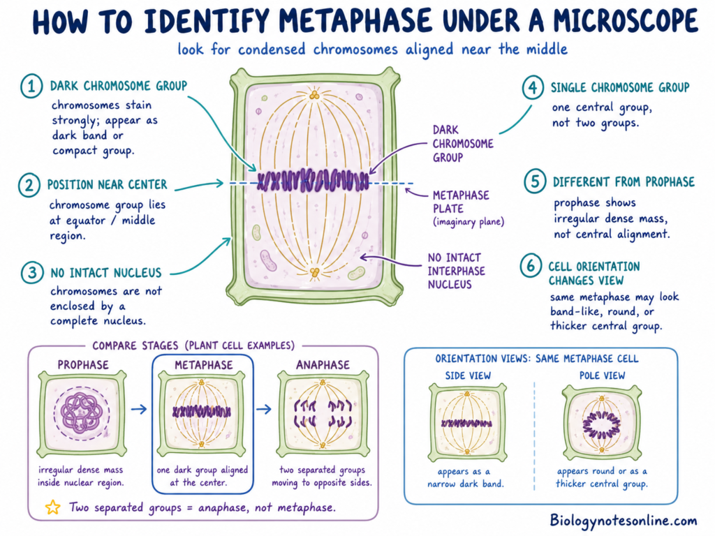
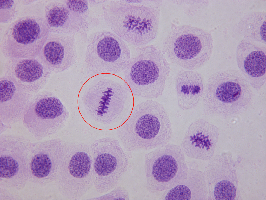
Metaphase under microscope is identified mainly from the position of condensed chromosomes. The chromosome material is present as a dark band or concentrated group near the middle region of cell. In a common school slide, every individual chromosome may not be separately clear.
The following points are used to identify metaphase-
- Dark chromosome group – Chromosomes are highly condensed and take the stain strongly. They are seen as a dark band, short line or compact group. Metaphase chromosomes are condensed enough for observation by light microscope, but chromosome overlapping and slide quality may hide the separate chromosome structures.
- Position near the centre – The dark chromosome group is found around the central or equatorial region of the cell. This region is referred to as the metaphase plate. It is an imaginary plane, not an actual plate-like cell structure.
- No intact interphase nucleus – A round and complete nucleus like interphase is not seen. In cells having open mitosis, the nuclear envelope has already disassembled before metaphase. The chromosomes therefore appear without an enclosing intact nucleus.
- Single chromosome group – Metaphase generally shows one chromosome group near the equatorial region. When two clearly separated chromosome groups are seen moving towards opposite sides, the cell is in anaphase, not metaphase.
- Difference from prophase – During prophase, condensed chromosome material is usually present as a dense and irregular mass inside the nuclear region. The chromosomes are not properly arranged at the middle. This irregular appearance may also be found during early prometaphase.
- Cell orientation changes its appearance – A side view of metaphase can show chromosomes as a narrow dark band. From another direction, the same chromosome arrangement may look round, ring-like or as a thicker central group. The three-dimensional spindle is being viewed as a flat microscope image, therefore all metaphase cells do not show same shape.
Metaphase in an Onion Root Tip
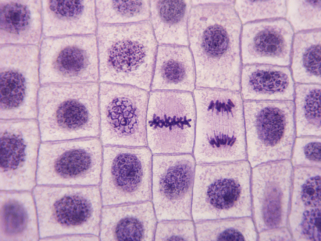
Root tips of onion (Allium cepa) are commonly used for observation of mitosis. The region behind the root cap contains meristematic cells, which are actively dividing. Onion also has comparatively large chromosomes and a high number of dividing cells in its root-tip region.
The following features can be observed-
- Rectangular cells – Onion root-tip cells usually have visible cell walls and appear rectangular or box-like. Different cells in the same field can be present in different stages of cell cycle.
- Stained chromosomes – The root-tip cells are stained and viewed under a light microscope. The stain gives contrast to chromosome material, which appears darker than the surrounding cytoplasm. Different chromosome stains have been used for observing the mitotic stages of Allium cepa.
- Equatorial arrangement – A metaphase cell contains a dark chromosome band or group near its middle part. The chromosomes have reached the equatorial region, but they are not divided into two groups.
- Individual chromosomes – Some good slides show short and dark chromosome structures separately. In other preparations, the chromosomes overlap and form one thick band. Therefore, counting every chromosome is not required only for identifying the stage.
- Comparison with prophase – A prophase cell shows a rounded or irregular mass of condensed chromatin. It does not show the clear middle arrangement found in metaphase.
- Comparison with anaphase – Anaphase shows two dark chromosome groups with a space developing between them. Metaphase has one central group. This is one of the easier differences under the microscope.
Metaphase in Animal Cells
Animal cells have no rigid cell wall. Their external shape and chromosome appearance can therefore be different from onion root-tip cells.
- Rounded mitotic cell – Many animal cells become rounded when entering mitosis. This rounded form is maintained during metaphase in several animal cell types, mainly cells dividing within tissues or culture.
- Chromosomes at central region – The condensed chromosomes become arranged across the middle part of cell. They can appear as a dark central band, a compact row or a rounded chromosome group depending on the viewing direction.
- No intact nucleus – A normal interphase-type nucleus is absent. The chromosomes are separately condensed and present in the spindle region.
- Spindle poles may not be visible – Routine chromosome staining mainly shows the chromosomes. The spindle microtubules and spindle poles may remain unclear or completely unseen. Special fluorescence, antibody staining or other microscope methods are generally used when spindle structure has to be observed.
- Textbook view is not always obtained – A perfect side view showing one straight metaphase line is only one possible appearance. A tilted cell or a view from near the spindle pole can show a broad group or ring-like arrangement. Such a cell can still be in metaphase when chromosomes are present around one central equatorial region.
Chromosome Number, Chromatids and DNA Content During Metaphase
Chromosome number, number of chromatids and amount of DNA are not same. They change differently during mitosis and meiosis.
The following points are important for counting-
- Chromosome number is generally counted from the centromeres. A duplicated chromosome has two sister chromatids, but before separation it is counted as one chromosome. When sister chromatids separate during anaphase, each one is now counted as a chromosome.
- DNA replication takes place during S phase. DNA amount becomes double, but chromosome number does not become double. A chromosome only changes from one chromatid form into a two sister chromatid form.
- The symbol n indicates the number of chromosome sets. C indicates the amount of DNA in one haploid, unreplicated set. Thus, a diploid G1 cell is written as 2n, 2C. After S phase, it remains 2n but contains 4C DNA.
The chromosome condition in a hypothetical cell having 2n = 4 is given below-
| Stage | Chromosome sets | Chromosome number | Chromatids or DNA molecules | DNA content | Main condition |
|---|---|---|---|---|---|
| G1 | 2n | 4 | 4 | 2C | Four unreplicated chromosomes are present. Each chromosome has one chromatid. |
| Mitotic metaphase | 2n | 4 | 8 | 4C | Chromosome number remains same as G1. Each of the four chromosomes now contains two sister chromatids. |
| Mitotic anaphase | 2n at each future pole | 8 in the complete undivided cell, 4 at each pole | 8 separated daughter chromosomes | 4C in complete cell, 2C at each pole | Sister centromeres have separated. For this reason, chromosome number temporarily becomes eight within the single cell. |
| Metaphase I | 2n | 4 | 8 | 4C | Two homologous pairs are present as two bivalents. All four chromosomes remain duplicated. |
| After meiosis I | n | 2 in each cell | 4 in each cell | 2C | Homologous chromosomes have separated. Each chromosome still contains two sister chromatids. |
| Metaphase II | n | 2 in each cell | 4 in each cell | 2C | The cell is haploid, but chromosomes are still duplicated. They become arranged separately at the spindle equator. |
| After meiosis II | n | 2 in each cell | 2 DNA molecules | 1C | Sister chromatids have separated. Each chromosome now consists of one chromatid, producing four haploid meiotic products. |
Mitotic metaphase has replicated DNA, but chromosome number is unchanged from G1. Thus, the hypothetical cell has four chromosomes in both G1 and metaphase. The difference is that G1 has four chromatids and metaphase has eight chromatids.
During metaphase I, the cell is still diploid in homologous chromosome-set terms. It contains 2n chromosomes and 4C DNA. Reduction of chromosome-set number occurs after homologous chromosomes separate in meiosis I.
Metaphase II cells are haploid. But each chromosome is not yet single. It still has two sister chromatids and the DNA content is 2C. Separation during anaphase II finally gives the 1C condition.
The temporary count of eight chromosomes during mitotic anaphase does not mean a stable tetraploid cell has been produced. Four daughter chromosomes move towards each pole, and each daughter nucleus normally receives the original 2n = 4 number.
Applications of Metaphase
Metaphase is used in different fields including Cytogenetics, Medical diagnosis, Cancer studies and chromosome research. Some of its applications include:
- Used for counting of chromosomes and preparation of a karyotype.
- Used to detect numerical chromosome abnormalities such as trisomy, monosomy and polyploidy.
- Used for detection of large structural changes of chromosomes including deletion, duplication, inversion and translocation.
- Used in prenatal diagnosis of different chromosome abnormalities.
- Used to study the chromosome condition of cancer cells, mainly in leukemia and lymphoma.
- Used in Fluorescence in situ hybridization (FISH) for detection and location of a particular DNA sequence.
- Used for mapping of genes and other sequences on chromosomes.
- Used to compare chromosomes of different species and to study their evolutionary changes.
- Used to check the chromosome stability of cell lines and stem cells during culture.
- Used for detection of chromosome changes produced by ionizing radiation.
- Used in genotoxicity studies for observing chromosome breaks, chromosome loss and disturbed metaphase.
- Used to study plant chromosomes from actively dividing root-tip cells such as Allium cepa.
- Used to study the spindle fibres, kinetochore attachment and chromosome alignment.
- Used for isolation and sorting of chromosomes with the help of flow cytometry.
Common Misconceptions About Metaphase
There are different misconceptions related to metaphase. The common misconceptions and their correct facts are given below-
- The metaphase plate is a physical plate – This is not correct. The metaphase plate is an imaginary equatorial plane of the spindle, where chromosomes become arranged. No solid plate or membrane is formed there.
- The cell plate forms during metaphase – The cell plate does not form in metaphase. It develops later during cytokinesis of plant cells. Phragmoplast helps in carrying vesicles to the cell middle, from which the cell plate is made.
- DNA replicates during metaphase – DNA replication is already completed before metaphase. It takes place during the S phase of interphase. The DNA amount becomes double, but chromosome separation is still not started.
- Chromosomes first become visible only in metaphase – Chromosome condensation starts earlier. During prophase, chromatin becomes condensed and individual chromosome structures can begin to be seen under light microscope. They become more compact by metaphase.
- Spindle fibres first appear during metaphase – Formation of mitotic spindle starts before metaphase, mainly during prophase. In cells having open mitosis, kinetochore capture is initiated after nuclear-envelope breakdown in prometaphase. Metaphase contains an already formed bipolar spindle.
- Spindle fibres attach directly to centromeric DNA – Spindle microtubules do not bind directly with naked centromeric DNA. They attach with the kinetochore, a protein complex assembled on centromeric chromatin. The kinetochore forms the chromosome-microtubule attachment site.
- Crossing over occurs during metaphase I – Crossing over normally takes place earlier during prophase I, between nonsister chromatids of paired homologous chromosomes. At metaphase I, the homologues may remain connected through chiasmata formed from the earlier crossover events. New crossing over is not the main event of metaphase I.
- Homologous chromosomes pair during mitotic metaphase – Homologous chromosome pairing is not a normal feature of mitotic metaphase. Duplicated chromosomes arrange individually at the spindle equator. Pairing of homologues into bivalents takes place in meiosis I.
- Sister chromatids separate during metaphase – Sister chromatids remain joined during metaphase. Their kinetochores become connected towards opposite spindle poles, but separation does not occur. They move apart after the beginning of anaphase.
- Haploid metaphase II chromosomes contain only one chromatid – A metaphase II cell is haploid because it has one member of each homologous chromosome pair. But each chromosome is still duplicated and contains two sister chromatids. These chromatids separate during anaphase II.
- All plant cells require centrioles for spindle formation – Most flowering plant cells do not contain the typical centrosomes having centrioles. Still, a bipolar spindle is formed by organized microtubules. Plant mitotic spindle is therefore generally described as an acentrosomal spindle.
- Metaphase is always the longest stage of mitosis – Metaphase is not fixed as the longest stage in every cell. Duration of mitotic stages varies with organism, cell type and cellular condition. Metaphase can also become longer when spindle attachment problems maintain the checkpoint.
- Every organism has an identical metaphase mechanism – The basic requirement of chromosome attachment and segregation is widely conserved. But exact organization is not same in all eukaryotes. Open, partially open and closed mitosis are present, and spindles may be centrosomal or acentrosomal. Some organisms also do not form a prominent metaphase plate like the common textbook figure.
Frequently Asked Questions
What is metaphase?
Metaphase is a stage of nuclear division in which condensed chromosomes become arranged near the equatorial region of spindle. It occurs in mitosis and both meiotic divisions.
What happens during metaphase?
Chromosomes become aligned near the spindle equator. Their kinetochores remain attached with spindle microtubules, and chromosome separation has not started.
What are the main characteristics of metaphase?
Chromosomes are highly condensed. A bipolar spindle is present, chromosomes occupy the middle region and sister chromatids remain joined.
Where do chromosomes line up during metaphase?
Chromosomes line up near the equatorial region between the two spindle poles. This position is referred to as the metaphase plate.
What is the metaphase plate?
The metaphase plate is an imaginary plane around the spindle equator. Chromosomes become arranged near this plane during metaphase.
Is the metaphase plate a real cellular structure?
No. It is not a membrane or any solid plate. It is only a positional region used for describing chromosome arrangement.
Which stage comes before metaphase?
In the five-stage model of mitosis, prometaphase comes before metaphase. Chromosome attachment and movement towards the equator mainly begin in this stage.
Which stage comes after metaphase?
Anaphase comes after metaphase. Sister chromatids begin separating during this stage.
What do spindle fibres attach to?
Spindle microtubules attach with the kinetochores of chromosomes. They do not attach directly with naked centromeric DNA.
What is the function of the kinetochore?
The kinetochore is a protein structure formed on centromeric chromatin. It provides attachment for spindle microtubules, supports chromosome movement and also takes part in spindle-checkpoint signalling.
What is chromosome bi-orientation?
Bi-orientation is the attachment of a duplicated chromosome towards both spindle poles. In mitosis, one sister kinetochore faces one pole and the other faces the opposite pole.
What does the spindle assembly checkpoint do?
The spindle assembly checkpoint (SAC) monitors kinetochore-spindle attachment. It delays anaphase when an unattached kinetochore or certain attachment problems are present.
How does metaphase change into anaphase?
After the checkpoint is satisfied, APC/C becomes active. Securin is degraded, separase becomes active and sister chromatid cohesion is removed. The chromatids now start separating.
What is the difference between prometaphase and metaphase?
During prometaphase, initial kinetochore capture and chromosome congression are going on. In metaphase, chromosomes are mostly arranged around the spindle equator. The change between them is gradual, not perfectly abrupt.
What is the difference between metaphase and anaphase?
Metaphase has joined sister chromatids arranged near one equatorial region. During anaphase, the sister chromatids separate and form two poleward-moving chromosome groups.
How does metaphase I differ from metaphase II?
In metaphase I, homologous chromosome pairs become aligned as bivalents. During metaphase II, chromosomes arrange individually and their sister kinetochores face opposite poles.
How does mitotic metaphase differ from metaphase I?
Mitotic metaphase contains individually arranged duplicated chromosomes. Metaphase I contains paired homologous chromosomes, and the two homologues are prepared for separation.
Does crossing over occur during metaphase I?
No. Crossing over normally takes place during prophase I. The chiasmata produced from earlier crossing over help to keep homologous chromosomes connected at metaphase I.
Is metaphase II haploid or diploid?
A metaphase II cell is haploid. It has one chromosome from each original homologous pair, but every chromosome still contains two sister chromatids.
Are chromosomes duplicated during metaphase?
Yes. DNA replication has already occurred during S phase. Therefore, each metaphase chromosome normally consists of two replicated sister chromatids.
How many chromatids does a chromosome have during metaphase?
A duplicated metaphase chromosome has two sister chromatids. It is still counted as one chromosome because the sister centromeric regions have not separated.
Why are metaphase chromosomes used for karyotyping?
They are highly condensed and can be seen separately. Chromosome number and large structural abnormalities can therefore be examined from metaphase chromosome preparations.
How can metaphase be identified under a microscope?
A metaphase cell commonly shows one dark chromosome band or compact group near its middle region. Two clearly separated chromosome groups indicate anaphase, not metaphase.
What happens if a chromosome fails to align?
An unattached chromosome generally maintains the spindle-checkpoint signal and delays anaphase. If an attachment error escapes correction, chromosome mis-segregation may occur.
Does metaphase occur in plant cells?
Yes. Plant cells also form a spindle and arrange chromosomes near its equatorial region during metaphase.
Do plant cells need centrioles to form a spindle?
No. Most higher plant cells lack the typical centrosomes containing centrioles. Still, they can organize microtubules and form a bipolar spindle.
What is metaphase arrest?
Metaphase arrest is a temporary or prolonged failure of a cell to progress into anaphase. It may be a normal developmental condition or it can result from spindle disruption and sustained checkpoint activity.
Why are mammalian oocytes arrested at metaphase II?
Mature mammalian oocytes remain arrested at metaphase II until fertilization. This keeps completion of meiosis II connected with sperm entry. Fertilization releases the arrest and allows sister chromatids to separate.
References
- Agarwal, S., & Varma, D. (2015). How the SAC gets the axe: Integrating kinetochore microtubule attachments with spindle assembly checkpoint signaling. BioArchitecture, 5(1–2), 1–12. https://doi.org/10.1080/19490992.2015.1090669
- Akera, T., & Watanabe, Y. (2016). The spindle assembly checkpoint promotes chromosome bi-orientation: A novel Mad1 role in chromosome alignment. Cell Cycle, 15(4), 493–497. https://doi.org/10.1080/15384101.2015.1128596
- Alfieri, C., Chang, L., Zhang, Z., Yang, J., Maslen, S., Skehel, M., & Barford, D. (2016). Molecular basis of APC/C regulation by the spindle assembly checkpoint. Nature, 536(7617), 431–436. https://doi.org/10.1038/nature19083
- Bharadwaj, R., & Yu, H. (2004). The spindle checkpoint, aneuploidy, and cancer. Oncogene, 23(11), 2016–2027. https://doi.org/10.1038/sj.onc.1207374
- Cairo, G., & Lacefield, S. (2020). Establishing correct kinetochore–microtubule attachments in mitosis and meiosis. Essays in Biochemistry, 64(2), 277–287. https://doi.org/10.1042/EBC20190072
- Chaurasia, S., & Lehner, C. F. (2018). Dynamics and control of sister kinetochore behavior during the meiotic divisions in Drosophila spermatocytes. PLoS Genetics, 14(5), e1007372. https://doi.org/10.1371/journal.pgen.1007372
- Civelekoglu-Scholey, G., & Cimini, D. (2014). Modelling chromosome dynamics in mitosis: A historical perspective on models of metaphase and anaphase in eukaryotic cells. Interface Focus, 4(3), 20130073. https://doi.org/10.1098/rsfs.2013.0073
- Claussen, U., Michel, S., Mühlig, P., Westermann, M., Grummt, U.-W., Kromeyer-Hauschild, K., & Liehr, T. (2002). Demystifying chromosome preparation and the implications for the concept of chromosome condensation during mitosis. Cytogenetic and Genome Research, 98(2–3), 136–146. https://doi.org/10.1159/000069817
- Clift, D., & Schuh, M. (2013). Restarting life: Fertilization and the transition from meiosis to mitosis. Nature Reviews Molecular Cell Biology, 14(9), 549–562. https://doi.org/10.1038/nrm3643
- Compton, D. A. (2011). Mechanisms of aneuploidy. Current Opinion in Cell Biology, 23(1), 109–113. https://doi.org/10.1016/j.ceb.2010.08.007
- Daum, J. R., Potapova, T. A., Sivakumar, S., Daniel, J. J., Flynn, J. N., Rankin, S., & Gorbsky, G. J. (2011). Cohesion fatigue induces chromatid separation in cells delayed at metaphase. Current Biology, 21(12), 1018–1024. https://doi.org/10.1016/j.cub.2011.05.032
- Dewar, H., Tanaka, K., Nasmyth, K., & Tanaka, T. U. (2004). Tension between two kinetochores suffices for their bi-orientation on the mitotic spindle. Nature, 428(6978), 93–97. https://doi.org/10.1038/nature02328
- Dou, Z., Prifti, D. K., Gui, P., Liu, X., Elowe, S., & Yao, X. (2019). Recent progress on the localization of the spindle assembly checkpoint machinery to kinetochores. Cells, 8(3), 278. https://doi.org/10.3390/cells8030278
- Duro, E., & Marston, A. L. (2015). From equator to pole: Splitting chromosomes in mitosis and meiosis. Genes & Development, 29(2), 109–122. https://doi.org/10.1101/gad.255554.114
- Fischer, E. S. (2023). Kinetochore-catalyzed MCC formation: A structural perspective. IUBMB Life, 75(4), 289–310. https://doi.org/10.1002/iub.2697
- Gregan, J., Polakova, S., Zhang, L., Tolić-Nørrelykke, I. M., & Cimini, D. (2011). Merotelic kinetochore attachment: Causes and effects. Trends in Cell Biology, 21(6), 374–381. https://doi.org/10.1016/j.tcb.2011.01.003
- Hirano, T. (2015). Chromosome dynamics during mitosis. Cold Spring Harbor Perspectives in Biology, 7(6), a015792. https://doi.org/10.1101/cshperspect.a015792
- Hofmann, N. R. (2012). Augmin’s role in microtubule generation in plants. The Plant Cell, 24(4), 1304. https://doi.org/10.1105/tpc.112.240411
- Hotta, T., Kong, Z., Ho, C.-M. K., Zeng, C. J. T., Horio, T., Fong, S., Vuong, T., Lee, Y.-R. J., & Liu, B. (2012). Characterization of the Arabidopsis augmin complex uncovers its critical function in the assembly of the acentrosomal spindle and phragmoplast microtubule arrays. The Plant Cell, 24(4), 1494–1509. https://doi.org/10.1105/tpc.112.096610
- Howe, B., Umrigar, A., & Tsien, F. (2014). Chromosome preparation from cultured cells. Journal of Visualized Experiments, 83, e50203. https://doi.org/10.3791/50203
- Iemura, K., & Tanaka, K. (2015). Chromokinesin Kid and kinetochore kinesin CENP-E differentially support chromosome congression without end-on attachment to microtubules. Nature Communications, 6, 6447. https://doi.org/10.1038/ncomms7447
- Ishiguro, K.-I. (2019). The cohesin complex in mammalian meiosis. Genes to Cells, 24(1), 6–30. https://doi.org/10.1111/gtc.12652
- Kapoor, T. M. (2017). Metaphase spindle assembly. Biology, 6(1), 8. https://doi.org/10.3390/biology6010008
- Kelly, A. E., & Funabiki, H. (2009). Correcting aberrant kinetochore microtubule attachments: An Aurora B-centric view. Current Opinion in Cell Biology, 21(1), 51–58. https://doi.org/10.1016/j.ceb.2009.01.004
- Kleinfeld, R. G., & Sisken, J. E. (1966). Morphological and kinetic aspects of mitotic arrest by and recovery from colcemid. Journal of Cell Biology, 31(3), 369–379. https://doi.org/10.1083/jcb.31.3.369
- Kops, G. J. P. L., Saurin, A. T., & Meraldi, P. (2010). Finding the middle ground: How kinetochores power chromosome congression. Cellular and Molecular Life Sciences, 67(13), 2145–2161. https://doi.org/10.1007/s00018-010-0321-y
- Kramer, E. M., Tayjasanant, P. A., & Cordone, B. (2021). Scaling laws for mitotic chromosomes. Frontiers in Cell and Developmental Biology, 9, 684278. https://doi.org/10.3389/fcell.2021.684278
- Krupina, K., Goginashvili, A., & Cleveland, D. W. (2021). Causes and consequences of micronuclei. Current Opinion in Cell Biology, 70, 91–99. https://doi.org/10.1016/j.ceb.2021.01.004
- Li, M., & Pinkel, D. (2006). Clinical cytogenetics and molecular cytogenetics. Journal of Zhejiang University Science B, 7(2), 162–163. https://doi.org/10.1631/jzus.2006.B0162
- Li, S., Sun, T., & Ren, H. (2015). The functions of the cytoskeleton and associated proteins during mitosis and cytokinesis in plant cells. Frontiers in Plant Science, 6, 282. https://doi.org/10.3389/fpls.2015.00282
- Lok, T. M., Wang, Y., Xu, W. K., Xie, S., Ma, H. T., & Poon, R. Y. C. (2020). Mitotic slippage is determined by p31^comet^ and the weakening of the spindle-assembly checkpoint. Oncogene, 39(13), 2819–2834. https://doi.org/10.1038/s41388-020-1187-6
- Madgwick, S., & Jones, K. T. (2007). How eggs arrest at metaphase II: MPF stabilisation plus APC/C inhibition equals cytostatic factor. Cell Division, 2, 4. https://doi.org/10.1186/1747-1028-2-4
- Maiato, H., Gomes, A. M., Sousa, F., & Barisic, M. (2017). Mechanisms of chromosome congression during mitosis. Biology, 6(1), 13. https://doi.org/10.3390/biology6010013
- Matson, D. R., & Stukenberg, P. T. (2011). Spindle poisons and cell fate: A tale of two pathways. Molecular Interventions, 11(2), 141–150. https://doi.org/10.1124/mi.11.2.12
- McAinsh, A. D., & Kops, G. J. P. L. (2023). Principles and dynamics of spindle assembly checkpoint signalling. Nature Reviews Molecular Cell Biology, 24(8), 543–559. https://doi.org/10.1038/s41580-023-00593-z
- McIntosh, J. R., Molodtsov, M. I., & Ataullakhanov, F. I. (2012). Biophysics of mitosis. Quarterly Reviews of Biophysics, 45(2), 147–207. https://doi.org/10.1017/S0033583512000017
- McNally, F. J. (2013). Mechanisms of spindle positioning. Journal of Cell Biology, 200(2), 131–140. https://doi.org/10.1083/jcb.201210007
- Merdes, A., & Cleveland, D. W. (1997). Pathways of spindle pole formation: Different mechanisms; conserved components. Journal of Cell Biology, 138(5), 953–956. https://doi.org/10.1083/jcb.138.5.953
- Meyer, R. E., & Dawson, D. S. (2013). Attaching to spindles before they form: Do early incorrect chromosome–microtubule attachments promote meiotic segregation fidelity? Cell Cycle, 12(13), 2011–2015. https://doi.org/10.4161/cc.25252
- Miller, M. P., Amon, A., & Ünal, E. (2013). Meiosis I: When chromosomes undergo extreme makeover. Current Opinion in Cell Biology, 25(6), 687–696. https://doi.org/10.1016/j.ceb.2013.07.009
- Mukhopadhyay, R., Varshitha, D. V., Telford, W. G., Sanders, C. K., & Chakraborty, U. (2023). Mammalian chromosome analysis and sorting by flow cytometry. Current Protocols, 3(5), e785. https://doi.org/10.1002/cpz1.785
- Nagaki, K., Yamamoto, M., Yamaji, N., Mukai, Y., & Murata, M. (2012). Chromosome dynamics visualized with an anti-centromeric histone H3 antibody in Allium. PLoS ONE, 7(12), e51315. https://doi.org/10.1371/journal.pone.0051315
- Nasmyth, K. (2015). A meiotic mystery: How sister kinetochores avoid being pulled in opposite directions during the first division. BioEssays, 37(6), 657–665. https://doi.org/10.1002/bies.201500006
- Nishimura, K., Johmura, Y., Deguchi, K., Jiang, Z., Uchida, K. S. K., Suzuki, N., Shimada, M., Chiba, Y., Hirota, T., Yoshimura, S. H., Kono, K., & Nakanishi, M. (2019). Cdk1-mediated DIAPH1 phosphorylation maintains metaphase cortical tension and inactivates the spindle assembly checkpoint at anaphase. Nature Communications, 10, 981. https://doi.org/10.1038/s41467-019-08957-w
- Orr, B., Godek, K. M., & Compton, D. A. (2015). Aneuploidy. Current Biology, 25(13), R538–R542. https://doi.org/10.1016/j.cub.2015.05.010
- Paliulis, L. V., & Nicklas, R. B. (2000). The reduction of chromosome number in meiosis is determined by properties built into the chromosomes. Journal of Cell Biology, 150(6), 1223–1232. https://doi.org/10.1083/jcb.150.6.1223
- Potapova, T., & Gorbsky, G. J. (2017). The consequences of chromosome segregation errors in mitosis and meiosis. Biology, 6(1), 12. https://doi.org/10.3390/biology6010012
- Silva, P., Barbosa, J., Nascimento, A. V., Faria, J., Reis, R., & Bousbaa, H. (2011). Monitoring the fidelity of mitotic chromosome segregation by the spindle assembly checkpoint. Cell Proliferation, 44(5), 391–400. https://doi.org/10.1111/j.1365-2184.2011.00767.x
- Sinclair, A. (2002). Genetics 101: Cytogenetics and FISH. CMAJ, 167(4), 373–374.
- Sparks, T. N., & Dugoff, L. (2023). How to choose a test for prenatal genetic diagnosis: A practical overview. American Journal of Obstetrics and Gynecology, 228(2), 178–186. https://doi.org/10.1016/j.ajog.2022.08.039
- Tanaka, T. U., & Desai, A. (2008). Kinetochore–microtubule interactions: The means to the end. Current Opinion in Cell Biology, 20(1), 53–63. https://doi.org/10.1016/j.ceb.2007.11.005
- Wadsworth, P., Lee, W.-L., Murata, T., & Baskin, T. I. (2011). Variations on theme: Spindle assembly in diverse cells. Protoplasma, 248(3), 439–446. https://doi.org/10.1007/s00709-010-0205-x
- Walczak, C. E., Cai, S., & Khodjakov, A. (2010). Mechanisms of chromosome behaviour during mitosis. Nature Reviews Molecular Cell Biology, 11(2), 91–102. https://doi.org/10.1038/nrm2832
- Wan, T. S. K. (2014). Cancer cytogenetics: Methodology revisited. Annals of Laboratory Medicine, 34(6), 413–425. https://doi.org/10.3343/alm.2014.34.6.413
- Weber, T. S., Jaehnert, I., Schichor, C., Or-Guil, M., & Carneiro, J. (2014). Quantifying the length and variance of the eukaryotic cell cycle phases by a stochastic model and dual nucleoside pulse labelling. PLoS Computational Biology, 10(7), e1003616. https://doi.org/10.1371/journal.pcbi.1003616
- Weise, A., Bhatt, S., Piaszinski, K., Kosyakova, N., Fan, X., Altendorf-Hofmann, A., Tanomtong, A., Chaveerach, A., de Cioffi, M. B., de Oliveira, E., Walther, J.-U., Liehr, T., & Chaudhuri, J. P. (2016). Chromosomes in a genome-wise order: Evidence for metaphase architecture. Molecular Cytogenetics, 9, 36. https://doi.org/10.1186/s13039-016-0243-y
- Wijeyaratne, W. M. D. N., & Wadasinghe, L. G. Y. J. G. (2019). Allium cepa bio assay to assess the water and sediment cytogenotoxicity in a tropical stream subjected to multiple point and nonpoint source pollutants. Journal of Toxicology, 2019, 5420124. https://doi.org/10.1155/2019/5420124
- Yanowitz, J. (2010). Meiosis: Making a break for it. Current Opinion in Cell Biology, 22(6), 744–751. https://doi.org/10.1016/j.ceb.2010.08.016
- Zickler, D., & Kleckner, N. (2015). Recombination, pairing, and synapsis of homologs during meiosis. Cold Spring Harbor Perspectives in Biology, 7(6), a016626. https://doi.org/10.1101/cshperspect.a016626
- Books and Book Chapters
- Alberts, B., Johnson, A., Lewis, J., Raff, M., Roberts, K., & Walter, P. (2002). Molecular biology of the cell (4th ed.). Garland Science.
- Brown, T. A. (2002). Genomes (2nd ed.). Wiley-Liss.
- Clark, M. A., Douglas, M., & Choi, J. (2018). Biology 2e. OpenStax.
- Cooper, G. M. (2000). The cell: A molecular approach (2nd ed.). Sinauer Associates.
- Dorée, M., Le Peuch, C., & Morin, N. (1995). Onset of chromosome segregation at the metaphase to anaphase transition of the cell cycle. In L. Meijer, S. Guidet, & L. Vogel (Eds.), Progress in cell cycle research (Vol. 1, pp. 309–318). Springer. https://doi.org/10.1007/978-1-4615-1809-9_25
- Fowler, S., Roush, R., & Wise, J. (2013). Concepts of biology. OpenStax.
- Genetic Alliance, & District of Columbia Department of Health. (2010). Understanding genetics: A District of Columbia guide for patients and health professionals. Genetic Alliance.
- Gottlieb, S. F., Gulani, A., & Tegay, D. H. (2023). Genetics, meiosis. In StatPearls. StatPearls Publishing.
- Griffiths, A. J. F., Miller, J. H., Suzuki, D. T., Lewontin, R. C., & Gelbart, W. M. (2000). An introduction to genetic analysis (7th ed.). W. H. Freeman.
- Institute for Quality and Efficiency in Health Care. (2023). How do most cells divide (mitosis)? In InformedHealth.org. Institute for Quality and Efficiency in Health Care.
- Lodish, H., Berk, A., Zipursky, S. L., Matsudaira, P., Baltimore, D., & Darnell, J. (2000). Molecular cell biology (4th ed.). W. H. Freeman.
- Rehman, I., Farooq, M., & Simpson, B. (2023). Genetics, mitosis. In StatPearls. StatPearls Publishing.
- Simpson, B., Tupper, C., & Al Aboud, N. M. (2023). Genetics, DNA packaging. In StatPearls. StatPearls Publishing.