

Prometaphase is a stage of mitosis that occurs between prophase and metaphase. During this stage, the nuclear envelope breaks down. The microtubules of the mitotic spindle then attach to the chromosomes at their kinetochores.
After the attachment, chromosomes begin to move towards the centre of the cell. They gradually get arranged at the metaphase plate. This arrangement is completed as the cell enters metaphase.
What Is Prometaphase?
Prometaphase is the second stage of mitosis which occurs between prophase and metaphase. It begins with the breakdown of the nuclear envelope.
During this stage, the microtubules of the mitotic spindle attach to the kinetochores of condensed chromosomes. The chromosomes then start moving towards the centre of the cell and gradually arrange themselves at the metaphase plate. Complete alignment of chromosomes occurs during metaphase.
In prophase, chromosome condensation and spindle formation begin. Prometaphase involves the breakdown of the nuclear envelope and attachment of spindle microtubules to the chromosomes. Some textbooks consider prometaphase as a part of late prophase rather than a separate stage, because these events occur as a continuous process.
When prometaphase is considered separately, mitosis consists of five stages which are as follows-
Prophase → Prometaphase → Metaphase → Anaphase → Telophase
Major Events of Prometaphase
Prometaphase involves a series of cellular events, starting with the breakdown of the nuclear envelope, followed by spindle attachment and chromosome movement towards the metaphase plate.
The major events of prometaphase are as follows-
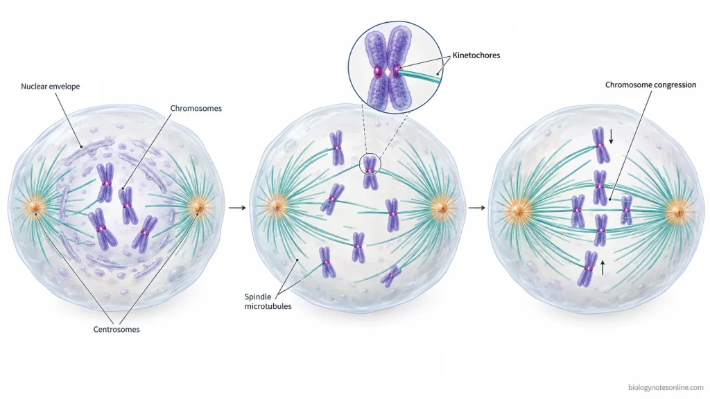
1. Breakdown of the Nuclear Envelope
The first event of prometaphase involves the disassembly of the nuclear lamina and breakdown of the nuclear envelope surrounding the chromosomes.
After the nuclear envelope breaks apart, the condensed chromosomes become accessible to the spindle microtubules.
2. Development of the Mitotic Spindle
During this stage, the mitotic spindle continues to develop. In animal cells, the centrosomes move towards the opposite poles.
The spindle microtubules extend into the region previously occupied by the nucleus, where they take part in the attachment and movement of chromosomes.
3. Attachment of Spindle Microtubules to Chromosomes
The attachment of spindle microtubules occurs at specialized protein structures called kinetochores. Each sister chromatid has a kinetochore present in its centromeric region.
Growing spindle microtubules interact with these kinetochores and form connections between the chromosomes and mitotic spindle.
4. Movement of Chromosomes
After attachment to the spindle microtubules, the chromosomes start moving within the cell. Their movement is regulated by the microtubules and associated motor proteins.
The chromosomes may move either towards or away from the spindle poles before reaching the central region.
5. Bipolar Attachment of Chromosomes
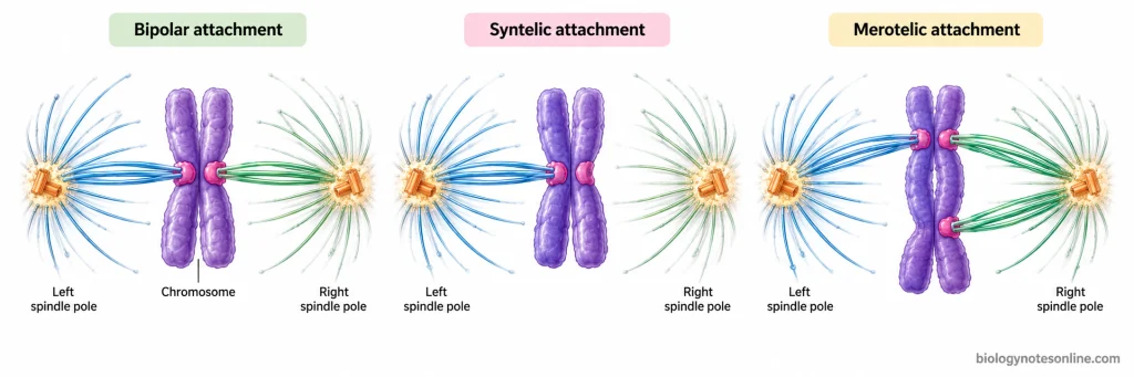
In this step, spindle microtubules coming from opposite poles attach to the kinetochores of sister chromatids. This type of attachment is referred to as bipolar attachment or bi-orientation.
Sometimes, incorrect attachments are formed. These are corrected through mechanisms which regulate the stability of kinetochore-microtubule interactions.
6. Alignment of Chromosomes at the Metaphase Plate
The movement of attached chromosomes towards the equatorial region of the mitotic spindle is known as chromosome congression.
Different spindle forces and motor proteins take part in bringing the chromosomes towards the centre of the cell. When all chromosomes are aligned at the metaphase plate, the cell enters metaphase.
Spindle Microtubules During Prometaphase
In animal cells, the mitotic spindle consists of three major types of microtubules. These are highly dynamic structures which undergo continuous growth and shrinkage. During prometaphase, the spindle microtubules attach to chromosomes and help in their movement towards the metaphase plate.
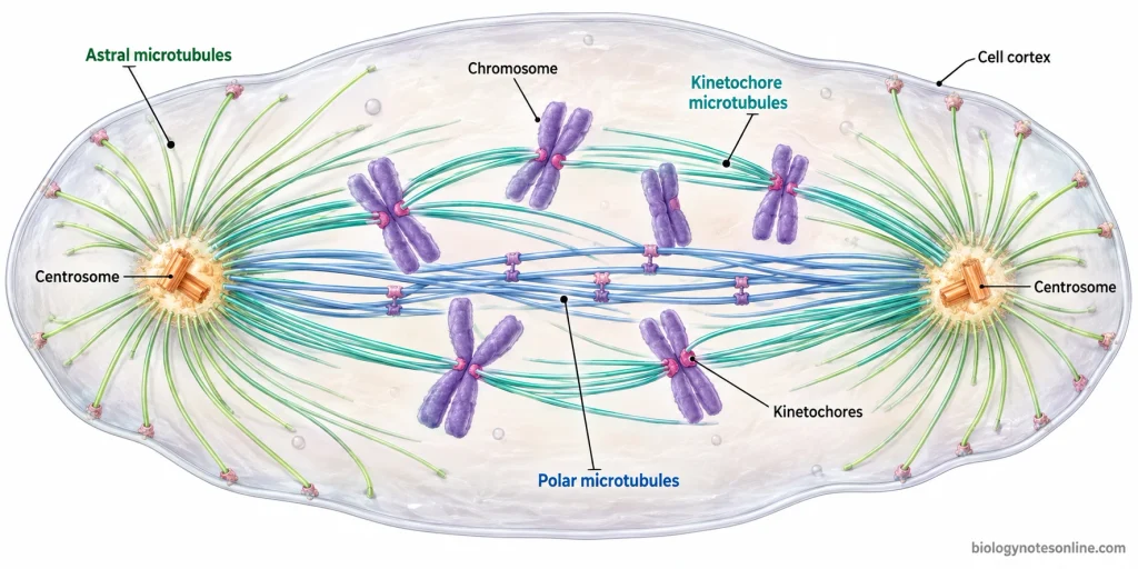
The three types of spindle microtubules and their functions are as follows-
- Kinetochore Microtubules- These microtubules attach to the chromosomes at specialized protein structures called kinetochores, which are present in the centromeric region of each sister chromatid. The growing microtubules interact with these structures during prometaphase and establish their attachment to the chromosomes. Gradually, the sister kinetochores become attached to microtubules coming from opposite spindle poles. They are responsible for chromosome movement and their arrangement at the metaphase plate.
- Polar Microtubules- These are also known as interpolar microtubules. They extend from the opposite spindle poles towards the central region of the cell, where the microtubules from opposite poles overlap with each other. No direct attachment with the kinetochores of chromosomes occurs. Motor proteins interact with the overlapping microtubules, producing forces which help in maintaining the bipolar structure of the mitotic spindle. The separation of spindle poles and mechanical stability of the developing spindle are also supported by these interactions.
- Astral Microtubules- In animal cells, astral microtubules radiate outwards from the centrosomes towards the cell periphery. They do not attach directly to chromosomes. Some of these microtubules interact with motor proteins located at the cell cortex. During this process, pulling forces are generated on the spindle poles, which help in the positioning and orientation of the mitotic spindle within the cell. These microtubules are not found in all types of mitotic spindles. They are absent in higher plants.
Prometaphase in Meiosis
Meiosis involves two prometaphase stages, prometaphase I and prometaphase II, occurring in the first and second meiotic divisions respectively.
In both stages, spindle microtubules attach to the kinetochores and participate in chromosome movement. The arrangement of chromosomes and their mode of spindle attachment, however, are different.
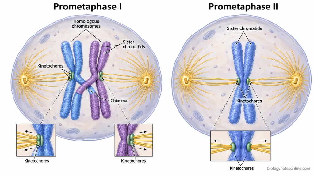
1. Prometaphase I
It is the stage between prophase I and metaphase I. By this stage, the nuclear envelope has broken down. The spindle microtubules can now reach the chromosomes and attach to their kinetochores.
Homologous chromosomes are still paired, held together at the chiasmata. Each pair is made up of four chromatids. This structure is referred to as a bivalent or tetrad.
The sister kinetochores of each homologous chromosome are oriented towards the same spindle pole, while the two homologous chromosomes establish kinetochore attachments with microtubules coming from opposite poles.
During this process, the homologous pairs move towards the equatorial region of the cell. Alignment of these chromosomes at the metaphase plate occurs in metaphase I.
2. Prometaphase II
The second prometaphase occurs in the haploid cells produced after the first meiotic division. It takes place between prophase II and metaphase II.
Each chromosome still has two sister chromatids. They remain attached at their centromeric regions.
In cells where the nuclear envelope is reformed after meiosis I, it breaks down again before the spindle microtubules establish chromosome attachments.
Unlike the first meiotic division, the sister kinetochores in prometaphase II establish attachments with microtubules coming from opposite spindle poles.
The chromosomes now move towards the centre of the cell. Their alignment at the metaphase plate is completed during metaphase II.
The attachments formed in prometaphase II are required for the separation of sister chromatids during anaphase II.
Prometaphase vs Prophase
Prophase and prometaphase are two consecutive stages of mitosis. During prophase, the chromosomes begin to condense, and the mitotic spindle starts to form. Prometaphase begins with the breakdown of the nuclear envelope, allowing the spindle microtubules to attach to chromosomes at their kinetochores. The chromosomes then begin to move towards the metaphase plate.
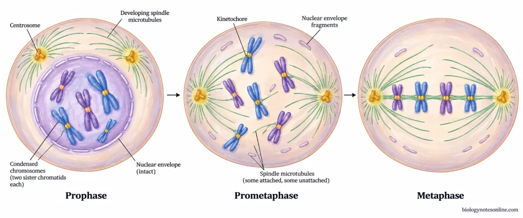
The major differences between prophase and prometaphase are given below.
| Parameter | Prophase | Prometaphase |
|---|---|---|
| Position during mitosis | It is the first stage of mitosis. | It is the second stage, which occurs between prophase and metaphase. |
| Chromosome condensation | Chromatin begins to condense and forms visible chromosomes, each consisting of two sister chromatids. | Chromosomes become more condensed and appear shorter and thicker. |
| Nuclear envelope | The nuclear envelope remains largely intact during early prophase. Its breakdown may begin towards the end of this stage. | The nuclear envelope breaks down, allowing spindle microtubules to access the chromosomes. |
| Mitotic spindle | The mitotic spindle begins to develop, and the centrosomes start moving apart in animal cells. | Spindle development continues. Microtubules extend into the former nuclear region and establish connections with chromosomes. |
| Kinetochore attachment | Kinetochores assemble, but spindle microtubules have not yet established attachments to the chromosomes. | Spindle microtubules attach to kinetochores, and sister chromatids progressively establish connections with opposite spindle poles. |
| Chromosome movement | Chromosomes undergo condensation and changes in their position but have not started spindle-mediated congression. | The attached chromosomes undergo movement and gradually move towards the metaphase plate. |
Note: Some textbooks consider prometaphase as a separate stage of mitosis, while others include it within late prophase. This variation occurs because chromosome condensation, spindle development and nuclear envelope breakdown are continuous processes. In the five-stage classification, prometaphase begins with nuclear envelope breakdown and ends when the chromosomes have aligned at the metaphase plate.
Prometaphase vs Metaphase
Prometaphase and metaphase are two consecutive stages of mitosis. During prometaphase, spindle microtubules attach to the kinetochores of chromosomes, which begin to move towards the centre of the cell. In metaphase, the chromosomes are aligned at the metaphase plate, with their sister kinetochores attached to microtubules from opposite spindle poles.
The major differences between prometaphase and metaphase are given below.
| Parameter | Prometaphase | Metaphase |
|---|---|---|
| Position during mitosis | It is the second stage of mitosis, occurring after prophase. | It is the third stage of mitosis, occurring after prometaphase. |
| Nuclear envelope | The nuclear envelope breaks down at the beginning of this stage. | The nuclear envelope remains disassembled. |
| Chromosome condensation | Chromosomes are highly condensed, and condensation may continue. | Chromosomes remain highly condensed and are clearly visible. |
| Mitotic spindle | The mitotic spindle continues to develop, and microtubules establish attachments with chromosomes. | A bipolar mitotic spindle is established, with microtubules attached to the chromosomes. |
| Kinetochore attachment | Spindle microtubules attach to kinetochores. Some chromosomes may initially attach to microtubules from only one spindle pole or form incorrect attachments. | Sister kinetochores are normally attached to spindle microtubules from opposite poles, establishing bipolar attachment. |
| Chromosome movement | Chromosomes move towards and away from the spindle poles while gradually moving towards the metaphase plate. | Chromosomes remain aligned near the metaphase plate but may undergo small oscillatory movements. |
| Chromosome alignment | The chromosomes are not yet completely aligned. They gradually move towards the equatorial region of the spindle. | All chromosomes become aligned at the metaphase plate. |
| Spindle assembly checkpoint | The checkpoint prevents premature chromosome separation while unattached kinetochores remain. | When all chromosomes have established appropriate attachments, checkpoint inhibition is removed, allowing progression towards anaphase. |
| Major events | Nuclear envelope breakdown, kinetochore attachment, chromosome movement and chromosome congression. | Maintenance of chromosome alignment and completion of the requirements for progression into anaphase. |
| Next stage | The cell enters metaphase after chromosome alignment. | The cell enters anaphase, during which the sister chromatids separate and move towards opposite spindle poles. |
Importance of Prometaphase
Prometaphase is an important stage of mitosis in which chromosomes establish their attachment with the mitotic spindle. During this stage, several events take place which are necessary for chromosome alignment and their proper separation during anaphase.
- Breakdown of the Nuclear Envelope- The nuclear envelope breaks down during prometaphase, allowing the spindle microtubules to gain access to the condensed chromosomes. The chromosomes can then establish their attachment with the mitotic spindle.
- Attachment of Chromosomes- The spindle microtubules attach to the chromosomes at specialized structures called kinetochores. These attachments connect the chromosomes with the spindle poles. It is necessary for the movement and subsequent separation of chromosomes.
- Bipolar Attachment- During prometaphase, sister kinetochores gradually establish attachments with microtubules coming from opposite spindle poles. This arrangement is required for the separation of sister chromatids towards opposite poles in anaphase.
- Chromosome Alignment- The attached chromosomes start moving towards the equatorial region of the cell. Their movement and positioning during prometaphase prepare the chromosomes for alignment at the metaphase plate.
- Correction of Improper Attachments- Sometimes, incorrect attachments are formed between the kinetochores and spindle microtubules. These are regulated and corrected by different cellular mechanisms. It reduces the chances of chromosome segregation errors during mitosis.
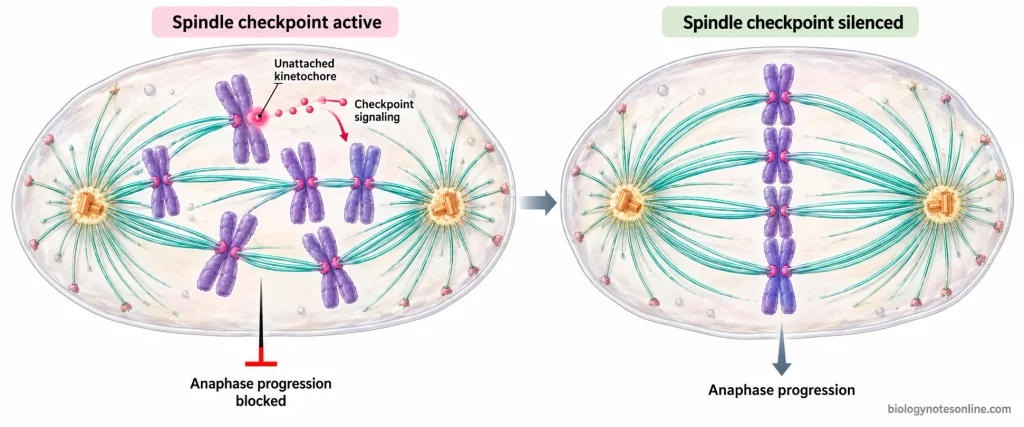
- Activation of the Spindle Assembly Checkpoint- The spindle assembly checkpoint is activated by unattached kinetochores. It prevents the cell from entering anaphase before the chromosomes establish their necessary attachments with the mitotic spindle. Until these attachments are formed, the checkpoint remains active.
Consequences of Prometaphase Dysfunction
Abnormalities during prometaphase may affect the formation of mitotic spindle, kinetochore attachment and proper separation of chromosomes. Some of the major consequences are as follows-
- Abnormal Spindle Formation- Failure of proper separation of centrosomes may result in the formation of a monopolar spindle. In some cells, the presence of additional centrosomes can lead to multipolar spindle formation. Even when these multipolar spindles become bipolar later, the chromosomes may already have developed incorrect attachments with spindle microtubules.
- Incorrect Kinetochore Attachment- In some chromosomes, both sister kinetochores may get attached to microtubules coming from the same spindle pole. This is referred to as “syntelic attachment”. Another type is merotelic attachment, in which a single kinetochore gets attached to spindle microtubules originating from both opposite poles. If these incorrect attachments are not corrected before anaphase, the chromosomes may fail to separate properly.
- Failure of Chromosome Alignment- Some chromosomes fail to reach the equatorial region due to improper attachment of spindle microtubules to the kinetochores. These chromosomes may remain near the spindle poles, resulting in delayed formation of the metaphase plate.
- Spindle Assembly Checkpoint Dysfunction- The spindle assembly checkpoint (SAC) prevents the cell from entering anaphase until the kinetochores establish proper microtubule attachments. If this checkpoint fails, the cell may enter anaphase prematurely, even in the presence of unattached chromosomes. In other cases, the checkpoint remains activated due to unattached kinetochores, delaying the progression of mitosis.
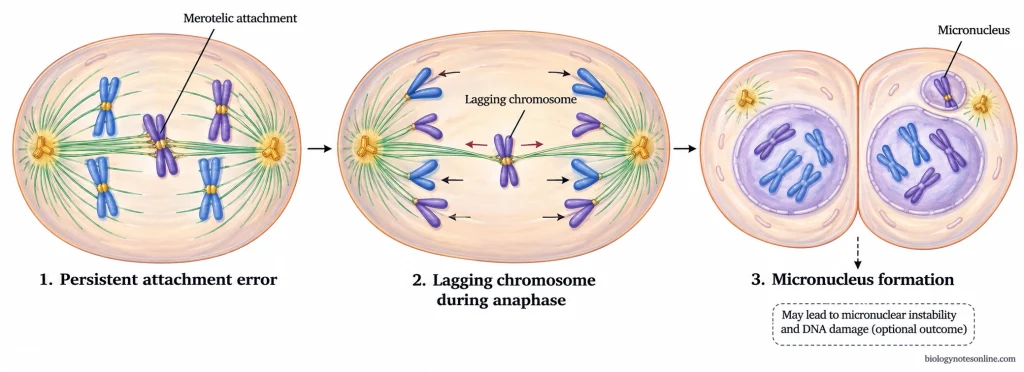
- Aneuploidy- In the case of merotelic attachment, the incorrect attachment may remain undetected by the spindle assembly checkpoint. During anaphase, these chromosomes may lag behind the other chromosomes or enter the wrong daughter cell. The daughter cells may contain extra or missing chromosomes. This condition is referred to as “aneuploidy”.
- Formation of Micronuclei- Sometimes, the lagging chromosomes fail to enter the main daughter nuclei during the reformation of nuclear envelope. These chromosomes may get enclosed separately in small nuclear structures called “micronuclei”. The DNA present inside the micronuclei may undergo defective replication and accumulate DNA damage. In certain cases, extensive chromosome fragmentation and rearrangement may also take place.
- Mitotic Arrest and Cell Death- Severe abnormalities in spindle formation may prevent the cell from completing prometaphase. The cell remains arrested in mitosis for a prolonged period, which may lead to programmed cell death (apoptosis). However, some cells may leave mitosis without proper chromosome separation and cytokinesis. This process is called “mitotic slippage”. Such cells may become tetraploid, containing twice the normal diploid chromosome complement.
Prometaphase at a Glance
The following table contains the important characteristics and major events of prometaphase during mitosis.
| Parameter | Description |
|---|---|
| Definition | Prometaphase is the stage of mitosis during which the nuclear envelope breaks down and spindle microtubules attach to chromosomes. |
| Position in mitosis | Second stage, occurring between prophase and metaphase in the five-stage classification. |
| Beginning of prometaphase | It begins with the breakdown of the nuclear envelope. |
| Nuclear envelope | The nuclear envelope disassembles, allowing spindle microtubules to access chromosomes. |
| Chromosome structure | Chromosomes are highly condensed. Each chromosome consists of two sister chromatids joined at the centromeric region. |
| Mitotic spindle | The mitotic spindle continues to develop, and its microtubules interact with chromosomes. |
| Types of spindle microtubules | Kinetochore, polar and astral microtubules are the three major types found in typical animal cells. |
| Kinetochore attachment | Spindle microtubules attach to chromosomes at specialized protein structures called kinetochores. |
| Bipolar attachment | Sister kinetochores progressively establish attachments with microtubules from opposite spindle poles. |
| Chromosome movement | The chromosomes move within the spindle and gradually approach the equatorial region of the cell. |
| Chromosome congression | The movement of chromosomes towards the spindle equator, where they become arranged at the metaphase plate. |
| Correction of improper attachments | Incorrect kinetochore-microtubule attachments may form and are corrected by cellular regulatory mechanisms. |
| Spindle assembly checkpoint | Unattached kinetochores activate the checkpoint, which prevents premature entry into anaphase. |
| Importance | Establishes chromosome-spindle attachments and prepares chromosomes for their proper alignment and subsequent separation. |
| End of prometaphase | It ends when all chromosomes have aligned at the metaphase plate, marking the transition to metaphase. |
| Textbook classification | Some textbooks describe prometaphase as a separate stage, while others include it within late prophase. |
References
- Alberts, B., Johnson, A., Lewis, J., Raff, M., Roberts, K., & Walter, P. (2002). Molecular biology of the cell (4th ed.). Garland Science. https://www.ncbi.nlm.nih.gov/books/NBK21054/
- Cheeseman, I. M. (2014). The kinetochore. Cold Spring Harbor Perspectives in Biology, 6(7), a015826. https://doi.org/10.1101/cshperspect.a015826
- Cimini, D. (2023). Twenty years of merotelic kinetochore attachments: A historical perspective. Chromosome Research, 31(3), Article 18. https://doi.org/10.1007/s10577-023-09727-7
- Clark, M. A., Douglas, M., & Choi, J. (2018). Biology 2e. OpenStax. https://openstax.org/books/biology-2e/pages/1-introduction
- Cooper, G. M. (2000). The cell: A molecular approach (2nd ed.). Sinauer Associates. https://www.ncbi.nlm.nih.gov/books/NBK9839/
- Devillers, R., dos Santos, A., Destombes, Q., Laplante, M., & Elowe, S. (2024). Recent insights into the causes and consequences of chromosome mis-segregation. Oncogene, 43, 3139–3150. https://doi.org/10.1038/s41388-024-03163-5
- Ferreira, L. T., & Maiato, H. (2021). Prometaphase. Seminars in Cell & Developmental Biology, 117, 52–61. https://doi.org/10.1016/j.semcdb.2021.06.004
- Foley, E. A., & Kapoor, T. M. (2013). Microtubule attachment and spindle assembly checkpoint signalling at the kinetochore. Nature Reviews Molecular Cell Biology, 14(1), 25–37. https://doi.org/10.1038/nrm3494
- Gregan, J., Polakova, S., Zhang, L., Tolić-Nørrelykke, I. M., & Cimini, D. (2011). Merotelic kinetochore attachment: Causes and effects. Trends in Cell Biology, 21(6), 374–381. https://doi.org/10.1016/j.tcb.2011.01.003
- Lischetti, T., & Nilsson, J. (2015). Regulation of mitotic progression by the spindle assembly checkpoint. Molecular & Cellular Oncology, 2(1), e970484. https://doi.org/10.4161/23723548.2014.970484
- Maiato, H., Gomes, A. M., Sousa, F., & Barisic, M. (2017). Mechanisms of chromosome congression during mitosis. Biology, 6(1), Article 13. https://doi.org/10.3390/biology6010013
- Matson, D. R., & Stukenberg, P. T. (2011). Spindle poisons and cell fate: A tale of two pathways. Molecular Interventions, 11(2), 141–150. https://doi.org/10.1124/mi.11.2.12
- McAinsh, A. D., & Kops, G. J. P. L. (2023). Principles and dynamics of spindle assembly checkpoint signalling. Nature Reviews Molecular Cell Biology, 24, 543–559. https://doi.org/10.1038/s41580-023-00593-z
- McNally, F. J. (2013). Mechanisms of spindle positioning. Journal of Cell Biology, 200(2), 131–140. https://doi.org/10.1083/jcb.201210007
- Miller, M. P., Amon, A., & Ünal, E. (2013). Meiosis I: When chromosomes undergo extreme makeover. Current Opinion in Cell Biology, 25(6), 687–696. https://doi.org/10.1016/j.ceb.2013.07.009
- Musacchio, A., & Salmon, E. D. (2007). The spindle-assembly checkpoint in space and time. Nature Reviews Molecular Cell Biology, 8(5), 379–393. https://doi.org/10.1038/nrm2163
- Tanaka, K. (2013). Regulatory mechanisms of kinetochore–microtubule interaction in mitosis. Cellular and Molecular Life Sciences, 70(4), 559–579. https://doi.org/10.1007/s00018-012-1057-7
- Tolić, I. M. (2018). Mitotic spindle: Kinetochore fibers hold on tight to interpolar bundles. European Biophysics Journal, 47(3), 191–203. https://doi.org/10.1007/s00249-017-1244-4
- Vader, G., Maia, A. F., & Lens, S. M. A. (2008). The chromosomal passenger complex and the spindle assembly checkpoint: Kinetochore-microtubule error correction and beyond. Cell Division, 3, Article 10. https://doi.org/10.1186/1747-1028-3-10
- Vukušić, K., & Tolić, I. M. (2022). Polar chromosomes—Challenges of a risky path. Cells, 11(9), Article 1531. https://doi.org/10.3390/cells11091531
- Zhang, C.-Z., Spektor, A., Cornils, H., Francis, J. M., Jackson, E. K., Liu, S., Meyerson, M., & Pellman, D. (2015). Chromothripsis from DNA damage in micronuclei. Nature, 522(7555), 179–184. https://doi.org/10.1038/nature14493