

What Is a Liposome?
A liposome is an artificially prepared spherical lipid vesicle having an aqueous compartment enclosed by one or more phospholipid bilayers. It is a closed membrane structure.
When phospholipids are dispersed in an aqueous medium, they arrange themselves in such a way that their hydrophilic heads remain towards water while the hydrophobic tails face each other. This forms a lipid bilayer. A similar arrangement of phospholipids is also found in biological cell membranes.
Based on the number of lipid bilayers, liposomes are of two types:
- Unilamellar liposomes- These contain a single lipid bilayer.
- Multilamellar liposomes- They are made up of several concentric lipid bilayers.
The size of liposomes varies. Depending on the method of preparation, their lipid composition, surface charge and membrane permeability may also differ.
Water-soluble substances can be enclosed within the aqueous compartment of liposomes. In the case of lipid-soluble substances, they are incorporated into the lipid bilayer.
Liposomes are generally considered as artificial vesicles because they are prepared outside living cells using selected lipids. These lipids may also be obtained from natural sources.
Extracellular vesicles (EVs), unlike liposomes, are naturally produced and released by living cells. Exosomes and microvesicles are some of the examples. Their membranes contain cell-derived lipids and proteins. Apart from these, EVs may also carry other cellular materials like proteins and nucleic acids (DNA and RNA).
Structure of a Liposome
The structure of a liposome consists of the following components.
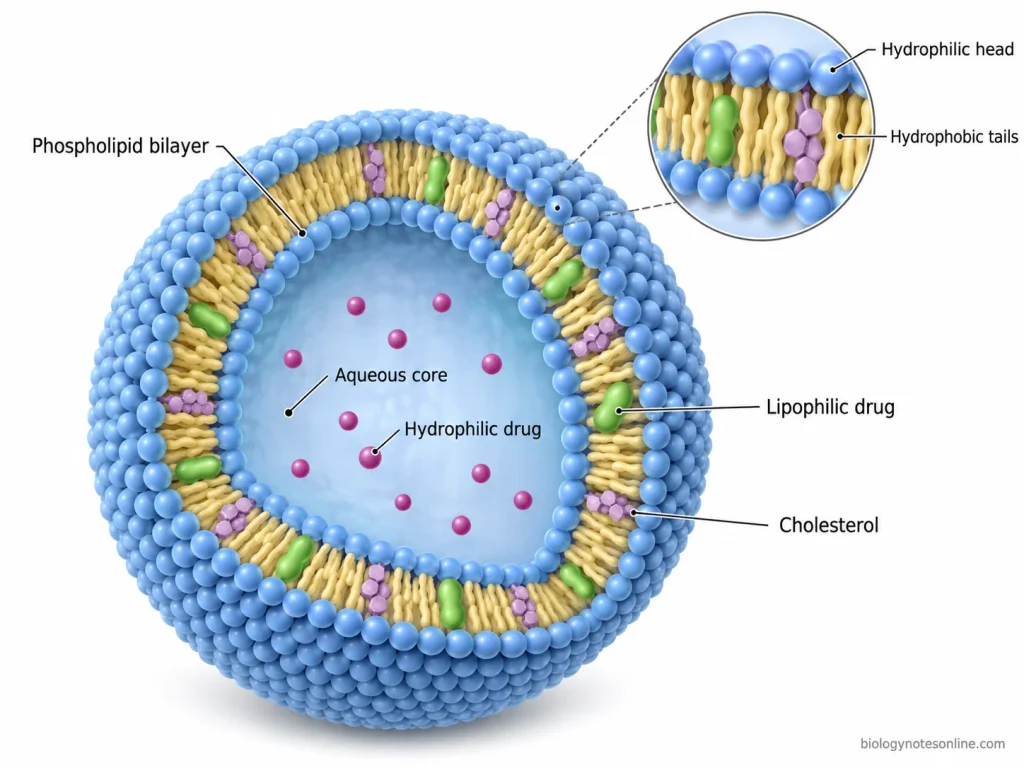
- Spherical structure- Liposome is a spherical vesicle, which is made up of one or more phospholipid bilayers. These bilayers enclose an internal aqueous compartment called the “aqueous core”.
- Phospholipids- The major structural components of liposomes are phospholipids. Each phospholipid molecule contains a polar “hydrophilic head” and non-polar “hydrophobic tails”. They are amphiphilic in nature. In an aqueous medium, these phospholipids arrange themselves to form a bilayer.
- Phospholipid bilayer- It is formed by the arrangement of phospholipid molecules in two layers. The hydrophilic heads are directed towards the internal and external aqueous environments. Whereas, the hydrophobic tails face each other and form the inner region of the bilayer.
- Aqueous core- It is a water-filled compartment present inside the liposome. Hydrophilic drugs and other water-soluble substances can be entrapped in this region.
- Hydrophobic region- The fatty acid chains of phospholipids form a hydrophobic region between the two layers of hydrophilic heads. Lipophilic drugs can be incorporated into this region. The incorporation of these drugs depends on their lipid solubility and membrane composition.
- Cholesterol- Cholesterol is commonly added to the phospholipid bilayer. It gets incorporated between the phospholipid molecules and regulates the membrane fluidity, lipid packing and permeability. It can also increase the stability of liposomal membrane. However, it is not an essential component of every liposomal formulation.
- Surface modification- The outer surface of liposomes may contain some additional components. These include polyethylene glycol (PEG) chains and targeting ligands. They can be attached to the lipid membrane in modified liposomes. These components are not present in the basic structure of conventional liposomes.
- Lamellar structure- Based on the number and arrangement of phospholipid bilayers, liposomes are divided into different structural forms. The following are the major types.
- Unilamellar vesicles (ULVs)- These liposomes contain only one phospholipid bilayer. An aqueous core is enclosed by this bilayer.
- Oligolamellar vesicles (OLVs)- They consist of a few concentric phospholipid bilayers, which are separated by aqueous layers.
- Multilamellar vesicles (MLVs)- These vesicles contain multiple concentric phospholipid bilayers. The bilayers are separated from each other by aqueous layers. This arrangement gives an “onion-like structure”.
- Multivesicular vesicles (MVVs)- In these vesicles, several smaller and separate vesicles are enclosed within a larger vesicle.
Composition of Liposomes
Liposomes are mainly composed of phospholipids, which form the lipid bilayer. Other components, such as cholesterol, charged lipids and surface-modifying agents, may also be added during their preparation. The major components of liposomes are as follows-
- Phospholipids– These are the main lipid components used in the preparation of liposomes. They may be obtained from natural sources, such as egg yolk and soybean, or prepared synthetically. Phosphatidylcholine (PC) is one of the most commonly used phospholipids. Other phospholipids include phosphatidylethanolamine (PE), phosphatidylserine (PS), phosphatidylglycerol (PG) and sphingomyelin. The type of phospholipid and its fatty acid composition affect the membrane fluidity, permeability and stability of liposomes.
- Cholesterol- It is a sterol, commonly added to liposomal formulations to modify the properties of the lipid bilayer. Cholesterol gets incorporated between the phospholipid molecules. Its presence affects lipid packing and membrane fluidity. It also helps to reduce the permeability of the membrane and improve its stability.
- Charged lipids- Some liposomes contain positively or negatively charged lipids, depending on their requirements. Negatively charged phospholipids include phosphatidylserine and phosphatidylglycerol. For the preparation of positively charged liposomes, cationic lipids such as DOTAP may be used. These lipids are used to modify the surface charge of liposomes and their interaction with other molecules.
- Polyethylene glycol (PEG)- In some liposomal preparations, PEG is attached to the phospholipids and incorporated into the membrane. The PEG chains extend from the outer surface of the liposome. They are used to reduce aggregation and interaction with plasma proteins, which may increase the circulation time of liposomes in the blood. Such liposomes are commonly referred to as “PEGylated liposomes”.
- Aqueous medium- Water or an aqueous buffer is used during the preparation of liposomes. It forms the aqueous compartment enclosed by the lipid bilayer.
- Encapsulated substances- Depending on the purpose of preparation, liposomes may also contain different substances, such as drugs, proteins or nucleic acids. Water-soluble substances are generally enclosed within the aqueous compartment, whereas lipid-soluble substances can be incorporated into the lipid bilayer.
Types of Liposomes
Liposomes are classified into different types based on their size, number of phospholipid bilayers and composition. They can also be classified according to their surface modification. The following are the major types of liposomes.
1. Based on Size and Number of Bilayers
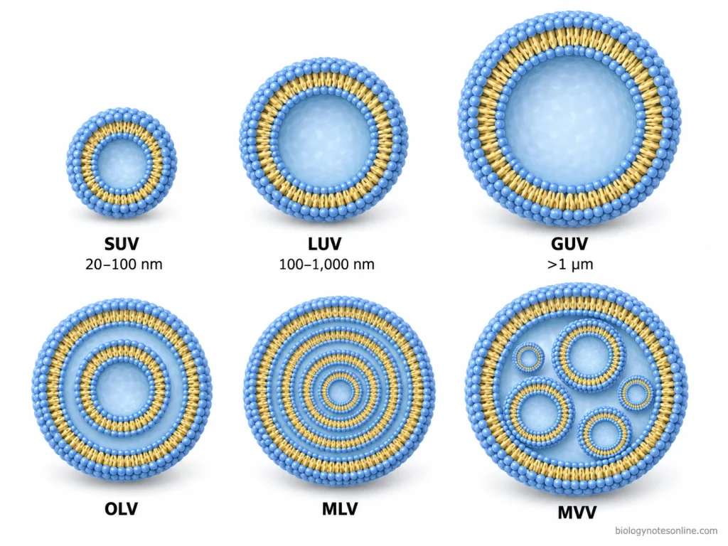
- Unilamellar vesicles (ULVs)- These liposomes contain a single phospholipid bilayer, which encloses an internal aqueous compartment. Based on their size, unilamellar vesicles are further divided into three types.
- Small unilamellar vesicles (SUVs)- These are small liposomes having a diameter of approximately 20-100 nm. They contain only one phospholipid bilayer and have a relatively small aqueous compartment.
- Large unilamellar vesicles (LUVs)- The diameter of these vesicles generally ranges from 100-1000 nm. Like SUVs, they also contain a single phospholipid bilayer. However, their larger size provides more internal aqueous space.
- Giant unilamellar vesicles (GUVs)- These liposomes are larger than 1 µm in diameter. They consist of a single phospholipid bilayer surrounding a large aqueous compartment.
- Oligolamellar vesicles (OLVs)- They contain a few concentric phospholipid bilayers, generally 2-5 layers. The bilayers are separated from each other by aqueous compartments. Their size commonly ranges from 100-1000 nm.
- Multilamellar vesicles (MLVs)- These liposomes are made up of multiple concentric phospholipid bilayers. An aqueous layer is present between two successive bilayers. This arrangement gives an “onion-like structure”. They are generally larger than 500 nm in diameter.
- Multivesicular vesicles (MVVs)- In these liposomes, several smaller vesicles are enclosed within a larger vesicle. The internal vesicles are arranged in a non-concentric manner. Unlike multilamellar vesicles, they contain multiple separate aqueous compartments. Their diameter is generally greater than 1 µm.
2. Based on Composition and Surface Modification
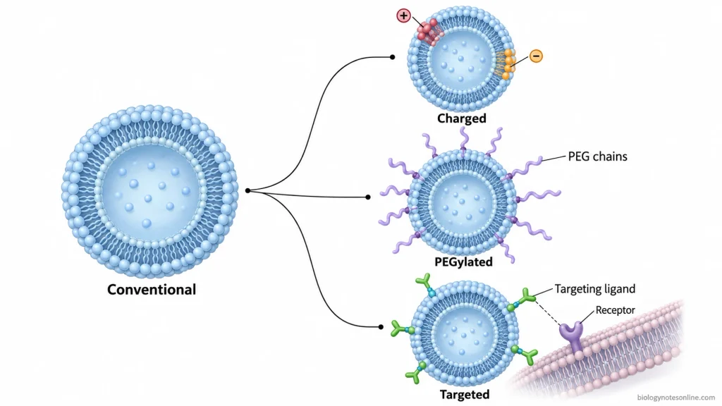
- Conventional liposomes- These are the basic type of liposomes, which are mainly composed of phospholipids with or without cholesterol. They do not contain any special surface modification. Conventional liposomes are also referred to as “first-generation liposomes”.
- Charged liposomes- These liposomes are prepared by incorporating positively or negatively charged lipids into the phospholipid bilayer. Based on their surface charge, they are divided into two types.
- Cationic liposomes- They contain positively charged lipids such as DOTAP or stearylamine. These liposomes are commonly studied for the delivery of nucleic acids.
- Anionic liposomes- Negatively charged lipids such as phosphatidylserine (PS) and phosphatidylglycerol (PG) are used for their preparation. They have a negative surface charge.
- Stealth liposomes- These are surface-modified liposomes, which are commonly prepared by attaching polyethylene glycol (PEG) to the phospholipids. The PEG chains form a hydrophilic layer over the liposomal surface. This modification reduces their recognition and uptake by the mononuclear phagocyte system. As a result, these liposomes can remain in the blood circulation for a longer period. They are also called “PEGylated liposomes”.
- Targeted liposomes- In this type, specific targeting ligands are attached to the outer surface of liposomes. These ligands may include antibodies, peptides, proteins or carbohydrates. They are used to promote the interaction of liposomes with specific receptors present on the target cells. Liposomes containing antibodies as targeting ligands are known as “immunoliposomes”.
- Stimuli-responsive liposomes- These liposomes are designed to release their encapsulated drugs in response to certain internal or external stimuli. The release mechanism depends on their lipid composition and membrane properties. Some of the important types are as follows.
- pH-sensitive liposomes- They are formulated to release the entrapped drug in response to a change in pH. Their membrane becomes unstable under specific pH conditions.
- Temperature-sensitive liposomes- These are also called “thermosensitive liposomes”. They are prepared using temperature-sensitive lipid compositions. When the temperature reaches a specific range, membrane permeability increases and promotes the release of encapsulated drugs.
How Liposomes Work (Step-by-Step Mechanism)
Liposomes are used as drug carriers, which can entrap both hydrophilic and lipophilic drugs. They carry the encapsulated drug and release it depending on their composition and interaction with the biological environment. The following are the major steps involved in the working mechanism of liposomes.
1. Drug Encapsulation
In this step, the drug is incorporated into the liposome either during or after its preparation. The location of drug mainly depends on its solubility and chemical properties.
- Hydrophilic drugs- These are mainly entrapped inside the aqueous compartment of liposomes.
- Lipophilic drugs- They are incorporated into the hydrophobic region of the phospholipid bilayer.
The phospholipid membrane acts as a barrier between the entrapped drug and the external aqueous environment.
2. Administration and Drug Protection
After administration, liposomes carry the encapsulated drug through the biological environment. In case of intravenous administration, they directly enter the bloodstream.
During this process, the phospholipid membrane separates the entrapped drug from the surrounding biological components. It can also prevent the immediate release of the drug. However, the protection mainly depends on the stability and composition of liposomal membrane.
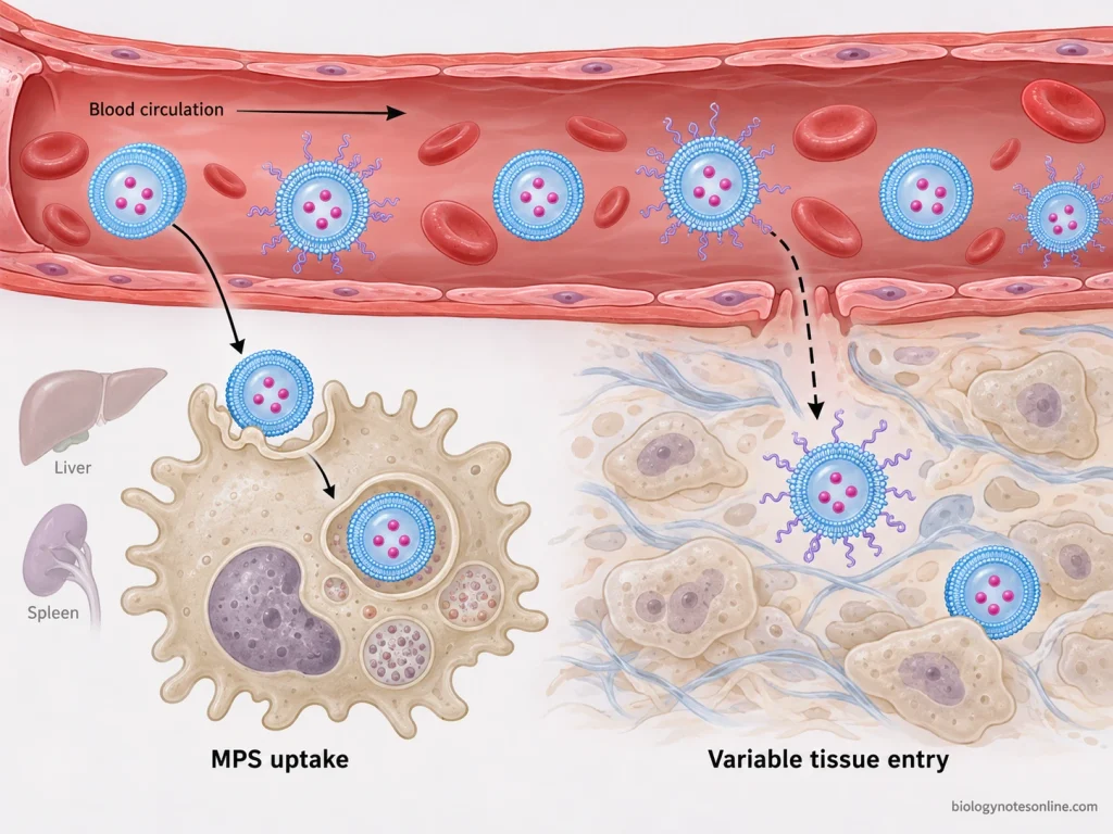
In some formulations, polyethylene glycol (PEG) is attached to the liposomal surface. These are called “PEGylated liposomes”. The PEG modification can reduce their uptake by the mononuclear phagocyte system (MPS) and increase their circulation time.
3. Distribution and Accumulation
The distribution of liposomes depends on their size, surface charge, lipid composition and route of administration. After entering the bloodstream, they interact with different biological components.
Conventional liposomes are commonly taken up by macrophages, particularly in the liver and spleen. Whereas, surface-modified liposomes may remain in the blood circulation for a longer period.
For the delivery of drugs to specific tissues, two types of targeting approaches are commonly studied.
- Passive targeting- It is based on the accumulation of liposomes in certain tissues due to their physiological and pathological characteristics. For example, some liposomes can accumulate in tumour tissues through the “enhanced permeability and retention” (EPR) effect. However, this accumulation may vary between different tumours.
- Active targeting- In this type, specific targeting ligands are attached to the liposomal surface. These ligands can bind to their corresponding receptors present on the target cells. Some commonly used ligands include antibodies, peptides and other receptor-specific molecules.
4. Interaction with the Cell Membrane
When liposomes reach the cells, they may interact with the cell membrane. The interaction mainly depends on the surface properties of liposomes and the characteristics of cell membrane.
In case of targeted liposomes, the attached ligands can recognise and bind to specific receptors. Positively charged liposomes may also interact with the negatively charged components present on the cell surface.
After binding, the liposomes may be taken up by the cells through different mechanisms.
5. Cellular Uptake
In this step, liposomes enter the cells through different pathways. The mechanism of cellular uptake depends on the lipid composition, type of liposome and properties of the target cells. Some of the important mechanisms are as follows.
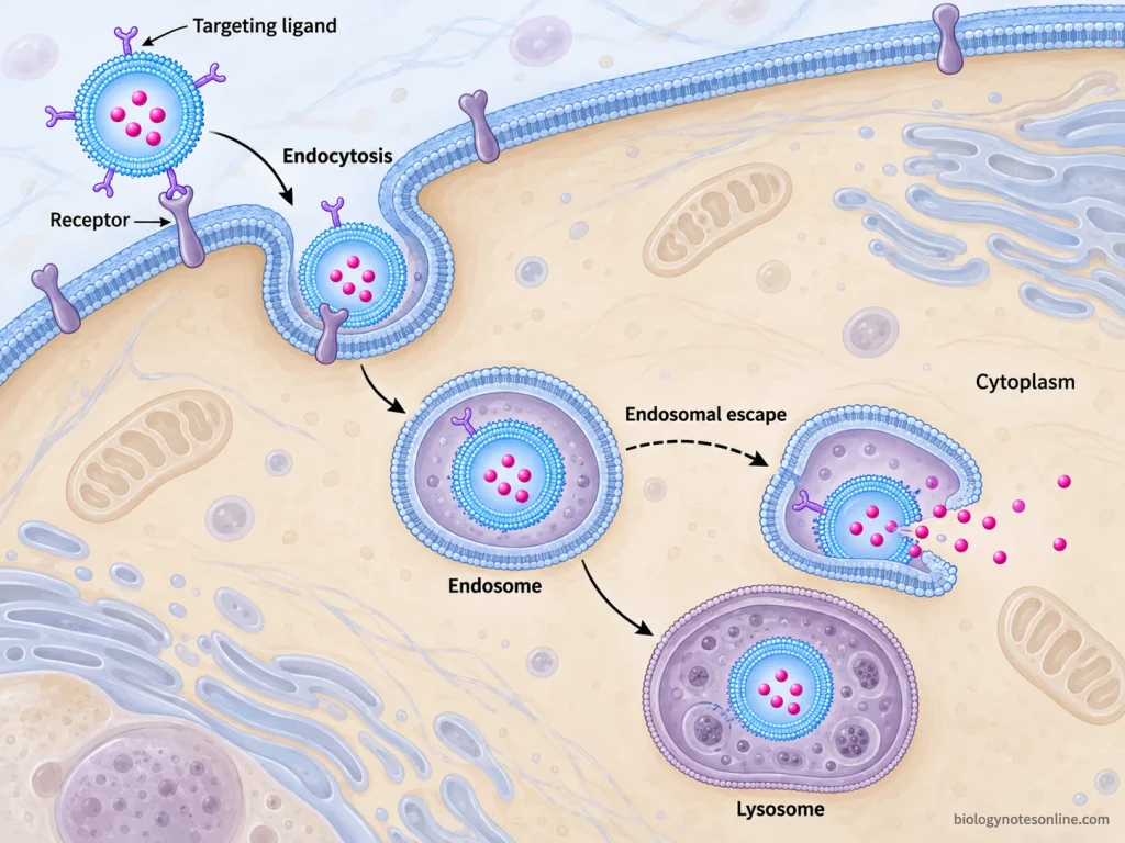
- Endocytosis– It is one of the major mechanisms involved in the cellular uptake of liposomes. During this process, the cell membrane surrounds the liposome and takes it inside the cell. The internalised liposome becomes enclosed within a membrane-bound compartment called the “endosome”.
- Membrane fusion- In this mechanism, certain liposomal formulations can directly fuse with the cell membrane. The phospholipid bilayer of liposome merges with the cellular membrane. During this process, the encapsulated material may be released into the cell.
- Receptor-mediated endocytosis- This mechanism is mainly associated with certain targeted liposomes. The targeting ligand first binds to its corresponding receptor present on the cell membrane. After binding, the liposome-receptor complex can be taken up by the cell through endocytosis.
6. Drug Release
The encapsulated drug may be released from liposomes either outside or inside the cells. The release mechanism mainly depends on the liposomal formulation and membrane properties.
In some formulations, the drug is gradually released through the phospholipid membrane. After release, the drug can enter the surrounding cells depending on its chemical properties.
For liposomes taken up through endocytosis, the drug initially remains enclosed within the endosomal compartment. During endosomal maturation, the internal environment gradually becomes acidic. The liposomal membrane may become unstable or undergo degradation after reaching the lysosomes.
However, certain drugs require their release directly into the cytoplasm. For this purpose, specially designed liposomes can disrupt the endosomal membrane and release the entrapped substances into the cytoplasm. This process is referred to as “endosomal escape”.
Some liposomes are also prepared to release their drugs under specific conditions, such as changes in pH or temperature. These are known as “stimuli-responsive liposomes”.
7. Drug Action
After release, the drug becomes available to interact with its biological target. The mechanism and site of action depend on the nature of drug.
Some drugs act on receptors or other targets present on the cell surface. Whereas, intracellular drugs need to reach their specific location inside the cell. The released drug may interact with cellular enzymes, proteins or nucleic acids according to its mechanism of action.
Preparation of Liposomes
Liposomes are prepared by using different laboratory techniques. The method of preparation mainly depends on the required size, number of phospholipid bilayers and drug encapsulation. Some methods are used for the formation of liposomes, while others are used to modify their size and lamellar structure.
1. Common Methods of Liposome Preparation
The following are the commonly used methods for the preparation of liposomes.
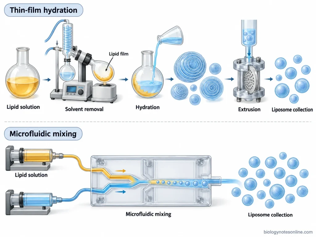
- Thin-film hydration method- It is one of the most commonly used techniques for liposome preparation. This method is also known as the “Bangham method”. In this process, phospholipids and other lipid components are dissolved in a suitable organic solvent. The solvent is then removed using a rotary evaporator, which forms a thin lipid film on the inner surface of the flask.After solvent removal, the dried lipid film is hydrated with water or an aqueous buffer. During hydration, the lipid layers swell and separate from each other to form liposomes. This method generally produces multilamellar vesicles (MLVs). For obtaining smaller liposomes, further treatment with sonication or extrusion is required.
- Ethanol injection method- In this method, phospholipids are first dissolved in ethanol. The lipid solution is then injected into an aqueous medium under continuous stirring. When ethanol mixes with the aqueous phase, the lipid molecules start to assemble and form liposomes.The size of liposomes depends on several factors, such as lipid concentration, injection rate and mixing conditions. This method is commonly used for the preparation of small unilamellar vesicles (SUVs). The remaining ethanol is removed from the liposomal suspension after preparation.
- Reverse-phase evaporation method- It is mainly used for the preparation of liposomes with a relatively large internal aqueous compartment. In this method, the lipid components are dissolved in an organic solvent and mixed with an aqueous solution. The mixture is then subjected to mechanical agitation or sonication, which forms a water-in-oil emulsion.During this process, the aqueous droplets become surrounded by lipid molecules. The organic solvent is gradually removed under reduced pressure. Initially, a viscous gel is formed. With further removal of the solvent, the lipid molecules rearrange themselves and form liposomes.This method can produce large unilamellar vesicles (LUVs) with relatively high entrapment of water-soluble substances. However, it may not be suitable for drugs that are sensitive to organic solvents.
- Detergent removal method- This method is based on the formation of mixed micelles containing phospholipids and detergent molecules. Initially, the lipid components are dissolved using a suitable detergent. It forms a mixture of lipid-detergent micelles.The detergent is then gradually removed from the mixture. During this process, the phospholipid molecules rearrange themselves to form closed lipid bilayers. Unilamellar vesicles can be obtained by this method.Some commonly used methods for detergent removal include dialysis, gel filtration and adsorption using suitable materials. The size of liposomes depends on the detergent concentration, lipid composition and rate of detergent removal.
- Microfluidic method- In this technique, the lipid solution and aqueous medium are passed through small channels of a microfluidic device. Both solutions are mixed under controlled conditions. During mixing, the organic solvent gets diluted and the lipid molecules assemble to form liposomes.The flow rate and mixing ratio are adjusted according to the required liposomal size. It can be used to prepare liposomes with a relatively narrow size distribution. The residual organic solvent must be removed after preparation.
- Electroformation method- It is mainly used for the preparation of giant unilamellar vesicles (GUVs). In this method, a thin lipid film is deposited on a suitable electrode surface. After removing the organic solvent, the lipid film is hydrated with an aqueous solution.An alternating electric field is applied during hydration. It promotes the swelling and separation of lipid bilayers, which gradually form large vesicles. This method is commonly used in laboratory studies involving lipid membranes and their properties.
2. Methods Used for Size Reduction and Structural Modification
The liposomes obtained from certain preparation methods may contain vesicles of different sizes and lamellar structures. For obtaining liposomes with the required structural properties, additional processing methods are used.
- Sonication– It is commonly used to reduce the size of previously prepared liposomes. In this method, the liposomal suspension is subjected to ultrasonic energy. The ultrasonic waves disrupt the larger vesicles and promote the formation of smaller liposomes.Two types of sonication techniques are commonly used.
- Probe sonication- The tip of an ultrasonic probe is directly immersed in the liposomal suspension. It produces high ultrasonic energy and is used for the preparation of small vesicles. However, excessive heating and contamination from the probe tip may occur.
- Bath sonication- In this method, the sample container is placed inside an ultrasonic water bath. The ultrasonic energy passes through the water and acts on the liposomal suspension. It provides a relatively gentle treatment compared to probe sonication.
- Membrane extrusion- In this method, the prepared liposomal suspension is passed through a membrane containing pores of a selected size. Polycarbonate membranes are commonly used for this purpose.During extrusion, the larger vesicles are disrupted and rearranged while passing through the membrane pores. The process may be repeated several times to obtain the required size and size distribution. It is commonly used for the preparation of relatively uniform unilamellar vesicles.
- Freeze-thaw method- The prepared liposomal suspension is repeatedly subjected to freezing and thawing. During freezing, the formation of ice crystals can disturb the lipid bilayers. When the suspension is thawed, the lipid membranes undergo rearrangement and may fuse with each other.This process can reduce the number of lipid bilayers and increase the entrapment of certain water-soluble substances. After freeze-thaw treatment, extrusion may be carried out to obtain liposomes with the required size.
- High-pressure homogenization– It is another method used to reduce the size of liposomes. In this process, the liposomal suspension is passed through a narrow opening under high pressure. The generated shear forces and turbulence break down larger vesicles into smaller ones.The process can be repeated depending on the required particle size. It is also used for the preparation of liposomes on a larger scale.
3. Purification and Characterization
After preparation, liposomes may contain free drugs, residual solvents and other unentrapped substances. These are removed using suitable purification methods, such as dialysis, gel filtration or centrifugation.
The prepared liposomes are then examined to determine their structural and physicochemical properties. Some of the important parameters are as follows.
- Particle size and size distribution- These are commonly determined using dynamic light scattering (DLS). The polydispersity index (PDI) is used to describe the width of the particle size distribution.
- Lamellarity and morphology- Transmission electron microscopy (TEM) and cryogenic transmission electron microscopy (Cryo-TEM) can be used to examine the shape, size and number of lipid bilayers.
- Surface charge- It is commonly determined by measuring the zeta potential of the liposomal suspension.
- Encapsulation efficiency- It is determined by measuring the amount of drug incorporated into the liposomes relative to the amount used during preparation. This measurement is carried out after separating the unentrapped drug from the liposomal suspension.
Applications of Liposomes
Liposomes are widely used in pharmaceutical and biomedical fields, mainly as drug delivery systems. Some of the important applications are as follows.
- Cancer treatment- Liposomes are used to deliver anticancer drugs and modify their distribution in the body. They can also reduce drug exposure to certain healthy tissues. For example, liposomal doxorubicin is used for the treatment of different types of cancer.
- Treatment of fungal infections- Liposomes are used to deliver antifungal drugs with reduced toxicity. One commonly used example is liposomal amphotericin B, which is used for the treatment of severe systemic fungal infections. It reduces the kidney toxicity associated with conventional amphotericin B.
- Antibacterial drug delivery- Certain antibiotics can be incorporated into liposomes to modify their distribution and release. For example, inhaled liposomal amikacin is used for the treatment of certain Mycobacterium avium complex (MAC) lung infections. They are also being studied for bacterial biofilms and intracellular infections.
- Vaccine development- In this application, liposomes are used as carriers for vaccine antigens and adjuvants. The antigens may be incorporated into the liposomes or attached to their surface. Some liposomal formulations can also deliver antigens and adjuvants together to improve the immune response.
- Gene delivery- Liposomes are used to carry genetic materials such as DNA and RNA. Positively charged liposomes can form complexes with negatively charged nucleic acids, which are called “lipoplexes”. These are mainly studied for non-viral gene delivery, although efficient intracellular delivery remains a challenge.
- Controlled drug delivery- Liposomes can be prepared for the gradual release of encapsulated drugs. The drug release mainly depends on the lipid composition and stability of liposomal membrane. Some formulations are also designed to release drugs in response to changes in pH or temperature.
- Topical drug delivery- They are used for the delivery of drugs to the skin. Liposomes can carry both water-soluble and lipid-soluble substances, and may improve drug deposition in the upper skin layers. They are also incorporated into certain cosmetic formulations.
- Ophthalmic drug delivery- Liposomes are used to modify drug release and improve drug retention in the eye. For example, liposomal verteporfin is used in photodynamic therapy for certain ocular conditions. Other formulations are being studied for drug delivery across ocular barriers.
- Pain management- Multivesicular liposomes are particularly used for prolonged drug release. One example is liposomal bupivacaine, which provides extended local pain relief after certain surgical procedures.
- Diagnostic applications- Liposomes can also be used as carriers for imaging agents, such as fluorescent compounds, radioactive labels and magnetic resonance imaging (MRI) contrast agents. Some experimental formulations contain both therapeutic drugs and imaging agents. These are referred to as “theranostic liposomes”.
Advantages of Liposomes
Some of the important advantages of liposomes are as follows-
- Liposomes can carry both water-soluble and lipid-soluble drugs. The water-soluble substances are enclosed within the aqueous compartment, while lipid-soluble substances are incorporated into the lipid bilayer.
- They are generally biocompatible and biodegradable, as many liposomes are prepared from natural phospholipids.
- The encapsulated drugs can be protected from premature degradation and inactivation, depending on the stability of the liposomal membrane.
- Liposomes can reduce the toxicity of certain drugs by altering their distribution in the body. Liposomal amphotericin B is an example, which has lower kidney toxicity than conventional amphotericin B.
- They help in the formulation and delivery of certain poorly water-soluble drugs.
- The release of drugs can be modified by changing the lipid composition and membrane permeability. Some liposomal preparations are developed for sustained and controlled drug release.
- Liposomes coated with polyethylene glycol (PEG) can remain in blood circulation for a longer period by reducing their rapid clearance.
- For targeted drug delivery, the surface of liposomes can be modified with antibodies and other targeting molecules. Such preparations are being investigated for delivering drugs to particular cells.
- The size, surface charge, lipid composition and number of bilayers can be modified according to the requirements of drug delivery.
- Different liposomal formulations can be prepared for intravenous, inhaled and topical administration, depending on the drug and its intended application.
Limitations of Liposomes
Some of the important limitations of liposomes are as follows-
- Liposomes have relatively low physical and chemical stability. Their structure and properties may change during preparation, storage and administration.
- The phospholipids present in the liposomal membrane are susceptible to oxidation and hydrolysis, which can affect the stability of liposomes.
- There is a possibility of leakage of encapsulated drugs due to changes in membrane permeability. Liposomes may also undergo aggregation and fusion, resulting in changes in their size and structure.
- The encapsulation efficiency of some drugs is relatively low, particularly for certain water-soluble substances. A considerable amount of drug may remain unencapsulated during preparation.
- Conventional liposomes may have a short circulation half-life. They are rapidly recognized and removed by the macrophages, mainly in the liver and spleen.
- For targeted drug delivery, the accumulation of liposomes in the desired tissues is not always efficient. Their penetration and distribution within solid tumours may also be limited.
- Some liposomal preparations can activate the complement system and cause hypersensitivity reactions. Repeated administration of certain PEGylated liposomes may also lead to “accelerated blood clearance” (ABC).
- The release of encapsulated drugs into the cytoplasm is another limitation. Drugs taken up through endocytosis may remain trapped within the endosomes or undergo degradation.
- Large-scale production of liposomes is relatively complex and expensive. It requires strict control of lipid composition, particle size, encapsulation efficiency and batch-to-batch reproducibility.
- Some preparation methods involve the use of organic solvents. The residual solvents must be removed from the final liposomal preparations.
- Sterilization of liposomes is difficult, as conventional heat and radiation sterilization methods may damage the phospholipid membrane or cause leakage of encapsulated drugs.
Liposomes Compared with Other Lipid Carriers
Liposomes, micelles and lipid nanoparticles are commonly used as drug delivery systems. However, they differ in their structural arrangement, internal compartments and drug-loading properties.
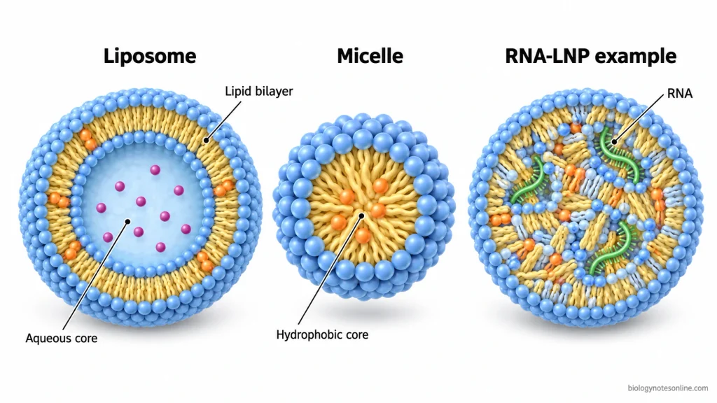
Liposome vs Micelle
Liposomes and micelles are formed by the arrangement of amphiphilic molecules. However, liposomes contain a lipid bilayer, whereas conventional micelles contain a single layer of amphiphilic molecules surrounding a hydrophobic core.
The major differences are as follows.
| Properties | Liposomes | Micelles |
|---|---|---|
| Structure | Spherical vesicles containing one or more lipid bilayers. | Small aggregates formed by a single layer of amphiphilic molecules. |
| Lipid arrangement | Hydrophilic heads face the aqueous environments, while hydrophobic tails face each other. | Hydrophilic heads face the external aqueous medium, whereas hydrophobic tails are directed towards the centre. |
| Internal compartment | They contain an internal aqueous compartment surrounded by a lipid bilayer. | They have a hydrophobic core without an enclosed aqueous compartment. |
| Drug encapsulation | Both hydrophilic and lipophilic drugs can be incorporated into liposomes. | Mainly used for the incorporation of lipophilic drugs into the hydrophobic core. |
| Drug location | Hydrophilic drugs are entrapped in the aqueous core. Whereas, lipophilic drugs are incorporated into the lipid bilayer. | Lipophilic drugs are mainly incorporated into the hydrophobic core. |
Liposome vs Lipid Nanoparticle
Lipid nanoparticles (LNPs) are a broad category of lipid-based carriers, which also includes nanosized liposomes. However, the term “LNP” is commonly used for specially formulated nanoparticles developed for nucleic acid delivery.
Unlike conventional liposomes, these LNPs may contain a more complex internal lipid arrangement. Their structure mainly depends on the lipid composition and encapsulated material.
The following are the major differences between conventional liposomes and commonly used nucleic-acid lipid nanoparticles.
| Properties | Liposomes | Lipid Nanoparticles (LNPs) |
|---|---|---|
| Structure | Spherical vesicles containing one or more phospholipid bilayers. | Lipid-based particles with different internal structures, depending on their formulation. |
| Composition | Mainly prepared from phospholipids, with or without cholesterol. | Commonly contain ionizable lipids, phospholipids, cholesterol and PEG-lipids. |
| Lipid arrangement | Phospholipids are arranged into closed lipid bilayers surrounding an aqueous compartment. | They may contain an outer lipid layer and complex internal lipid arrangements. Some formulations also show bilayer regions. |
| Internal compartment | A distinct aqueous compartment is enclosed by the phospholipid bilayer. | They generally have a complex internal structure containing lipids and nucleic acids, rather than a simple aqueous core. |
| Encapsulated substances | Can incorporate both hydrophilic and lipophilic drugs. | Commonly used for the encapsulation of nucleic acids, such as messenger RNA (mRNA) and small interfering RNA (siRNA). |
| Drug incorporation | Hydrophilic drugs are mainly entrapped in the aqueous core, while lipophilic drugs are incorporated into the bilayer. | Nucleic acids are incorporated within the particles and may remain associated with the internal lipids. |
| Drug delivery | Used for the delivery of different therapeutic substances, including anticancer and antifungal drugs. | Commonly used for nucleic acid delivery, particularly in mRNA vaccines and RNA-based therapeutics. |
References
- Akbarzadeh, A., Rezaei-Sadabady, R., Davaran, S., Joo, S. W., Zarghami, N., Hanifehpour, Y., Samiei, M., Kouhi, M., & Nejati-Koshki, K. (2013). Liposome: Classification, preparation, and applications. Nanoscale Research Letters, 8, Article 102. https://doi.org/10.1186/1556-276X-8-102
- Andra, V. V. S. N. L., Pammi, S. V. N., Bhatraju, L. V. K. P., & Ruddaraju, L. K. (2022). A comprehensive review on novel liposomal methodologies, commercial formulations, clinical trials and patents. BioNanoScience, 12(1), 274–291. https://doi.org/10.1007/s12668-022-00941-x
- Bozzuto, G., & Molinari, A. (2015). Liposomes as nanomedical devices. International Journal of Nanomedicine, 10, 975–999. https://doi.org/10.2147/IJN.S68861
- Bulbake, U., Doppalapudi, S., Kommineni, N., & Khan, W. (2017). Liposomal formulations in clinical use: An updated review. Pharmaceutics, 9(2), Article 12. https://doi.org/10.3390/pharmaceutics9020012
- Chaves, M. A., Ferreira, L. S., Baldino, L., Pinho, S. C., & Reverchon, E. (2023). Current applications of liposomes for the delivery of vitamins: A systematic review. Nanomaterials, 13(9), Article 1557. https://doi.org/10.3390/nano13091557
- De Mel, J. U., Gupta, S., Willner, L., & Schneider, G. J. (2021). Manipulating phospholipid vesicles at the nanoscale: A transformation from unilamellar to multilamellar by an n-alkyl-poly(ethylene oxide). Langmuir, 37(7), 2362–2375. https://doi.org/10.1021/acs.langmuir.0c03302
- Deshpande, P. P., Biswas, S., & Torchilin, V. P. (2013). Current trends in the use of liposomes for tumor targeting. Nanomedicine, 8(9), 1509–1528. https://doi.org/10.2217/nnm.13.118
- Eloy, J. O., de Souza, M. C., Petrilli, R., Barcellos, J. P. A., Lee, R. J., & Marchetti, J. M. (2014). Liposomes as carriers of hydrophilic small molecule drugs: Strategies to enhance encapsulation and delivery. Colloids and Surfaces B: Biointerfaces, 123, 345–363. https://doi.org/10.1016/j.colsurfb.2014.09.029
- Hou, X., Zaks, T., Langer, R., & Dong, Y. (2021). Lipid nanoparticles for mRNA delivery. Nature Reviews Materials, 6, 1078–1094. https://doi.org/10.1038/s41578-021-00358-0
- Immordino, M. L., Dosio, F., & Cattel, L. (2006). Stealth liposomes: Review of the basic science, rationale, and clinical applications, existing and potential. International Journal of Nanomedicine, 1(3), 297–315. https://pmc.ncbi.nlm.nih.gov/articles/PMC2426795/
- Kim, E.-M., & Jeong, H.-J. (2021). Liposomes: Biomedical applications. Chonnam Medical Journal, 57(1), 27–35. https://doi.org/10.4068/cmj.2021.57.1.27
- Lamichhane, N., Udayakumar, T. S., D’Souza, W. D., Simone, C. B., II, Raghavan, S. R., Polf, J., & Mahmood, J. (2018). Liposomes: Clinical applications and potential for image-guided drug delivery. Molecules, 23(2), Article 288. https://doi.org/10.3390/molecules23020288
- Le, N. T. T., Cao, V. D., Nguyen, T. N. Q., Le, T. T. H., Tran, T. T., & Hoang Thi, T. T. (2019). Soy lecithin-derived liposomal delivery systems: Surface modification and current applications. International Journal of Molecular Sciences, 20(19), Article 4706. https://doi.org/10.3390/ijms20194706
- Lee, M.-K. (2020). Liposomes for enhanced bioavailability of water-insoluble drugs: In vivo evidence and recent approaches. Pharmaceutics, 12(3), Article 264. https://doi.org/10.3390/pharmaceutics12030264
- Liu, P., Chen, G., & Zhang, J. (2022). A review of liposomes as a drug delivery system: Current status of approved products, regulatory environments, and future perspectives. Molecules, 27(4), Article 1372. https://doi.org/10.3390/molecules27041372
- Luiz, H., Pinho, J. O., & Gaspar, M. M. (2023). Advancing medicine with lipid-based nanosystems: The successful case of liposomes. Biomedicines, 11(2), Article 435. https://doi.org/10.3390/biomedicines11020435
- Mishra, G. P., Bagui, M., Tamboli, V., & Mitra, A. K. (2011). Recent applications of liposomes in ophthalmic drug delivery. Journal of Drug Delivery, 2011, Article 863734. https://doi.org/10.1155/2011/863734
- Monteiro, N., Martins, A., Reis, R. L., & Neves, N. M. (2014). Liposomes in tissue engineering and regenerative medicine. Journal of the Royal Society Interface, 11(101), Article 20140459. https://doi.org/10.1098/rsif.2014.0459
- Nsairat, H., Khater, D., Sayed, U., Odeh, F., Al Bawab, A., & Alshaer, W. (2022). Liposomes: Structure, composition, types, and clinical applications. Heliyon, 8(5), Article e09394. https://doi.org/10.1016/j.heliyon.2022.e09394
- Olusanya, T. O. B., Haj Ahmad, R. R., Ibegbu, D. M., Smith, J. R., & Elkordy, A. A. (2018). Liposomal drug delivery systems and anticancer drugs. Molecules, 23(4), Article 907. https://doi.org/10.3390/molecules23040907
- Sercombe, L., Veerati, T., Moheimani, F., Wu, S. Y., Sood, A. K., & Hua, S. (2015). Advances and challenges of liposome assisted drug delivery. Frontiers in Pharmacology, 6, Article 286. https://doi.org/10.3389/fphar.2015.00286
- Simões, S., Filipe, A., Faneca, H., Mano, M., Penacho, N., Düzgüneş, N., & de Lima, M. P. (2005). Cationic liposomes for gene delivery. Expert Opinion on Drug Delivery, 2(2), 237–254. https://doi.org/10.1517/17425247.2.2.237
- Šturm, L., & Poklar Ulrih, N. (2021). Basic methods for preparation of liposomes and studying their interactions with different compounds, with the emphasis on polyphenols. International Journal of Molecular Sciences, 22(12), Article 6547. https://doi.org/10.3390/ijms22126547
- Uzun, H. D., Tiris, Z., Czarnetzki, M., López-Marqués, R. L., & Pomorski, T. G. (2024). Electroformation of giant unilamellar vesicles from large liposomes. The European Physical Journal Special Topics, 233, 2931–2940. https://doi.org/10.1140/epjs/s11734-024-01104-7
- van der Koog, L., Gandek, T. B., & Nagelkerke, A. (2022). Liposomes and extracellular vesicles as drug delivery systems: A comparison of composition, pharmacokinetics, and functionalization. Advanced Healthcare Materials, 11(5), Article 2100639. https://doi.org/10.1002/adhm.202100639
- Wang, S., Chen, Y., Guo, J., & Huang, Q. (2023). Liposomes for tumor targeted therapy: A review. International Journal of Molecular Sciences, 24(3), Article 2643. https://doi.org/10.3390/ijms24032643
- Wang, Z., Li, W., Jiang, Y., Park, J., Gonzalez, K. M., Wu, X., Zhang, Q.-Y., & Lu, J. (2024). Cholesterol-modified sphingomyelin chimeric lipid bilayer for improved therapeutic delivery. Nature Communications, 15, Article 2073. https://doi.org/10.1038/s41467-024-46331-7
- Weiner, N., Lieb, L., Niemiec, S., Ramachandran, C., Hu, Z., & Egbaria, K. (1994). Liposomes: A novel topical delivery system for pharmaceutical and cosmetic applications. Journal of Drug Targeting, 2(5), 405–410. https://doi.org/10.3109/10611869408996816
- Yingchoncharoen, P., Kalinowski, D. S., & Richardson, D. R. (2016). Lipid-based drug delivery systems in cancer therapy: What is available and what is yet to come. Pharmacological Reviews, 68(3), 701–787. https://doi.org/10.1124/pr.115.012070