

Lysosomes are membrane-bound cell organelles containing different degradative enzymes. They are the major degradative compartments of animal cells. These organelles break down proteins, nucleic acids, carbohydrates, lipids and different cellular materials.
What Are Lysosomes?
Lysosomes are small membrane-bound organelles mainly present in animal cells, containing different hydrolytic enzymes for degradation of cellular materials. It is a part of the endomembrane or endolysosomal system of the cell.
Proteins, carbohydrates, lipids and nucleic acids are some of the macromolecules broken down in lysosomes. They also receive materials entering by endocytosis and phagocytosis. Damaged or old cell components brought through autophagy are degraded here as well.
The lysosomal interior is acidic with a pH generally around 4.5-5.0. This condition is suitable for the activity of its acid hydrolases. Proton pumps present in the membrane maintain the acidic condition inside.
The term lysosome originated from Greek words ‘lysis’ meaning dissolution or breakdown and ‘soma’ meaning body. The name was given based on its lytic activity.
Discovery and Naming
The discovery of lysosomes dates back to 1955 when Christian de Duve and his coworkers were studying enzymes of rat liver cells.
During cell fractionation experiments, an unusual behaviour was observed with acid phosphatase. Only a small amount of its total activity was initially detected. After damaging the particles, much higher enzyme activity became available.
This indicated that acid phosphatase was enclosed inside a membrane-limited particle. Further fractionation showed some other hydrolytic enzymes occurring with it, and the new group of particles were named lysosomes by de Duve in 1955.
Cell fractionation and differential centrifugation were important in this discovery. These methods allowed different cellular components to be separated and their enzyme activities studied.
In 1974, Christian de Duve along with Albert Claude and George E. Palade received the Nobel Prize in Physiology or Medicine for discoveries concerning structural and functional organization of the cell. De Duve’s studies of lysosomes formed an important part of these works.
Where Are Lysosomes Found?
Lysosomes occur in eukaryotic cells, especially in animal cells. The number is variable and depends on type and activity of the cell.
Macrophages and other phagocytic cells have a highly developed lysosomal system. These cells take up microorganisms, dead cells and other foreign materials. A large amount of this engulfed material is carried to lysosomal compartments and degraded.
Mature mammalian erythrocytes (RBCs) lack normal membrane-bound organelles. During their maturation, the nucleus and organelles are removed, hence lysosomes are also absent in mature mammalian RBCs.
Plant cells have lytic vacuoles (LVs) which perform many of the degradative functions carried out by lysosomes in animal cells. They contain hydrolytic enzymes and are involved in breakdown of different cellular materials. Plant lytic vacuoles are generally discussed separately from the typical lysosomes of animal cells.
Characteristics of Lysosomes
- Lysosomes are membrane-enclosed organelles having a single limiting membrane.
- Acidic condition is present inside the lysosome, generally having a pH of about 4.5-5.0.
- Different acid hydrolases are present in the lumen. Proteases, nucleases, glycosidases, lipases, phosphatases and sulfatases are some of them.
- Most of the lysosomal hydrolases have their better activity under acidic condition.
- V-type H⁺-ATPase (V-ATPase) is present in the lysosomal membrane which maintains the low internal pH by pumping H⁺ inside.
- Lysosomal membrane proteins are highly glycosylated in general. LAMP1 and LAMP2 are major proteins of this membrane.
- Different transport proteins occur in the lysosomal membrane for movement of digested materials across it.
- Size of lysosomes is not fixed. It differs according to the type and condition of cell.
- A definite shape is also not present in all lysosomes. Spherical and other variable forms are found.
- The number of lysosomes is not same in every cell. Cells having greater degradative activity can possess a more developed lysosomal system.
- Lysosomes are dynamic organelles, undergoing movement, fusion and fission within the cell.
- Their position in the cytoplasm can also change with cellular and nutritional condition.
- Lysosomes form a part of the endomembrane or endolysosomal system of eukaryotic cells.
Structure of a Lysosome
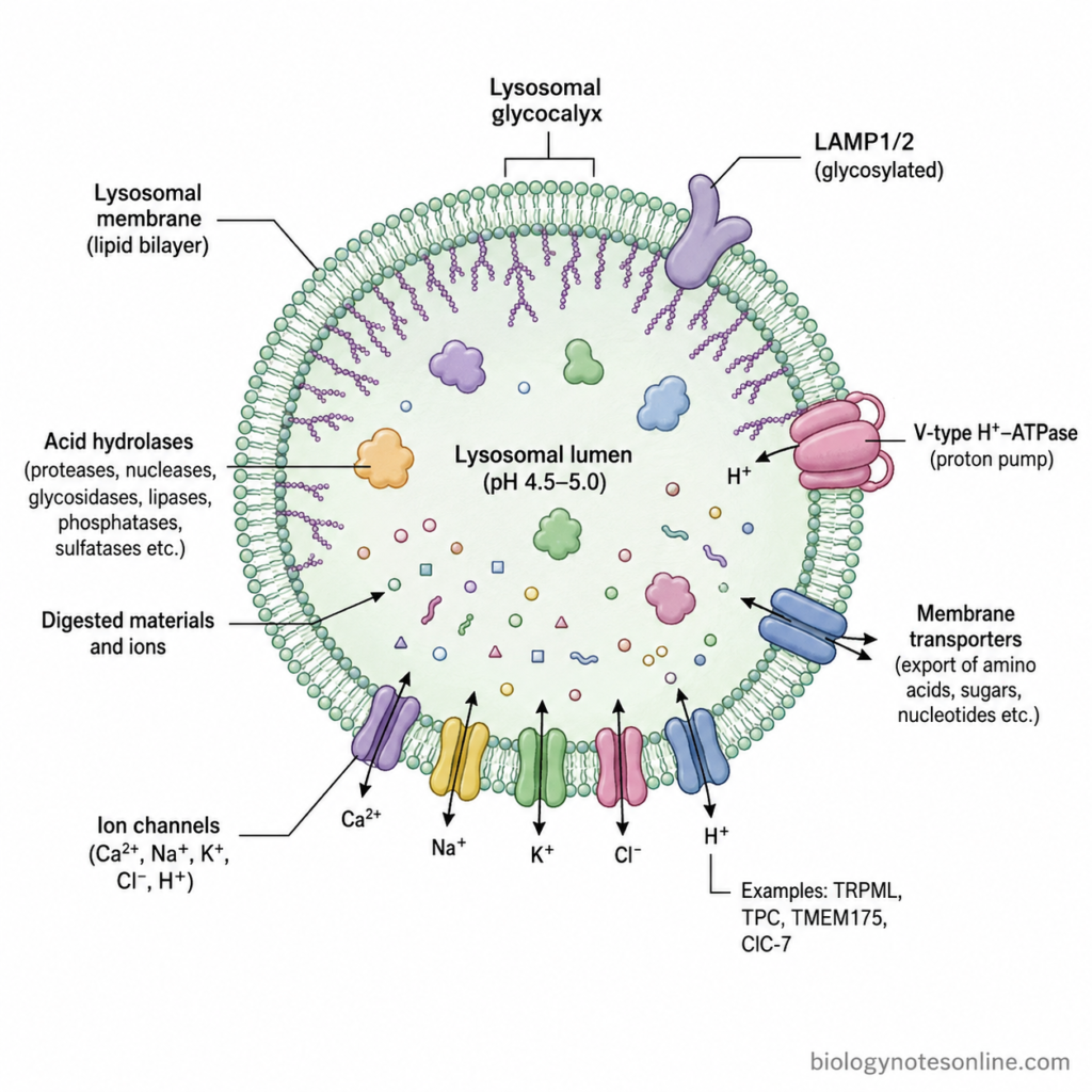
- Shape and size – Lysosomes are small vesicular structures and their shape is not always similar. Most appear spherical or oval, but irregular and tubular lysosomal structures can also occur. The size also changes depending on the cell type, materials present inside and state of the lysosome.
- Lysosomal membrane – It is surrounded by a single limiting membrane which separates the lysosomal contents from the surrounding cytoplasm. The membrane is made up of lipid bilayer together with different integral and peripheral membrane proteins. This membrane prevents the hydrolytic enzymes from freely entering into the cytoplasm.
- Lysosomal lumen – The inner space enclosed by the membrane is known as the lysosomal lumen. It contains soluble hydrolytic enzymes and different materials that are brought for digestion. Ions, degradation products and other soluble components are also present inside this region.
- Acidic internal environment – The lumen has an acidic condition with pH generally around 4.5-5.0. This low pH is an important structural condition of the lysosome as most of its hydrolytic enzymes require acidic medium for their activity.
- Acid hydrolases – A large number of digestive enzymes are enclosed within the lysosomal lumen. These include proteases, nucleases, glycosidases, lipases, phospholipases, phosphatases and sulfatases. They are collectively referred to as acid hydrolases because their better activity occurs under acidic conditions.
- Lysosomal glycocalyx – On the luminal side of lysosomal membrane, a thick carbohydrate-rich covering is present which is called the glycocalyx. It is formed mainly by the highly glycosylated regions of membrane proteins. This covering protects the inner membrane surface from degradation by the lysosomal hydrolases present close to it.
- LAMP proteins – LAMP1 and LAMP2 (Lysosome-associated membrane proteins) are abundant proteins of lysosomal membrane. A large glycosylated portion of these proteins is exposed towards the lumen and contributes to formation of the lysosomal glycocalyx. Other lysosomal integral membrane proteins are also present along with them.
- V-type H⁺-ATPase – The lysosomal membrane contains Vacuolar H⁺-ATPase (V-ATPase). It is an ATP-dependent proton pump that transports H⁺ from cytoplasm into the lysosomal lumen. In this way the acidic pH inside the lysosome is maintained.
- Membrane transporters – Different transport proteins are present throughout the lysosomal membrane. The products formed after digestion such as amino acids, sugars and nucleotides are carried across the membrane by specific transporters, from where these can enter into the cytosol.
- Ion channels – Lysosomal membrane also possesses a number of ion channels. Channels and transporters for Ca²⁺, Na⁺, K⁺, Cl⁻ and H⁺ help in maintaining the ionic condition of the lysosomal lumen. TRPML, TPC, TMEM175 and ClC-7 are some of the membrane proteins involved in these ion movements.
Lysosomal Enzymes
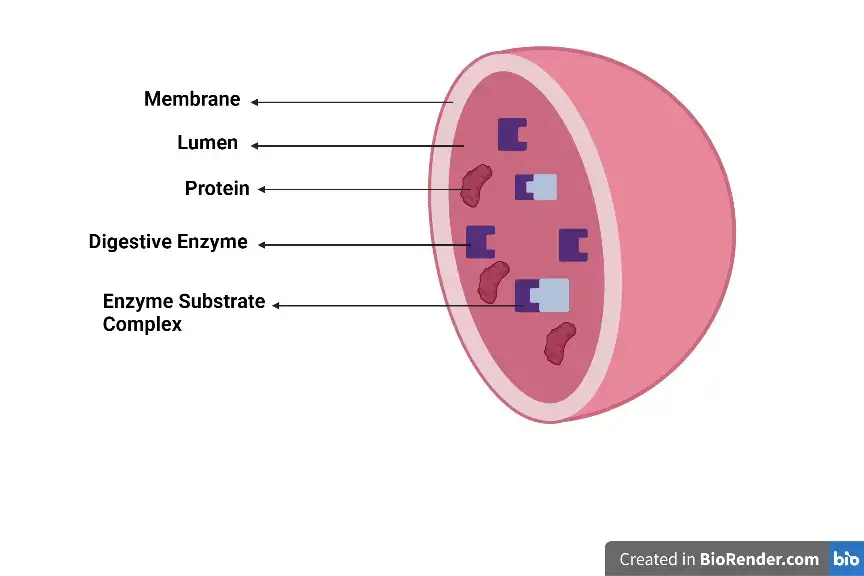
Lysosomes contain many hydrolytic enzymes. These are commonly referred to as acid hydrolases, because most of them show their optimum activity in acidic pH of the lysosome. More than 50 different enzymes are known.
The major lysosomal enzymes include-
- Proteases – The major protein degrading enzymes are cathepsins. Proteins and peptide materials brought into lysosome are hydrolyzed by them, finally producing smaller peptides and amino acids. Cathepsin B, D, H, K and L are some common forms.
- Glycosidases – These hydrolyze different carbohydrate-containing materials. β-galactosidase, β-hexosaminidase, α-glucosidase and β-glucuronidase are included in this group. They act during degradation of oligosaccharides, glycoproteins and glycolipids.
- Lipases – Lysosomal acid lipase (LAL) is an important lipid-degrading enzyme. Cholesteryl esters and triglycerides are hydrolyzed to free cholesterol and fatty acids.
- Phospholipases – Phospholipids present in cellular membrane materials are degraded by phospholipases.
- Nucleases – DNA and RNA entering into lysosome are broken down by different nucleases. DNase II is an important acidic enzyme for degradation of DNA. Ribonucleases act on RNA.
- Phosphatases – Acid phosphatase is one of the characteristic lysosomal enzymes. It hydrolyzes phosphate groups from phosphorylated compounds.
- Sulfatases – These enzymes act on sulfated compounds. Arylsulfatase A, arylsulfatase B and iduronate-2-sulfatase are some examples, mainly involved during breakdown of sulfated glycolipids and glycosaminoglycans.
- Peptidases – Smaller peptide molecules are further hydrolyzed by peptidases. Several cathepsins also show this type of activity.
- Other lysosomal hydrolases – Lysosomes also contain different enzymes for degradation of nucleotides, phosphodiesters and other complex cellular materials. The enzyme present depends upon the substrate which is to be degraded.
Why Do Lysosomal Enzymes Work at Low pH?
Most of the lysosomal enzymes are acid hydrolases. They work better at acidic pH. The reason is associated with the ionization of different amino acid residues present in the active site of enzyme, which are required for binding and catalytic activity.
A change in pH changes the charge of these amino acid groups. It may also change the arrangement of active site. At the proper acidic condition, the enzyme remains in a suitable form and the substrate can bind with it. The optimum pH is not exactly same for every lysosomal enzyme.
The inside of lysosome is maintained at about pH 4.5-5.0. V-type H⁺-ATPase (V-ATPase) is present in the lysosomal membrane. It uses ATP. During this process, H⁺ ions are pumped from the cytoplasm to the lumen, resulting in accumulation of protons inside lysosome and keeping it acidic.
Lysosomal enzymes are enclosed by the lysosomal membrane. Cytoplasm, on the other hand, has a pH close to neutral. Most acid hydrolases have much lower activity under this condition. Thus, enzymes which may escape in a small amount into the cytoplasm do not remain as active as they are inside the lysosome.
The membrane also keeps the enzymes separated from different cellular components. This compartmentalization limits direct contact of active lysosomal hydrolases with the remaining part of cell.
Why Don’t Lysosomes Digest Themselves?
Lysosomes contain many acid hydrolases. These enzymes can degrade proteins, lipids, carbohydrates and nucleic acids. The enzymes remain enclosed inside the lysosome.
A single membrane surrounds the lysosome. It separates the lysosomal lumen from cytoplasm and prevents free contact of the enzymes with other cellular materials.
The inner surface of lysosomal membrane is highly glycosylated. LAMP1 and LAMP2 are major membrane proteins having large carbohydrate regions towards the lumen. A carbohydrate-rich covering is formed on this surface.
This covering protects the luminal side of membrane from lysosomal enzymes. The membrane proteins themselves are also less easily attacked due to heavy glycosylation.
Lysosomal enzymes do not act on every type of molecule. Different enzymes have different substrates. Proteases act on proteins. Lipases hydrolyze lipids, and nucleases act on nucleic acids.
The enzymes are also kept within a separate compartment. Materials for degradation are brought into the lysosome, while most of the other cellular components remain outside.
Most lysosomal hydrolases have their better activity under acidic condition. The lysosomal lumen has a pH of about 4.5-5.0.
Cytoplasm has a nearly neutral pH. If a small amount of lysosomal enzyme enters into cytoplasm, its activity becomes much lower under this condition.
Formation and Biogenesis of Lysosomes
Lysosome formation involves the endoplasmic reticulum (ER), Golgi apparatus and endosomal system. Lysosomal enzymes and membrane proteins are first synthesized, and then transported to endosomal compartments. Late endosomes finally acquire the components required for formation of functional lysosomes.
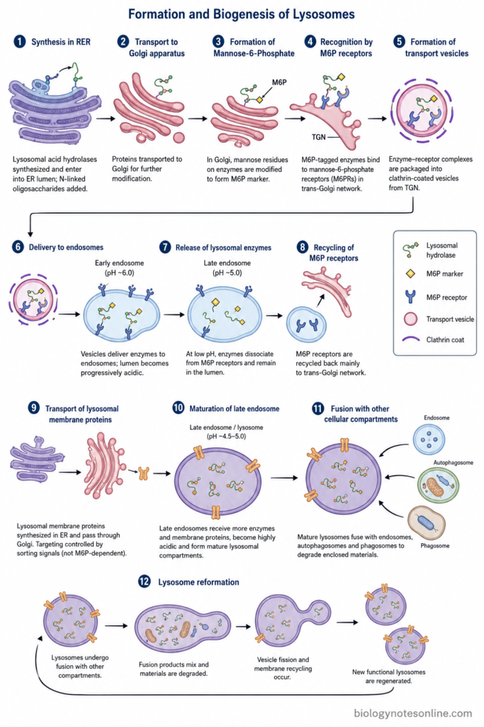
The major steps involved are as follows-
- Synthesis in Rough Endoplasmic Reticulum (RER) – Lysosomal acid hydrolases are first synthesized on the rough endoplasmic reticulum. They enter into the ER lumen during their synthesis. N-linked oligosaccharides are also added to these proteins.
- Transport to Golgi apparatus – From ER, the newly formed lysosomal enzymes are carried to the Golgi apparatus. Further modification of their carbohydrate chains takes place here.
- Formation of Mannose-6-Phosphate (M6P) – In the Golgi, selected mannose residues present on most lysosomal hydrolases are modified. A mannose-6-phosphate (M6P) recognition marker is formed. Not every lysosomal enzyme uses this pathway.
- Recognition by M6P receptors – The M6P-containing enzymes reach the trans-Golgi network (TGN). Here, they bind with mannose-6-phosphate receptors (M6PRs). Two types of these receptors are present in mammalian cells, the cation-dependent and cation-independent M6P receptors.
- Formation of transport vesicles – Enzyme-receptor complexes are packed into transport carriers from the trans-Golgi network. Clathrin-coated vesicles are involved in this transport of many lysosomal hydrolases. These vesicles move towards the endosomal compartment.
- Delivery to endosomes – The transport vesicles deliver lysosomal hydrolases to endosomes. The internal condition becomes progressively acidic during endosomal maturation.
- Release of lysosomal enzymes – At low pH of the endosomal compartment, lysosomal hydrolases separate from their M6P receptors. The enzymes remain within the endosomal lumen.
- Recycling of M6P receptors – M6P receptors are not normally degraded along with the enzymes. They are carried back mainly towards the trans-Golgi network, and can again participate in transport of lysosomal enzymes.
- Transport of lysosomal membrane proteins – Lysosomal membrane proteins are also synthesized in ER and pass through the Golgi. Their targeting is generally controlled by sorting signals present in the cytoplasmic region of the proteins. Their pathway is not dependent on M6P tagging in the same way as most soluble hydrolases.
- Maturation of late endosome – Late endosomes receive more lysosomal enzymes and membrane proteins. Their lumen becomes highly acidic and the degradative capacity increases. A mature lysosomal compartment is formed during this endosomal maturation and remodeling.
- Fusion with other cellular compartments – Mature lysosomes can fuse with endosomes, autophagosomes and phagosomes. Materials carried by these compartments are then exposed to lysosomal acid hydrolases for degradation.
- Lysosome reformation – Lysosomes are not formed only once and retained permanently. Their membranes and contents continuously undergo fusion, fission and recycling within the endolysosomal system. New functional lysosomes can again be generated from these compartments.
Types of Lysosomes
Lysosomes occur in different forms during their degradative cycle. On the basis of functional stage, the classical classification includes primary lysosomes, secondary lysosomes and residual bodies. Secondary lysosomes may be formed during heterophagy or autophagy.
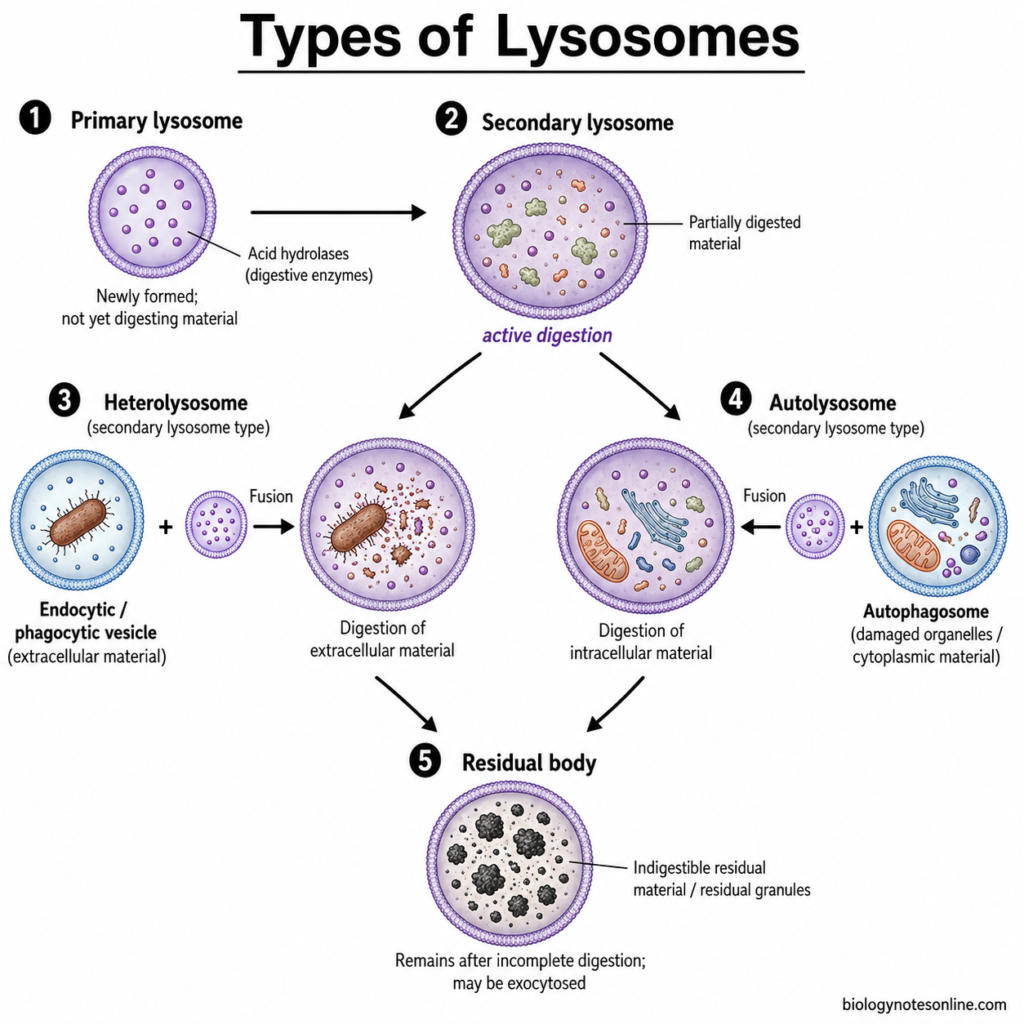
- Primary Lysosomes – These are newly formed lysosomal vesicles containing acid hydrolases. The enzymes have not yet been involved in active digestion of cellular materials. Primary lysosomes participate in fusion with endosomal, phagosomal or other degradative compartments.
- Secondary Lysosomes – A primary lysosome after interaction with a vesicle containing material for digestion forms a secondary degradative compartment. Hydrolysis of the enclosed material takes place here. Their size and internal appearance are variable because of having different materials inside.
- Heterolysosomes – These are secondary lysosomal compartments containing materials obtained from outside the cell. Bacteria, cell debris and other particles taken up by endocytosis or phagocytosis can reach this compartment. Digestion now takes place by lysosomal hydrolases.
- Autolysosomes – Intracellular materials are degraded in this type. An autophagosome carrying cytoplasmic components or damaged organelles fuses with lysosomal compartments, forming an autolysosome. The enclosed components are then broken down.
- Residual Bodies – Some materials cannot be completely degraded by lysosomal enzymes. These undigested materials remain enclosed inside membrane-bound structures called residual bodies. In some long-lived cells, such material can remain for a long period and may contribute to lipofuscin granules.
Functions of Lysosomes
Lysosomes perform several functions related to digestion and removal of different cellular materials. Some of the important functions are as follows-
- Intracellular digestion – Breaking down of proteins, lipids, carbohydrates and nucleic acids is one of the major functions of lysosomes. Different acid hydrolases are involved in it.
- Digestion of extracellular materials – Materials entering into the cell by endocytosis are finally brought to lysosomes. Here the materials are degraded.
- Phagocytic digestion – Bacteria, foreign particles and dead cellular materials engulfed by macrophages and other phagocytic cells are digested with the involvement of lysosomes. The phagosome containing these materials combines with lysosomal compartments.
- Autophagy – Old and damaged cell organelles are removed by this process. Mitochondria and portions of cytoplasm can be enclosed within an autophagosome, which is then delivered to the lysosomal system for degradation.
- Recycling of cellular materials – The products formed after lysosomal digestion are not always wasted. Amino acids, sugars and other smaller molecules can return to cytoplasm and are again used in different cellular processes.
- Removal of membrane receptors – Some cell-surface receptors after internalization are transported to lysosomes and degraded. It decreases their amount on the cell membrane.
- Nutrient sensing – Lysosome also has a role in sensing the nutritional condition of cell. mTORC1 and its associated proteins are present on the lysosomal surface during nutrient signalling.
- Plasma membrane repair – After injury to plasma membrane, lysosomes can move towards the damaged region and fuse with the membrane. Lysosomal exocytosis participates in this repair process.
- Antigen processing – In antigen-presenting cells, foreign proteins are degraded in acidic endosomal-lysosomal compartments. The peptide fragments formed are used during MHC class II antigen presentation.
- Storage and removal of indigestible materials – Some substances are not completely degraded by lysosomal enzymes. Such remaining materials can stay inside residual bodies, especially in long-lived cells.
Why Are Lysosomes Called “Suicidal Bags”?
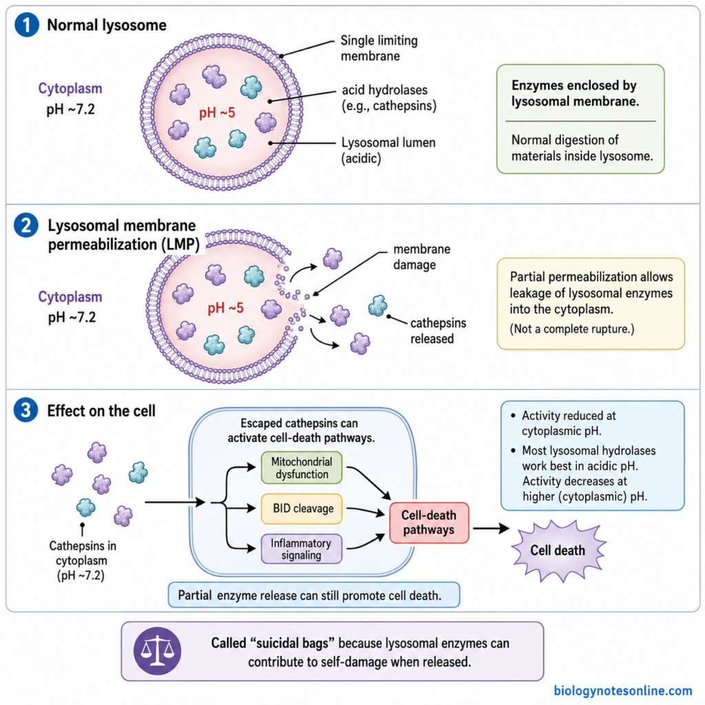
Lysosomes are sometimes referred to as the “suicidal bags” of the cell. The term is associated with their large number of hydrolytic enzymes. These enzymes are capable of degrading proteins, lipids, carbohydrates and nucleic acids.
Normally, the enzymes remain enclosed within the lysosomal membrane. They digest only those materials which are brought into the lysosomal compartment. The cytoplasmic components remain separated from them.
Damage to the lysosomal membrane can cause release of cathepsins and other hydrolases into the cytoplasm. This is referred to as lysosomal membrane permeabilization (LMP). In severe or controlled cellular conditions, released lysosomal enzymes can participate in pathways leading to cell death.
The early idea was that rupture of lysosomes could release all their digestive enzymes and result in self-digestion of the cell. This gave rise to the “suicidal bag” description. Lysosomal involvement in cell death is now known to be more complex. Complete rupture and simple digestion of the whole cell is not the normal mechanism.
Most lysosomal enzymes also require acidic condition for their better activity. The lysosomal lumen has a pH of about 5, while cytoplasm is near pH 7.2. When acid hydrolases enter into the cytoplasm, activity of many of them becomes much lower.
Partial release of lysosomal proteases can still affect the cell. Cathepsins released during LMP can activate or increase different cell-death pathways, depending upon the amount of lysosomal damage and condition of cell. Cytoplasmic protease inhibitors can also restrict some of these escaped enzymes.
Lysosomes in Plant Cells
In traditional cell biology terminology, lysosomes are mainly described in animal cells. Plant cells generally use vacuoles for many of these degradative functions. Different types of vacuoles can also be present in a plant cell.
The lytic vacuole is commonly regarded as the functional equivalent of the animal lysosome. It is a major degradative compartment of plant cells. The membrane surrounding it is called the tonoplast.
Lytic vacuoles contain different hydrolytic enzymes. Proteinases, nucleases, phosphatases, glycosidases and other acid hydrolases are found in this compartment. Many of these are similar in function to the hydrolytic enzymes of animal lysosomes.
Different cellular materials are broken down inside the lytic vacuole. Proteins, membrane components and damaged cellular materials can reach this compartment for degradation. Smaller products formed after breakdown may again take part in cellular metabolism.
Autophagy also delivers intracellular materials to the vacuole. Cytoplasmic materials and damaged organelles are enclosed within autophagosomes. These are then brought towards the lytic vacuole.
In plant cells, the autophagosome fuses with the tonoplast. Its inner membrane together with the cargo enters into the vacuolar lumen, forming an autophagic body. The cargo is degraded here by vacuolar lytic enzymes.
How Lysosomal Dysfunction Causes Disease
Most lysosomal storage diseases result from genetic defects affecting proteins required for normal lysosomal activity. The type of protein affected is not same in every disorder.
- Enzyme deficiency – A lysosomal hydrolase can be absent or have greatly reduced activity. The particular substrate is then not completely degraded and starts accumulating inside lysosomes.
- Membrane protein defects – Some disorders do not involve a digestive enzyme. Lysosomal membrane proteins can be affected, causing problems in movement of lipids, ions or other molecules. Niemann-Pick disease type C is an example involving the NPC1 or NPC2 proteins and abnormal lipid trafficking.
- Trafficking defects – Lysosomal enzymes must reach the lysosome after their synthesis. A defect in this targeting process can affect several enzymes at the same time. I-cell disease (mucolipidosis II) involves defective formation of the mannose-6-phosphate targeting signal.
- Accumulation of substrate – Undegraded or partly degraded materials remain within lysosomes. Glycolipids, glycogen, glycosaminoglycans and other substances can accumulate, depending upon the disorder.
- Loss of cellular homeostasis – Increasing lysosomal storage affects other processes of cell. Autophagy, membrane trafficking, signalling and normal metabolism can become disturbed. The effect becomes more extensive as abnormal materials continue accumulating.
Examples of Lysosomal Storage Disorders
| Disorder | Major defect/substance involved |
|---|---|
| Gaucher disease | Deficiency of glucocerebrosidase (β-glucocerebrosidase), causing accumulation mainly of glucosylceramide-related material. |
| Tay-Sachs disease | Deficiency of hexosaminidase A. GM2 ganglioside accumulates. |
| Fabry disease | Deficient α-galactosidase A activity, with accumulation mainly of globotriaosylceramide (Gb3). |
| Pompe disease | Deficiency of lysosomal acid α-glucosidase (GAA). Glycogen becomes accumulated inside lysosomes. |
| Niemann-Pick disease | The defect depends on type. Types A/B involve acid sphingomyelinase, while type C involves NPC1/NPC2-mediated lipid trafficking. |
| I-cell disease | Defect in GlcNAc-phosphotransferase and lysosomal enzyme targeting. Many hydrolases fail to receive the normal mannose-6-phosphate (M6P) signal. |
Diagnosis and Treatment Overview
Diagnosis depends upon the type of lysosomal disorder. Enzyme activity testing is commonly used where a specific enzyme deficiency is suspected. Blood cells, plasma or dried blood spots can be used for some assays.
Molecular or genetic testing is used to identify pathogenic variants and confirm many lysosomal storage disorders. It is especially important in disorders involving membrane or trafficking proteins, where a simple lysosomal enzyme assay may not provide the diagnosis.
Newborn screening is available for selected lysosomal diseases in some screening programmes. Measurement of lysosomal enzyme activity in dried blood spots is commonly used for disorders included in such programmes.
Enzyme replacement therapy (ERT) is used for certain disorders, including some forms of Gaucher, Fabry and Pompe disease. The missing or deficient enzyme is supplied in recombinant form.
Substrate-reduction therapy (SRT) is another approach for some lysosomal diseases. It reduces formation of the material which is being accumulated.
Hematopoietic stem-cell transplantation is used in selected lysosomal disorders. Gene-based approaches are also used or being developed for particular diseases, depending upon the disorder and affected tissues.
Lysosome vs Peroxisome vs Vacuole vs Endosome
| Features | Lysosome | Peroxisome | Vacuole | Endosome |
|---|---|---|---|---|
| Definition | Lysosome is a degradative organelle containing different acid hydrolases. | Peroxisome is an oxidative organelle having enzymes for fatty-acid oxidation and peroxide metabolism. | Vacuole is a membrane-bound compartment mainly associated with storage, degradation and osmotic functions. | Endosome is a membrane-bound sorting compartment of the endocytic pathway. |
| Membrane | Surrounded by a single membrane. | A single membrane is present. | It is also enclosed by a single membrane. In plants, this membrane is called tonoplast. | Endosomes have a single limiting membrane. |
| Internal pH | Acidic, generally about pH 4.5-5.0. | No strongly acidic lumen like that of lysosome. | Plant lytic vacuoles are acidic. The exact pH differs with vacuole type and condition. | Acidic condition is present. Early endosomes are less acidic, while further acidification takes place during maturation. |
| Major enzymes | Proteases, lipases, nucleases, glycosidases, phosphatases and other acid hydrolases. | Catalase, oxidases and different enzymes of fatty-acid oxidation are present. | Lytic vacuoles contain different hydrolytic enzymes. | Early endosomes have limited degradative enzymes. Hydrolytic activity increases in late endosomal compartments. |
| Main function | Intracellular digestion and recycling of cellular materials. | Oxidation of fatty acids and metabolism of hydrogen peroxide (H₂O₂) are important functions. | Storage is a major function. Lytic vacuoles also perform intracellular degradation. | Sorting of material entering by endocytosis. |
| Material handled | Proteins, lipids, carbohydrates, nucleic acids, damaged organelles and endocytosed materials are degraded. | Fatty acids and different oxidative substrates are acted upon here. | Water, ions, metabolites, pigments, proteins and waste materials can be present depending on vacuole type. | Internalized receptors, membrane proteins, nutrients and other endocytic cargo are present. |
| Hydrogen peroxide | H₂O₂ breakdown is not a characteristic lysosomal function. | H₂O₂ is produced during several oxidation reactions. Catalase converts it into water and oxygen. | It is not a major feature of vacuolar function. | Not a characteristic function of endosomes. |
| Protein targeting | Many lysosomal hydrolases pass through RER and Golgi apparatus. The mannose-6-phosphate (M6P) pathway is used by many of them. | Most matrix proteins are synthesized in cytosol and imported into peroxisome by specific targeting signals. | Vacuolar proteins generally enter the secretory pathway and are sorted towards vacuoles. | Endosomal proteins and cargo reach it through endocytic and intracellular trafficking pathways. |
| Relation with endomembrane system | Lysosome forms a part of the endolysosomal system. | It is not a classical component of the endomembrane trafficking system. | Vacuoles form a part of the endomembrane system. | It is an important component of the endomembrane and endocytic system. |
| Formation | Functional lysosomes develop through maturation and interaction of late endosomal-lysosomal compartments. | Peroxisomes grow and divide from pre-existing peroxisomes, with ER also contributing to their biogenesis. | Vacuoles develop through the endomembrane trafficking system. | Endosomes are formed during uptake and sorting of endocytosed materials, followed by maturation of endosomal compartments. |
| Major types/forms | Primary lysosomes, degradative lysosomal compartments and residual bodies are described in classical terminology. | Peroxisomal morphology differs with cell type and metabolic condition. | Lytic vacuoles and protein storage vacuoles are important plant forms. | Early endosome, recycling endosome and late endosome are major forms. |
| Autophagy | Autophagic cargo is finally degraded in lysosomal compartments. | Damaged peroxisomes themselves can be removed by pexophagy. | In plant cells, autophagic materials are delivered to the lytic vacuole for degradation. | Endosomal compartments interact with the autophagic and lysosomal pathways, but they are not the main final site of autophagic digestion. |
| Occurrence | Mainly described as the degradative organelle of animal cells. | Present in both animal and plant cells and many other eukaryotes. | Especially prominent in plant and fungal cells. | Present in eukaryotic cells having an endocytic system. |
| Plant-cell equivalent | Typical animal-type lysosome terminology is generally not used for the major plant degradative compartment. | Plant cells have peroxisomes and specialized forms such as glyoxysomes. | The lytic vacuole is commonly regarded as the functional equivalent of animal lysosome. | Plant cells also possess endosomal compartments involved in membrane and cargo trafficking. |
Frequently Asked Questions (FAQs)
1. What is a lysosome in simple words?
A lysosome is a small membrane-bound organelle containing digestive enzymes. It breaks down unwanted cellular materials, macromolecules and damaged cell components.
2. Where are lysosomes located in a cell?
Lysosomes are present in the cytoplasm of eukaryotic cells, especially animal cells. Their position is not fixed. They can move between peripheral and perinuclear regions of the cell.
3. What is the normal pH inside a lysosome?
The lysosomal lumen has an acidic pH, generally about 4.5-5.0. This condition is suitable for activity of most lysosomal acid hydrolases.
4. How many lysosomes can a cell contain?
The number is not same in every cell. A mammalian cell may contain several hundred lysosomes, while cells having greater degradative activity can have a more developed lysosomal system.
5. Why are lysosomes acidic?
V-type H⁺-ATPase (V-ATPase) present in the lysosomal membrane pumps H⁺ ions into the lumen. ATP is used during this process. Accumulation of H⁺ keeps the internal region acidic.
6. What keeps lysosomal enzymes from damaging the cytoplasm?
The enzymes remain enclosed by a single lysosomal membrane. Cytoplasm also has a nearly neutral pH, where many lysosomal acid hydrolases show much lower activity. A glycosylated covering on the luminal membrane surface provides further protection.
7. What happens to molecules after they are digested in lysosomes?
Large molecules are broken down into smaller products such as amino acids, sugars, fatty acids and nucleotides. Many of these are transported back to cytoplasm and can be used again by the cell.
8. Are lysosomes present in bacterial cells?
No. Bacteria are prokaryotic cells and lack membrane-bound organelles such as lysosomes. Degradative reactions in bacteria are carried out by enzymes present in other cellular locations.
9. Are lysosomes present in mature red blood cells?
Mature mammalian red blood cells (RBCs) do not contain lysosomes. During erythrocyte maturation, the nucleus and normal membrane-bound organelles are removed.
10. Are lysosomes single- or double-membrane organelles?
Lysosomes are single-membrane organelles. One lipid bilayer separates the acidic lysosomal lumen from the cytoplasm.
11. Which organelle produces lysosomal enzymes?
Most lysosomal enzymes are synthesized on the rough endoplasmic reticulum (RER). They are then transported to the Golgi apparatus, where further processing and sorting takes place before delivery towards lysosomal compartments.
12. What is a phagolysosome?
A phagolysosome is formed when a phagosome containing engulfed material interacts and fuses with lysosomal compartments. Bacteria and other large particles are degraded inside it.
13. What is an autolysosome?
An autolysosome is a degradative compartment formed after an autophagosome fuses with a lysosome. The cellular materials carried by the autophagosome are broken down here.
14. Is the sperm acrosome a lysosome?
The acrosome is not simply a conventional lysosome. It is a specialized lysosome-related organelle present in the sperm head, containing hydrolytic enzymes required during fertilization.
15. Can lysosomes repair the plasma membrane?
Yes. After some types of plasma membrane injury, lysosomes move towards the damaged region and fuse with the plasma membrane. Lysosomal exocytosis participates in the membrane repair process.
16. What happens when lysosomal function is defective?
Materials which normally undergo degradation can start accumulating inside lysosomes. Autophagy, membrane trafficking and other cellular processes may also become disturbed. Genetic defects affecting lysosomal enzymes, membrane proteins or trafficking pathways can produce lysosomal storage diseases.
References
- Appelqvist, H., Wäster, P., Kågedal, K., & Öllinger, K. (2013). The lysosome: From waste bag to potential therapeutic target. Journal of Molecular Cell Biology, 5(4), 214–226. https://doi.org/10.1093/jmcb/mjt022
- Ballabio, A. (2016). The awesome lysosome. EMBO Molecular Medicine, 8(2), 73–76. https://doi.org/10.15252/emmm.201505966
- Barral, D. C., Staiano, L., Almeida, C. G., Cutler, D. F., Eden, E. R., Futter, C. E., Galione, A., Marques, A. R. A., Medina, D. L., Napolitano, G., Settembre, C., Vieira, O. V., Aerts, J. M. F. G., Atakpa-Adaji, P., Bruno, G., Capuozzo, A., De Leonibus, E., Di Malta, C., Escrevente, C., . . . Seabra, M. C. (2022). Current methods to analyze lysosome morphology, positioning, motility and function. Traffic, 23(5), 238–269. https://doi.org/10.1111/tra.12839
- Boller, T., & Kende, H. (1979). Hydrolytic enzymes in the central vacuole of plant cells. Plant Physiology, 63(6), 1123–1132. https://doi.org/10.1104/pp.63.6.1123
- Bouhamdani, N., Comeau, D., & Turcotte, S. (2021). A compendium of information on the lysosome. Frontiers in Cell and Developmental Biology, 9, 798262. https://doi.org/10.3389/fcell.2021.798262
- Boya, P., & Kroemer, G. (2008). Lysosomal membrane permeabilization in cell death. Oncogene, 27(50), 6434–6451. https://doi.org/10.1038/onc.2008.310
- Coutinho, M. F., Prata, M. J., & Alves, S. (2012). Mannose-6-phosphate pathway: A review on its role in lysosomal function and dysfunction. Molecular Genetics and Metabolism, 105(4), 542–550. https://doi.org/10.1016/j.ymgme.2011.12.012
- Cui, Y., He, Y., Cao, W., Gao, J., & Jiang, L. (2018). The multivesicular body and autophagosome pathways in plants. Frontiers in Plant Science, 9, 1837. https://doi.org/10.3389/fpls.2018.01837
- de Duve, C. (2005). The lysosome turns fifty. Nature Cell Biology, 7(9), 847–849. https://doi.org/10.1038/ncb0905-847
- de Duve, C., Pressman, B. C., Gianetto, R., Wattiaux, R., & Appelmans, F. (1955). Tissue fractionation studies. 6. Intracellular distribution patterns of enzymes in rat-liver tissue. Biochemical Journal, 60(4), 604–617. https://doi.org/10.1042/bj0600604
- Futerman, A. H., & van Meer, G. (2004). The cell biology of lysosomal storage disorders. Nature Reviews Molecular Cell Biology, 5(7), 554–565. https://doi.org/10.1038/nrm1423
- Gelb, M. H., Scott, C. R., & Turecek, F. (2015). Newborn screening for lysosomal storage diseases. Clinical Chemistry, 61(2), 335–346. https://doi.org/10.1373/clinchem.2014.225771
- Ghosh, P., Dahms, N. M., & Kornfeld, S. (2003). Mannose 6-phosphate receptors: New twists in the tale. Nature Reviews Molecular Cell Biology, 4(3), 202–212. https://doi.org/10.1038/nrm1050
- Ishida, Y., Nayak, S., Mindell, J. A., & Grabe, M. (2013). A model of lysosomal pH regulation. Journal of General Physiology, 141(6), 705–720. https://doi.org/10.1085/jgp.201210930
- Kang, B.-H., Anderson, C. T., Arimura, S.-I., Bayer, E., Bezanilla, M., Botella, M. A., Brandizzi, F., Burch-Smith, T. M., Chapman, K. D., Dünser, K., Gu, Y., Jaillais, Y., Kirchhoff, H., Otegui, M. S., Rosado, A., Tang, Y., Kleine-Vehn, J., Wang, P., & Zolman, B. K. (2022). A glossary of plant cell structures: Current insights and future questions. The Plant Cell, 34(1), 10–52. https://doi.org/10.1093/plcell/koab247
- Lawrence, R. E., & Zoncu, R. (2019). The lysosome as a cellular centre for signalling, metabolism and quality control. Nature Cell Biology, 21(2), 133–142. https://doi.org/10.1038/s41556-018-0244-7
- Münz, C. (2012). Antigen processing for MHC class II presentation via autophagy. Frontiers in Immunology, 3, 9. https://doi.org/10.3389/fimmu.2012.00009
- Ney, P. A. (2011). Normal and disordered reticulocyte maturation. Current Opinion in Hematology, 18(3), 152–157. https://doi.org/10.1097/MOH.0b013e328345213e
- Nielsen, J. E., & McCammon, J. A. (2003). Calculating pKa values in enzyme active sites. Protein Science, 12(9), 1894–1901. https://doi.org/10.1110/ps.03114903
- Platt, F. M., d’Azzo, A., Davidson, B. L., Neufeld, E. F., & Tifft, C. J. (2018). Lysosomal storage diseases. Nature Reviews Disease Primers, 4, Article 27. https://doi.org/10.1038/s41572-018-0025-4
- Pu, J., Guardia, C. M., Keren-Kaplan, T., & Bonifacino, J. S. (2016). Mechanisms and functions of lysosome positioning. Journal of Cell Science, 129(23), 4329–4339. https://doi.org/10.1242/jcs.196287
- Seo, J., & Oh, D.-B. (2022). Mannose-6-phosphate glycan for lysosomal targeting: Various applications from enzyme replacement therapy to lysosome-targeting chimeras. Animal Cells and Systems, 26(3), 84–91. https://doi.org/10.1080/19768354.2022.2079719
- Settembre, C., Fraldi, A., Medina, D. L., & Ballabio, A. (2013). Signals for the lysosome: A control center for cellular clearance and energy metabolism. Nature Reviews Molecular Cell Biology, 14(5), 283–296. https://doi.org/10.1038/nrm3565
- Turk, B., & Turk, V. (2009). Lysosomes as “suicide bags” in cell death: Myth or reality? Journal of Biological Chemistry, 284(33), 21783–21787. https://doi.org/10.1074/jbc.R109.023820
- Xiong, J., & Zhu, M. X. (2016). Regulation of lysosomal ion homeostasis by channels and transporters. Science China Life Sciences, 59(8), 777–791. https://doi.org/10.1007/s11427-016-5090-x
- Xu, H., & Ren, D. (2015). Lysosomal physiology. Annual Review of Physiology, 77, 57–80. https://doi.org/10.1146/annurev-physiol-021014-071649
- Yang, C., & Wang, X. (2021). Lysosome biogenesis: Regulation and functions. Journal of Cell Biology, 220(6), e202102001. https://doi.org/10.1083/jcb.202102001
- Alberts, B., Johnson, A., Lewis, J., Raff, M., Roberts, K., & Walter, P. (2002). Molecular biology of the cell (4th ed.). Garland Science. https://www.ncbi.nlm.nih.gov/books/NBK21054/
- Cooper, G. M. (2000). The cell: A molecular approach (2nd ed.). Sinauer Associates. https://www.ncbi.nlm.nih.gov/books/NBK9839/
- Freeze, H. H. (2009). Genetic disorders of glycan degradation. In A. Varki, R. D. Cummings, J. D. Esko, H. H. Freeze, P. Stanley, C. R. Bertozzi, G. W. Hart, & M. E. Etzler (Eds.), Essentials of glycobiology (2nd ed.). Cold Spring Harbor Laboratory Press. https://www.ncbi.nlm.nih.gov/books/NBK1934/
- Freeze, H. H., Kinoshita, T., & Schnaar, R. L. (2017). Genetic disorders of glycan degradation. In A. Varki, R. D. Cummings, J. D. Esko, et al. (Eds.), Essentials of glycobiology (3rd ed.). Cold Spring Harbor Laboratory Press. https://www.ncbi.nlm.nih.gov/books/NBK453095/
- Mehta, A., Beck, M., & Sunder-Plassmann, G. (Eds.). (2006). Fabry disease: Perspectives from 5 years of FOS. Oxford PharmaGenesis. https://www.ncbi.nlm.nih.gov/books/NBK11586/
- Neufeld, E. F. (2006). Enzyme replacement therapy—A brief history. In A. Mehta, M. Beck, & G. Sunder-Plassmann (Eds.), Fabry disease: Perspectives from 5 years of FOS. Oxford PharmaGenesis. https://www.ncbi.nlm.nih.gov/books/NBK11588/
- Saftig, P. (2006). Physiology of the lysosome. In A. Mehta, M. Beck, & G. Sunder-Plassmann (Eds.), Fabry disease: Perspectives from 5 years of FOS. Oxford PharmaGenesis. https://www.ncbi.nlm.nih.gov/books/NBK11604/
- Siegel, G. J., Agranoff, B. W., Albers, R. W., Fisher, S. K., & Uhler, M. D. (Eds.). (1999). Basic neurochemistry: Molecular, cellular and medical aspects (6th ed.). Lippincott-Raven. https://www.ncbi.nlm.nih.gov/books/NBK20385/
- Suzuki, K., & Vanier, M. T. (1999). The lysosome. In G. J. Siegel, B. W. Agranoff, R. W. Albers, S. K. Fisher, & M. D. Uhler (Eds.), Basic neurochemistry: Molecular, cellular and medical aspects (6th ed.). Lippincott-Raven. https://www.ncbi.nlm.nih.gov/books/NBK28119/
- Suzuki, K., & Vanier, M. T. (1999). Lysosomal disease. In G. J. Siegel, B. W. Agranoff, R. W. Albers, S. K. Fisher, & M. D. Uhler (Eds.), Basic neurochemistry: Molecular, cellular and medical aspects (6th ed.). Lippincott-Raven. https://www.ncbi.nlm.nih.gov/books/NBK28215/
- Tokarev, A. A., Alfonso, A., & Segev, N. (2009). Overview of intracellular compartments and trafficking pathways. In N. Segev (Ed.), Trafficking inside cells: Pathways, mechanisms and regulation. Landes Bioscience/Springer. https://www.ncbi.nlm.nih.gov/books/NBK7286/
- Lysosomal Storage Disease Book Chapters
- Bremova-Ertl, T., & Patterson, M. C. (2025). Niemann-Pick disease type C. In M. P. Adam, S. Bick, G. M. Mirzaa, et al. (Eds.), GeneReviews. University of Washington, Seattle. https://www.ncbi.nlm.nih.gov/books/NBK1296/
- Mehta, A., & Hughes, D. A. (2024). Fabry disease. In M. P. Adam, S. Bick, G. M. Mirzaa, et al. (Eds.), GeneReviews. University of Washington, Seattle. https://www.ncbi.nlm.nih.gov/books/NBK1292/
- Sperry, E., Leslie, N., Berry, L., & Pena, L. (2025). Pompe disease. In M. P. Adam, S. Bick, G. M. Mirzaa, et al. (Eds.), GeneReviews. University of Washington, Seattle. https://www.ncbi.nlm.nih.gov/books/NBK1261/