

Plant cell is the basic structural and functional unit of plants. A typical plant cell is a eukaryotic cell, which means it contains a membrane-bound nucleus. Different membrane-bound cell organelles are also present in the cytoplasm. Plant cells are found in the members of kingdom Plantae.
The major structures that distinguish the plant cell include cell wall, plastids and a large vacuole. The cell wall is present outside the plasma membrane and it provides shape and mechanical support to the cell. Plastids are used in photosynthesis, pigment formation and storage, based on their type. The vacuole is a fluid-filled organelle. It is used for storage and also helps in maintaining the turgor condition of the cell.
The plant cell represented in an illustrated diagram is a generalized plant cell. It shows the major cell organelles together for understanding the cell structure. However, all plant cells do not have the same shape and organelle arrangement. These may vary according to the type, stage and function of the cell.
Study of plant cell structure is important for understanding the different activities carried out within a plant. It explains photosynthesis, storage of materials, cell growth, transport and mechanical support. These cellular activities are necessary for normal growth and development of the plant.
What Is a Plant Cell?
A plant cell is the smallest living structural and functional unit of plant tissues. This is the direct plant cell definition. It is a eukaryotic plant cell, which means it has a membrane-bound nucleus. The cell also contains cytoplasm and different cell organelles.
Every living plant cell has a plasma membrane. It surrounds the living content of the cell. The cytoplasm, nucleus and the other organelles form the protoplast. In most of the plant cells, a cell wall is present outside the plasma membrane and surrounds this protoplast.
The plant cell meaning should not be used only for an empty wall structure. A living plant cell has its protoplast. During the maturation of some cells, such as the xylem vessel elements, the nucleus and cytoplasmic contents are lost by programmed cell death. The thick cell wall and an empty lumen are left behind. This is a dead mature cell element, and not a living cell.
Plant cells are not similar in all the plant tissues. Their size, shape, wall structure and organelle contents may change according to the tissue, age and function of the cell. A young meristematic cell is different from a leaf mesophyll cell. The mature xylem element is again very different.
Main Characteristics of Plant Cells
The characteristics of plant cell are associated with its wall, plastids, vacuole and cellular organization. Some of the important features of plant cells are as follows-
- Eukaryotic nature- Plant cells are eukaryotic cells with a membrane-bound nucleus. The cytoplasm contains mitochondria, endoplasmic reticulum, Golgi bodies, ribosomes and other cell organelles.
- Cell wall- A cell wall is present outside the plasma membrane in most plant cells. It mainly contains cellulose, hemicellulose and pectin. The wall provides shape, protection and mechanical strength to the cell.
- Plastids- Plastids are characteristic organelles of plant cells. Chloroplasts perform photosynthesis, while chromoplasts contain pigments. Amyloplasts are mainly used for formation and storage of starch.
- Vacuole- Many mature plant cells contain a large central vacuole. It stores water, ions, sugars, pigments and different metabolites. The vacuole also takes part in degradation and maintenance of internal cell condition.
- Plasmodesmata- The neighbouring living cells are connected through small channels called plasmodesmata. These channels pass across the cell wall. They allow movement of substances and signals from one cell to another.
- Turgor support- Water present within the vacuole produces turgor pressure against the cell wall. This pressure helps in maintaining cell stiffness and supports the soft parts of plants. It is also important during cell enlargement.
- Cell plate- Cytokinesis in most plant cells takes place by formation of a cell plate. Vesicles gather in the middle region of the dividing cell and fuse together. This later develops into a new wall between two daughter cells.
- Starch storage- The major reserve carbohydrate of plants is starch. It is stored as granules within chloroplasts or amyloplasts. Plants can also contain soluble carbohydrates such as sucrose.
- Regeneration- Plant cells have a high capacity for differentiation. Under suitable conditions, many living cells can form new tissues or organs. However, this capacity is not equal in every cell type and plant species.
Size and Shape of Plant Cells
Plant cells show considerable differences in their size and shape. It depends on the tissue, stage of development and the function performed by the cell. Their size and shape also change during cell differentiation.
Typical Size
- Size variation- Plant cells do not have one fixed or universal size. Some cells remain very small, whereas some mature cells increase many times during their development. So, one size range cannot represent all types of plant cells.
- Meristematic cells- Meristematic cells are usually small and closely arranged. They contain dense cytoplasm, a prominent nucleus and small vacuolar compartments. These cells are actively involved in cell division.
- Mature cells- Many mature parenchyma cells become much larger than the meristematic cells. During this process, the vacuole expands and occupies a major portion of the cell. It allows the cell to increase its size without producing the same amount of new cytoplasm.
- Size range- A general size value may be given for a particular tissue or species. It should not be considered the normal size for every plant cell. Vessel elements, fibres, epidermal cells and storage cells may have very different dimensions.
Why Many Plant Cells Look Rectangular
- The cell wall is a firm structure surrounding the protoplast. It controls the direction of cell expansion and helps in maintaining the cell shape. Due to this wall, many plant cells show straight or angular boundaries.
- Water within the cell produces turgor pressure against the cell wall. The wall resists this pressure. Together, they maintain the size, shape and stiffness of living plant cells.
- Plant cells remain fixed and closely packed within tissues. Their walls are shared with the neighbouring cells. This arrangement often produces rectangular, polygonal or polyhedral forms rather than a free rounded form.
- A plant cell diagram is a simplified two-dimensional representation. The actual cell has a three-dimensional structure. A cell that looks rectangular in a section may have several faces and an irregular form when observed in three dimensions.
Shape Variation Among Specialized Cells
- Parenchyma- Parenchyma cells are commonly rounded, isodiametric or polygonal. Their exact form depends on the packing and presence of intercellular spaces. Some parenchyma cells are also elongated according to their location.
- Fibres- Plant fibres are narrow and highly elongated cells. Their ends are generally tapered. The thick secondary walls provide mechanical strength to the plant tissues.
- Vessel elements- Vessel elements are elongated and tubular in shape. They are joined end to end and form the water-conducting vessels. At maturity, their cytoplasmic contents are lost and a hollow conducting structure is produced.
- Guard cells- Guard cells are commonly kidney-shaped in eudicots and many non-grass plants. In grasses, they are usually dumbbell-shaped. Their shape changes with turgor and it controls the opening and closing of the stomatal pore.
- Root-hair cells- A root-hair cell forms a long tubular projection from the root epidermis. This projection increases the surface area of the cell. It is mainly used for absorption of water and mineral nutrients from the soil.
- Pavement cells- Leaf pavement cells often have an irregular and lobed shape. These cells appear like the pieces of a jigsaw puzzle. The degree of lobing differs between plant species, leaf regions and stages of cell development.
Labeled Diagram of a Typical Plant Cell
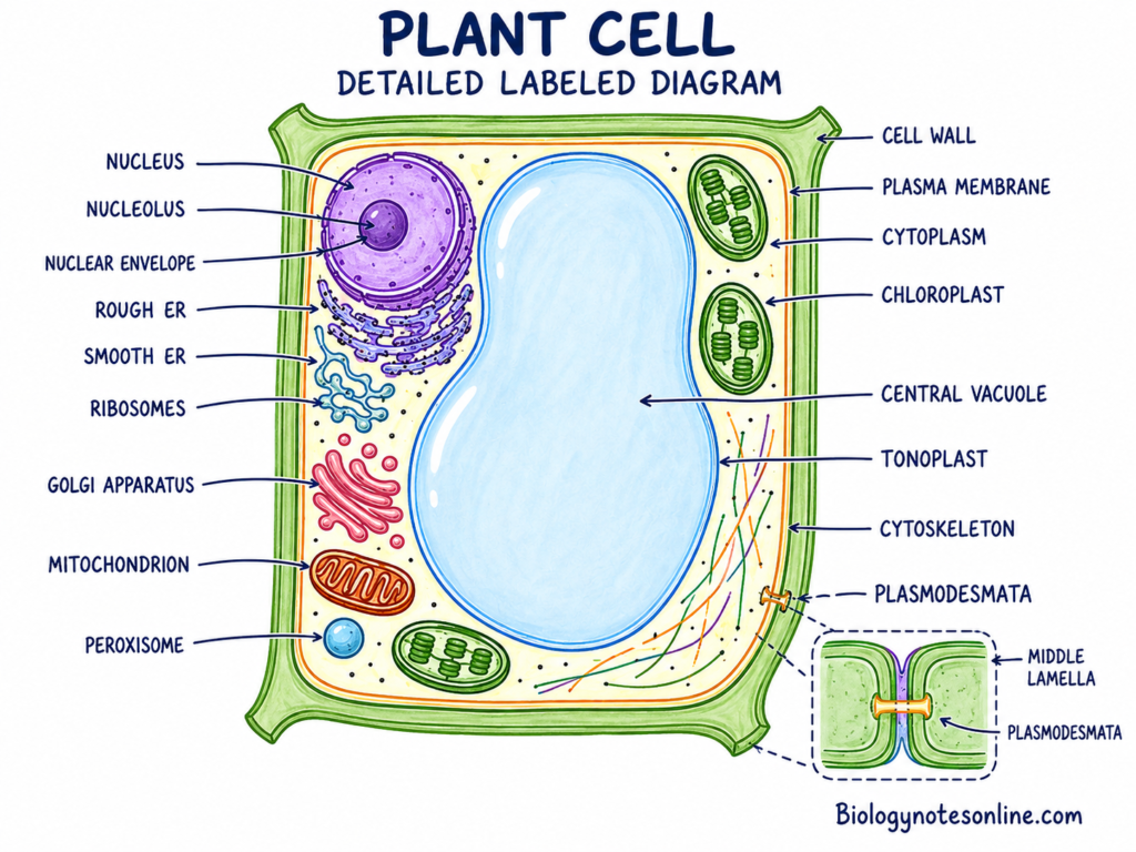
Plant Cell Organelles and Their Functions
Plant cells have different cell organelles and cellular structures. These organelles perform various specific functions to maintain cellular metabolism, growth and development. Some of them are characteristic structures of plant cells, while most of the organelles are also present in other eukaryotic cells.
The major plant cell organelles and their functions are summarized below-
| Organelle or structure | Membrane status | Main composition | Main function | Plant-specific or shared |
|---|---|---|---|---|
| Cell wall | Non-membranous | Cellulose, hemicelluloses and pectins | Protection, shape and mechanical support | Plant-characteristic |
| Plasma membrane | Single membrane | Phospholipids, sterols and proteins | Selective transport and signaling | Shared |
| Cytoplasm | No surrounding membrane | Cytosol, organelles and cytoskeleton | Site of different metabolic reactions | Shared |
| Nucleus | Double membrane | DNA, RNA, proteins and nucleoplasm | Storage and expression of genetic material | Shared |
| Ribosomes | Non-membranous | rRNA and proteins | Protein synthesis | Shared |
| Endoplasmic reticulum | Single membrane | Membrane tubules, cisternae and proteins | Protein and lipid synthesis | Shared |
| Golgi apparatus | Single membrane | Stacked cisternae and enzymes | Modification, sorting and secretion | Shared |
| Mitochondria | Double membrane | Membranes, enzymes, DNA and ribosomes | Cellular respiration and ATP production | Shared |
| Plastids | Double membrane | Membranes, enzymes, DNA and ribosomes | Photosynthesis, storage and biosynthesis | Plants and algae |
| Central vacuole | Single membrane | Cell sap enclosed by tonoplast | Storage, turgor and degradation | Prominent in plant cells |
| Peroxisomes | Single membrane | Oxidative enzymes and catalase | Oxidative metabolism | Shared |
| Cytoskeleton | Non-membranous | Actin filaments and microtubules | Movement, division and cell organization | Shared |
| Plasmodesmata | Membrane-lined channels | Plasma membrane, cytoplasm and desmotubule | Cell-to-cell transport | Plant-characteristic |
| Storage materials | Variable | Starch, oils, proteins, pigments and crystals | Storage and deposition | Shared, but forms vary |
1. Cell Wall
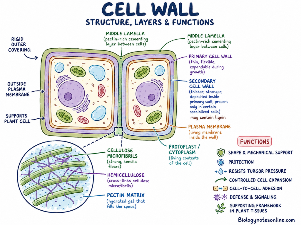
The cell wall is the rigid outer covering of the plant cell. It is present outside the plasma membrane and surrounds the protoplast. It is a major distinguishing structure between a plant cell and an animal cell.
The wall contains cellulose microfibrils, which are placed within a hydrated matrix. This matrix is mainly made up of hemicelluloses, pectins, proteins and water. The proportion and arrangement of these substances are not the same in all cell walls.
Cellulose microfibrils provide tensile strength to the wall. Hemicelluloses connect with the cellulose surface, while pectins help in wall hydration, porosity and adhesion between cells.
a. Middle Lamella
The middle lamella is present between the primary cell walls of two neighbouring cells. It is mainly rich in pectic substances.
This region acts as a cementing layer. It holds the neighbouring cells together and helps in the formation of a continuous plant tissue.
b. Primary Cell Wall
The primary cell wall is formed while the cell is growing. It is relatively thin and flexible as compared to a secondary cell wall.
The primary wall can expand when water enters the cell and the protoplast increases in size. Its cellulose microfibrils and matrix components are rearranged during this growth.
c. Secondary Cell Wall
The secondary cell wall is deposited inside the primary wall in certain specialized cells. Its formation generally starts after cell expansion has stopped or become greatly reduced.
It is commonly thicker and stronger than the primary wall. Secondary walls of xylem cells and fibres may contain lignin, which gives stiffness and resistance to compression.
Functions of the Cell Wall
- The cell wall gives a fixed form and mechanical support to the plant cell.
- It protects the protoplast from physical injury and some harmful environmental conditions.
- The wall resists the internal turgor pressure produced when water enters the cell. Due to this, the cell does not burst during normal water uptake.
- Primary walls allow controlled cell expansion. The arrangement of cellulose microfibrils also affects the direction of cell growth.
- Cell-wall components take part in cellular signaling. Changes in the wall can be detected by the plant cell during growth, injury or pathogen attack.
- The wall contains defensive compounds and wall-derived molecules, which may activate defense responses during an infection.
- It forms a continuous supporting framework in stems, leaves, roots and other plant parts.
2. Plasma Membrane
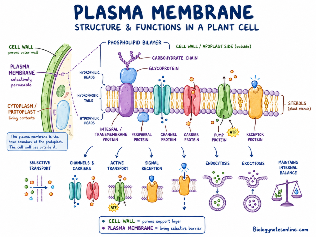
The plasma membrane is a thin living membrane present immediately inside the cell wall. It forms the actual boundary of the plant-cell protoplast.
It is mainly formed of a phospholipid bilayer. Different proteins, sterols and carbohydrate-containing molecules are present within or attached to this bilayer.
Some proteins extend completely across the membrane. Others are attached only to its inner or outer surface. These proteins act as channels, carriers, pumps, enzymes and receptors.
The plasma membrane is different from the cell wall. The wall matrix is porous and allows water and many small substances to move through it. The plasma membrane is selectively permeable and controls which substances enter or leave the living cell.
Functions of the Plasma Membrane
- The plasma membrane controls the movement of ions, water and molecules between the cytoplasm and the surrounding region.
- Membrane channels and carrier proteins allow selective transport of different substances.
- Pumps present in the membrane use energy to move ions against their concentration gradient.
- Receptor proteins detect plant hormones, pathogen molecules and different environmental signals.
- It takes part in cellular signaling and helps the cell to respond according to the signal received.
- The plasma membrane also participates in endocytosis, exocytosis and delivery of vesicle materials to the cell surface.
- It maintains the internal chemical condition of the plant cell.
3. Cytoplasm and Cytosol
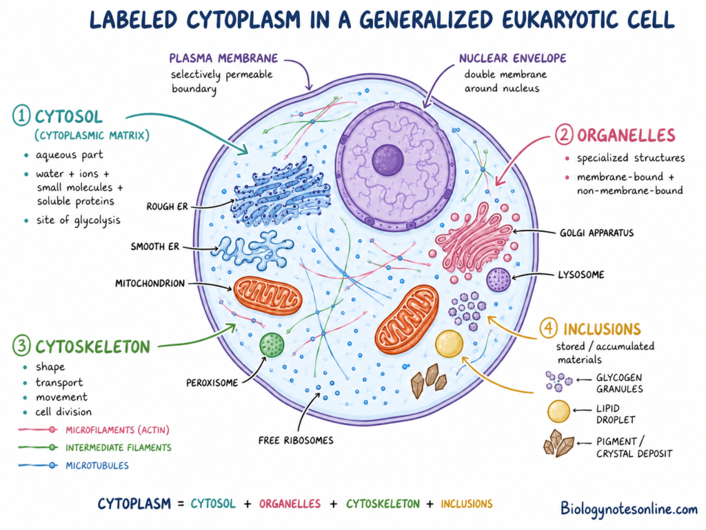
The cytoplasm is the cellular region present between the plasma membrane and nucleus. It includes cytosol, organelles, cytoskeleton and different suspended materials.
The cytosol is only the fluid portion of the cytoplasm. It is mainly made up of water, ions, soluble proteins, sugars, amino acids and different metabolites.
Therefore, cytoplasm and cytosol are not the same. Cytosol is one component of the cytoplasm.
Different organelles remain suspended or arranged within the cytoplasm. The cytoplasm also provides the medium through which many substances are transported from one part of the cell to another.
In many mature plant cells, the central vacuole occupies most of the inner space. Due to this, the cytoplasm remains as a thin layer near the plasma membrane, together with some cytoplasmic strands passing around the vacuole.
Functions of the Cytoplasm and Cytosol
- The cytosol is the site of several metabolic reactions. Glycolysis and many steps of carbohydrate, amino-acid and nucleotide metabolism take place here.
- Cytoplasm holds the cell organelles and provides a medium for their activities.
- It allows movement and distribution of metabolites, proteins and signaling molecules.
- Cytoplasmic streaming moves the cytoplasm and organelles within the cell. This process is especially clear in large plant cells.
- Streaming helps in the distribution of chloroplasts, mitochondria and other cellular materials.
- The cytoplasm also connects different regions of the cell and allows rapid exchange of substances between organelles.
4. Nucleus and Nucleolus
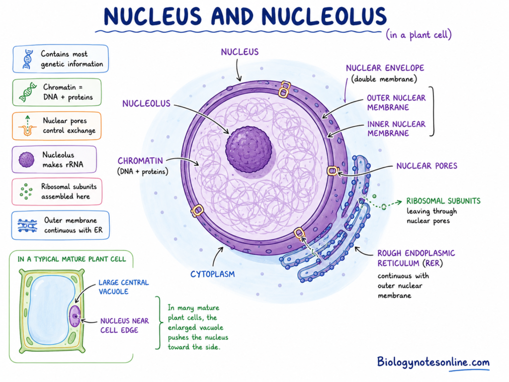
The nucleus is a membrane-bound organelle that contains most of the genetic information of a plant cell. It is generally one of the larger organelles present in the cytoplasm.
The nucleus is enclosed by a double membrane called the nuclear envelope. The outer nuclear membrane is continuous at some regions with the endoplasmic reticulum.
Several nuclear pores are present in the nuclear envelope. These pores control the movement of RNA, proteins, ribosomal subunits and other molecules between the nucleus and cytoplasm.
The genetic material remains in the form of chromatin, which is made up of DNA and associated proteins. During cell division, chromatin becomes highly condensed and forms visible chromosomes.
The position of nucleus is not fixed in every plant cell. In young cells, it may be present near the central region. When the central vacuole enlarges, the nucleus is commonly pushed towards the side of the cell.
Nucleolus
The nucleolus is a dense non-membranous region present within the nucleus. A nucleus may contain one or more nucleoli depending on the cell type and its activity.
Ribosomal RNA (rRNA) is produced and processed in the nucleolus. Ribosomal proteins made in the cytoplasm are transported into the nucleus and combine with rRNA.
The early formation of small and large ribosomal subunits takes place here. These subunits later move through the nuclear pores into the cytoplasm.
Functions of the Nucleus and Nucleolus
- The nucleus stores DNA and protects the genetic information of the plant cell.
- DNA replication takes place within the nucleus before cell division.
- The nucleus controls gene expression through the process of transcription.
- Messenger RNA, transfer RNA and different regulatory RNA molecules are produced from nuclear DNA.
- By controlling gene expression, the nucleus regulates growth, metabolism, differentiation and response of the cell.
- The nucleolus produces ribosomal RNA and takes part in the initial assembly of ribosomal subunits.
- Nuclear pores allow controlled exchange of proteins and RNA between the nucleus and cytoplasm.
5. Ribosomes
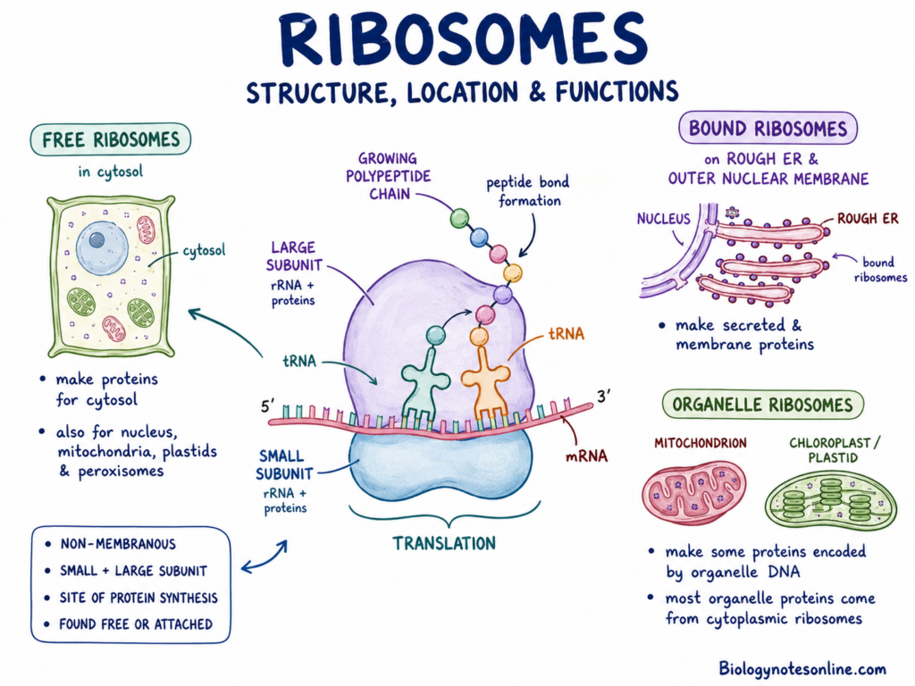
Ribosomes are small cellular structures responsible for protein synthesis. They are not surrounded by a membrane, hence ribosomes are not membrane-bound organelles.
They are made up of ribosomal RNA (rRNA) and proteins. Each ribosome contains a small subunit and a large subunit.
Free ribosomes are present within the cytosol. Some ribosomes become attached to the outer surface of the rough endoplasmic reticulum and nuclear envelope.
Mitochondria and plastids also contain their own ribosomes. These organelle ribosomes synthesize some proteins encoded by mitochondrial or plastid DNA.
Most of the mitochondrial and plastid proteins, however, are encoded by nuclear genes and produced by cytoplasmic ribosomes.
Functions of Ribosomes
- Ribosomes perform translation, in which the nucleotide sequence of messenger RNA is converted into an amino-acid sequence.
- Free ribosomes mainly synthesize proteins used within the cytosol and some proteins transported to the nucleus, mitochondria, plastids and peroxisomes.
- Ribosomes attached to the rough endoplasmic reticulum produce many secreted and membrane proteins.
- They join amino acids by peptide bonds and form a growing polypeptide chain.
- Mitochondrial and plastid ribosomes synthesize a limited number of proteins required by these organelles.
6. Endoplasmic Reticulum
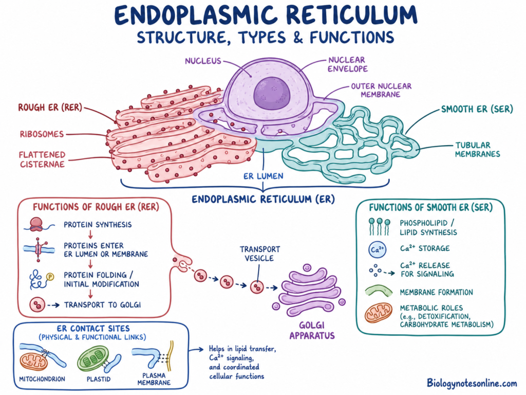
The endoplasmic reticulum (ER) is a continuous network of membrane tubules and flattened sacs present within the cytoplasm. It is connected with the outer membrane of the nuclear envelope.
The inner space of the ER is called the ER lumen. Materials can move through this lumen and through the membrane network.
There are two major regions of the endoplasmic reticulum. These are the rough endoplasmic reticulum and smooth endoplasmic reticulum.
a. Rough Endoplasmic Reticulum
The rough endoplasmic reticulum (RER) contains ribosomes attached to its cytosolic surface. Due to these ribosomes, the membrane has a rough appearance.
Rough ER is commonly formed of flattened membrane sacs called cisternae. Newly synthesized proteins enter the ER lumen or become inserted within the ER membrane.
Inside the ER, proteins undergo initial folding and some chemical modification. Incorrectly folded proteins can be retained and degraded.
b. Smooth Endoplasmic Reticulum
The smooth endoplasmic reticulum (SER) does not have ribosomes attached to its surface. It commonly contains more tubular membrane regions.
Smooth ER is involved in synthesis of phospholipids and different other lipids. The amount and activity of smooth ER vary in different plant tissues.
The ER also stores some calcium ions. Calcium can be released from the ER during signaling and other cellular responses.
Functions of the Endoplasmic Reticulum
- Rough ER synthesizes proteins that are secreted outside the cell or inserted into cellular membranes.
- It also produces proteins transported to the Golgi apparatus, vacuoles and other parts of the endomembrane system.
- Newly synthesized proteins are folded and initially modified within the ER.
- Smooth ER participates in the synthesis of phospholipids and other lipids.
- It takes part in calcium storage, calcium regulation and cellular signaling.
- The ER contributes to the formation of new cellular membranes.
- ER membranes form contact regions with the plasma membrane, mitochondria, plastids and other organelles.
- It also has metabolic roles which differ according to the type and condition of the plant cell.
7. Golgi Apparatus
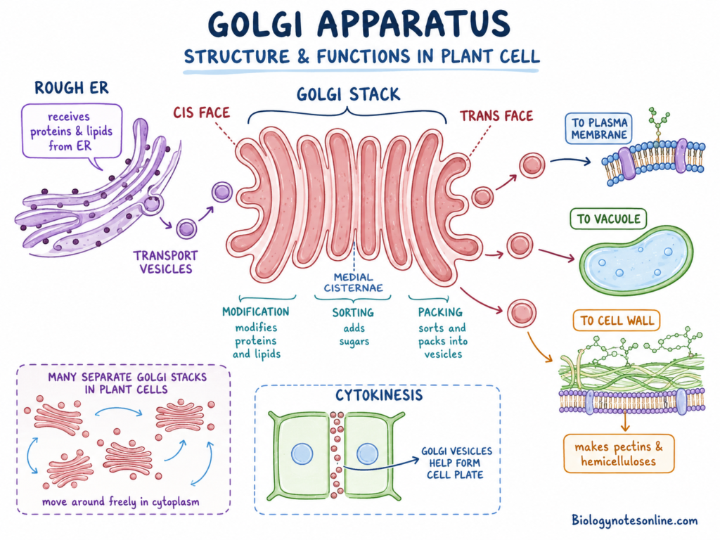
The Golgi apparatus, also called Golgi bodies or Golgi complex, is a membrane-bound organelle present within the cytoplasm. It is made up of flattened membrane sacs known as cisternae.
Plant cells generally contain many separate Golgi stacks. These stacks move within the cytoplasm along the cytoskeleton and are not present as one permanently fixed Golgi body.
A Golgi stack contains a cis region, medial region and trans region. The cis side receives materials from the endoplasmic reticulum, while the trans side sorts and sends the processed materials.
Proteins and lipids are transported to the Golgi within small membrane vesicles. These materials pass through different cisternae where they are modified.
Functions of the Golgi Apparatus
- Golgi bodies modify proteins and lipids received from the endoplasmic reticulum.
- Sugars and other chemical groups may be attached to proteins during their movement through the Golgi.
- The Golgi sorts the modified products and packs them into transport vesicles.
- These vesicles carry materials to the plasma membrane, vacuole, cell wall and other cellular regions.
- Many wall polysaccharides, especially pectins and hemicelluloses, are synthesized within the plant Golgi.
- The wall polysaccharides are packed into vesicles and secreted outside the plasma membrane.
- During cytokinesis, Golgi-derived vesicles move towards the central region of the dividing cell.
- These vesicles fuse together and take part in formation of the cell plate.
8. Mitochondria
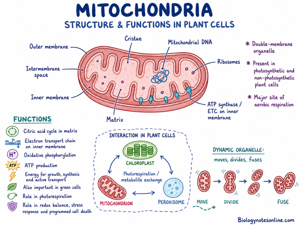
Mitochondria are double-membrane organelles present in photosynthetic and non-photosynthetic plant cells. They are important sites of aerobic cellular respiration.
A mitochondrion has an outer membrane and a highly folded inner membrane. The folds of the inner membrane are known as cristae.
The space enclosed by the inner membrane is called the mitochondrial matrix. Enzymes of the citric acid cycle and several other metabolic processes are present within this matrix.
The electron transport chain and ATP synthase are associated with the inner mitochondrial membrane.
Mitochondria contain their own DNA and ribosomes. However, most mitochondrial proteins are produced from nuclear genes and transported into the organelle.
Plant mitochondria are dynamic. They change shape, divide, fuse and move through the cytoplasm. Temporary contact can also occur between mitochondria, plastids, peroxisomes and ER.
Functions of Mitochondria
- Mitochondria are the major sites of aerobic cellular respiration.
- The citric acid cycle takes place mainly within the mitochondrial matrix.
- Electron transport and oxidative phosphorylation take place at the inner mitochondrial membrane.
- These reactions are used for the production of adenosine triphosphate (ATP).
- Mitochondria provide energy required for active transport, synthesis, growth and other cellular activities.
- They occur in green cells also. Photosynthesis does not replace the requirement of mitochondrial respiration.
- Plant mitochondria take part in photorespiration through exchange of metabolites with chloroplasts and peroxisomes.
- They are also involved in redox regulation, stress responses and programmed cell death.
9. Plastids
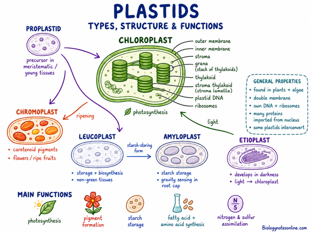
Plastids are double-membrane organelles found in plants and algae. These organelles are used in photosynthesis, pigment formation, storage and synthesis of different compounds.
All plastids develop from small precursor plastids called proplastids. Proplastids are mainly present in meristematic and young plant tissues.
Plastids contain their own DNA and ribosomes. But a large number of plastid proteins are encoded by nuclear genes and transported into the plastid.
Some plastid types can change into another plastid type. This interconversion depends on tissue development, light and other environmental conditions.
a. Chloroplasts
Chloroplasts are green plastids that perform photosynthesis. They are surrounded by an outer and inner envelope membrane.
The internal fluid region is called the stroma. Enzymes required for carbon fixation and different biosynthetic reactions are present within the stroma.
Chloroplasts contain an internal membrane system made of flattened sacs called thylakoids. Several thylakoids are arranged into stacks called grana.
The grana are connected by unstacked stroma thylakoids. Chlorophyll, electron carriers and photosynthetic protein complexes are present within the thylakoid membranes.
Chloroplasts are common in leaf mesophyll and other green tissues. They are not present in every plant cell. Most root cells and many internal non-green cells contain other plastid forms.
b. Chromoplasts
Chromoplasts are coloured plastids that synthesize and store carotenoid pigments. These pigments produce yellow, orange and red colours.
Chromoplasts are common in flowers, ripe fruits and some roots. They may develop from proplastids or by conversion of chloroplasts during tissue development.
The green chloroplasts of some fruits change into chromoplasts during ripening. Chlorophyll is lost and carotenoids become accumulated.
c. Leucoplasts and Amyloplasts
Leucoplasts are colourless plastids mainly found in non-photosynthetic tissues. They occur in roots, seeds, tubers and internal storage regions.
Leucoplasts take part in storage and biosynthesis. Different forms may store starch, oils or proteins.
Amyloplasts are specialized plastids used for starch synthesis and storage. Starch is deposited within these plastids as starch grains.
Some amyloplasts in root-cap cells also take part in gravity sensing because their dense starch grains settle according to gravity.
d. Etioplasts
Etioplasts develop in tissues grown under darkness. These plastids do not contain a fully developed photosynthetic thylakoid system.
They contain a characteristic membrane arrangement called the prolamellar body. Chlorophyll is not fully formed under dark conditions.
After exposure to light, etioplasts develop towards chloroplasts. Chlorophyll is produced and the thylakoid membrane system becomes organized.
Functions of Plastids
- Chloroplasts perform photosynthesis and convert light energy into chemical energy.
- Plastids synthesize fatty acids, some amino acids and several other cellular compounds.
- Chromoplasts synthesize and store carotenoid pigments.
- These pigments provide colour to flowers, fruits and some storage tissues.
- Leucoplasts take part in storage and biosynthesis within non-green tissues.
- Amyloplasts synthesize and store starch.
- Plastids also participate in nitrogen and sulfur assimilation.
- Some plastids can change their structure and function according to tissue development or environmental conditions.
10. Central Vacuole and Tonoplast
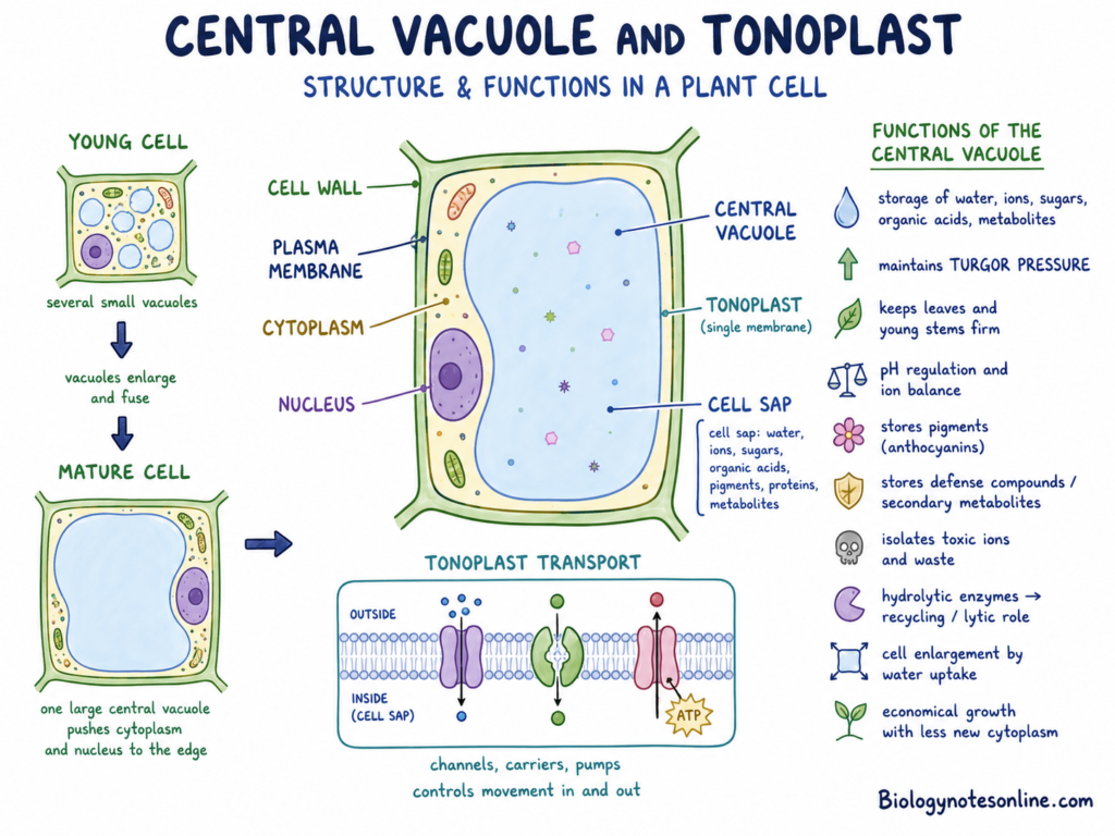
The central vacuole is a large fluid-filled organelle found in many mature plant cells. It may occupy a major part of the total cell volume.
The vacuole is surrounded by a single membrane known as the tonoplast. The fluid and dissolved substances present inside the vacuole are called cell sap.
Cell sap contains water, mineral ions, sugars, organic acids, pigments, proteins and different metabolites. Its composition varies greatly between cell types.
Young plant cells may contain several small vacuoles. During cell development, these vacuoles commonly enlarge and fuse to produce a large central vacuole.
Functions of the Central Vacuole and Tonoplast
- The vacuole stores water, ions, sugars, organic acids and different metabolites.
- It maintains turgor pressure when water enters the cell.
- Turgor pressure helps in maintaining the stiffness of leaves, young stems and other non-woody tissues.
- The tonoplast contains channels, carriers and pumps which control movement of substances into and outside the vacuole.
- The vacuole takes part in pH regulation and maintenance of ion balance.
- Pigments such as anthocyanins may be stored within the cell sap.
- Several secondary metabolites and defensive compounds are also accumulated inside the vacuole.
- Toxic ions, waste products and harmful compounds can be separated from the cytoplasm by vacuolar storage.
- Some vacuoles contain hydrolytic enzymes that break down proteins, nucleic acids and other macromolecules.
- Due to these enzymes, the vacuole also performs recycling and lytic functions.
- Vacuolar enlargement allows the cell to grow mainly by taking up water. The cell does not need to produce an equal amount of new cytoplasm.
- This makes the enlargement of plant cells more economical in terms of cellular materials and energy.
11. Peroxisomes and Glyoxysomes
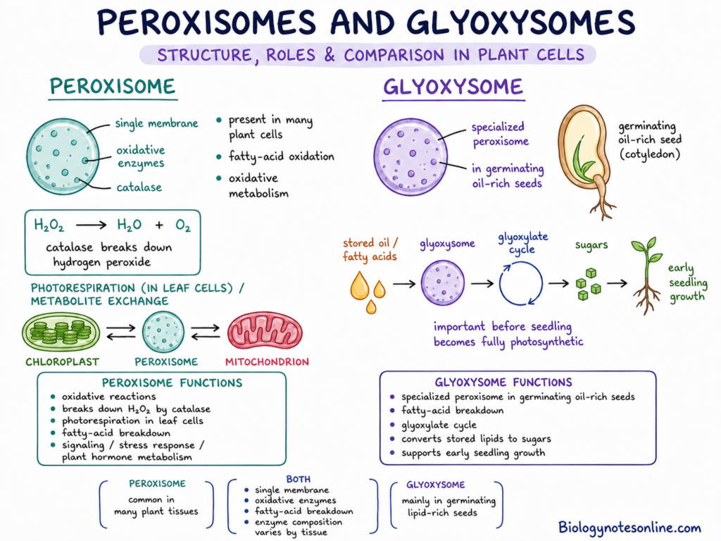
Peroxisomes are small single-membrane organelles present in different plant cells. They contain enzymes involved in oxidation of fatty acids and several other compounds.
Some reactions within peroxisomes produce hydrogen peroxide (H₂O₂). This compound can damage cellular components when present in high amount.
The enzyme catalase is commonly present in peroxisomes. It breaks hydrogen peroxide into water and molecular oxygen.
Plant peroxisomes are not exactly the same in every tissue. Their enzyme composition changes according to cell type, development and environmental condition.
Glyoxysomes
Glyoxysomes are specialized peroxisomes mainly present in lipid-rich seeds during germination. They contain enzymes for fatty-acid breakdown and the glyoxylate cycle.
During germination, stored oils are broken down and converted into compounds which can be used for sugar formation. This provides carbon and energy before the young seedling becomes fully photosynthetic.
Glyoxysomes are not present as a major organelle in every mature plant cell. They are especially associated with germinating oil-rich seeds and young tissues using stored lipids.
Functions of Peroxisomes and Glyoxysomes
- Peroxisomes perform different oxidative reactions within the plant cell.
- Catalase removes excess hydrogen peroxide and limits its harmful effect.
- Leaf peroxisomes have a major role in photorespiration.
- During photorespiration, metabolites move between chloroplasts, peroxisomes and mitochondria.
- Peroxisomes also take part in fatty-acid breakdown.
- Glyoxysomes convert stored lipids into compounds used during carbohydrate production.
- This process is important during the early germination of several oil-rich seeds.
- Plant peroxisomes are also involved in signaling, stress responses and metabolism of some plant hormones.
12. Cytoskeleton
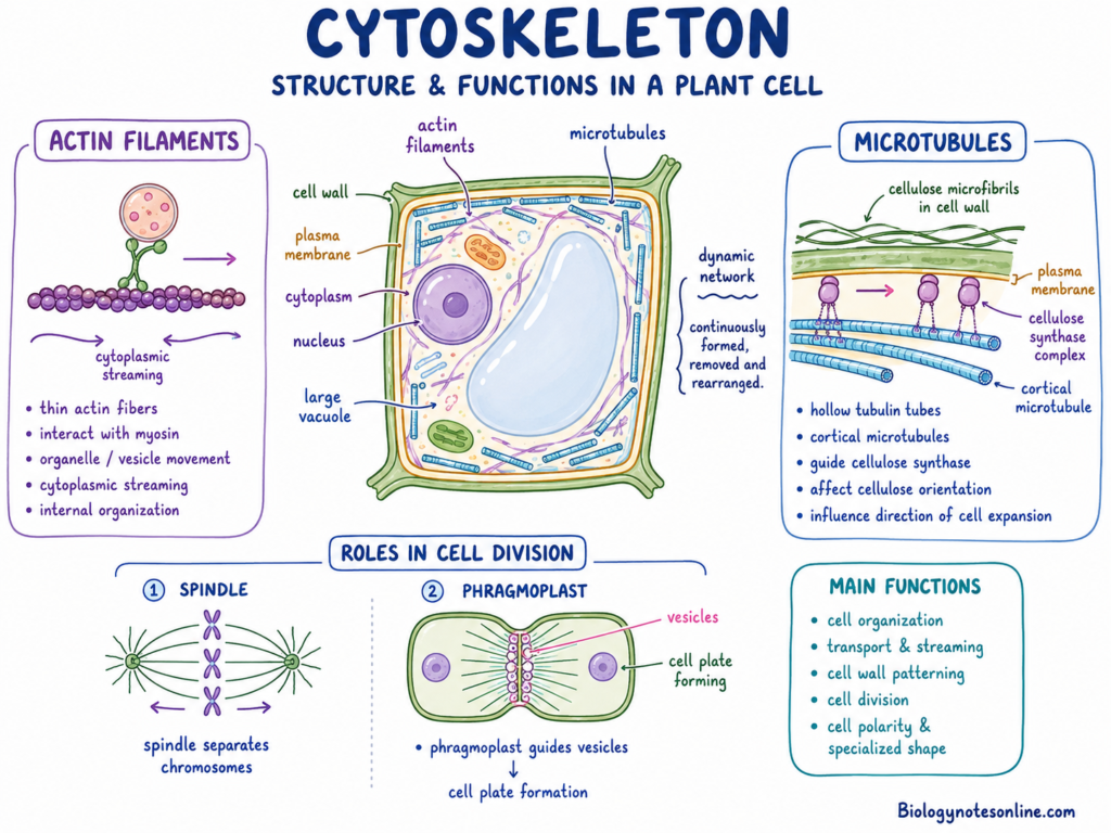
The cytoskeleton is a network of protein filaments present throughout the cytoplasm. Its major components are actin filaments and microtubules.
Actin filaments are thin protein fibres formed mainly of actin. They interact with motor proteins such as myosins.
Microtubules are hollow tubular structures formed of tubulin proteins. Their arrangement changes during cell growth, division and differentiation.
The cytoskeleton is not a fixed structure. Its filaments are continuously formed, removed and rearranged according to the need of the plant cell.
Functions of the Cytoskeleton
- The cytoskeleton provides internal organization to the plant-cell cytoplasm.
- Actin filaments are involved in organelle movement and transport of vesicles.
- They also play a major role in cytoplasmic streaming.
- Cortical microtubules guide the movement of cellulose-synthesizing complexes present in the plasma membrane.
- Due to this, microtubules affect cellulose orientation and organization of the cell wall.
- The arrangement of wall cellulose influences the direction of cell expansion.
- Microtubules form the spindle used for chromosome separation during cell division.
- They also form the phragmoplast, which guides vesicles towards the developing cell plate.
- The cytoskeleton takes part in cell polarity, tip growth and development of specialized cell shape.
13. Plasmodesmata
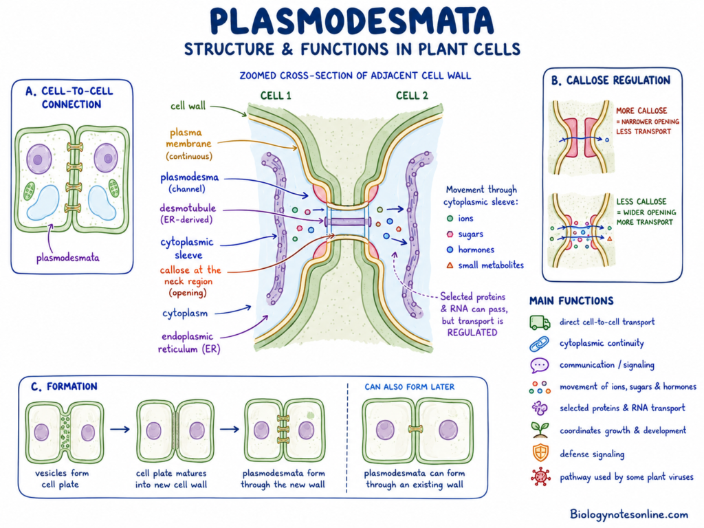
Plasmodesmata are small membrane-lined channels passing through the walls of neighbouring plant cells. They connect the cytoplasm of two adjacent living cells.
The plasma membrane lines the outer part of each plasmodesma. Thus, the plasma membrane remains continuous from one cell to another through this channel.
A narrow tube derived from the endoplasmic reticulum passes through many plasmodesmata. It is known as the desmotubule.
A cytoplasmic region is present between the desmotubule and the surrounding plasma membrane. Different substances can move through this region.
Plasmodesmata may be formed during cell division when the new cell plate is developing. Other plasmodesmata can also form later through an already existing cell wall.
Functions of Plasmodesmata
Plasmodesmata support direct cell-to-cell transport and communication across plant-cell walls.
- They provide a continuous cytoplasmic connection between neighbouring cells.
- Ions, sugars, hormones and different small metabolites can move through plasmodesmata.
- Selected proteins and RNA molecules may also pass from one cell to another.
- This movement is regulated and it is not freely open for every cellular molecule.
- Callose can be deposited around the opening of plasmodesmata.
- Increased callose deposition generally reduces the size of the transport pathway, while callose removal can increase movement.
- Plasmodesmata coordinate growth and development between neighbouring cells.
- They are also used during movement of developmental and defense signals.
- Plant viruses and some other pathogens may use plasmodesmata for spreading from one cell to the next.
14. Cellular Inclusions and Storage Materials
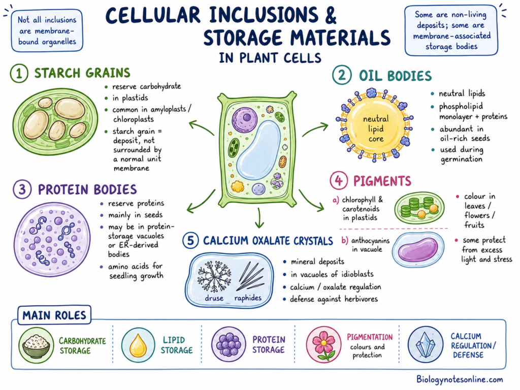
Plant cells contain different stored substances and deposited materials. These are commonly referred to as cellular inclusions or storage materials.
All cellular inclusions are not membrane-bound organelles. Some are non-living deposits formed within an organelle, while some storage bodies have their own membrane and associated proteins.
a. Starch Grains
Starch grains are deposits of reserve carbohydrate. They are formed within plastids such as chloroplasts and amyloplasts.
The starch grain itself is not surrounded by a normal unit membrane. It is an organized carbohydrate deposit present inside the plastid.
Starch may be used when the plant cell needs carbon and energy.
b. Oil Bodies
Oil bodies, also called lipid droplets, store neutral lipids. They are especially abundant in oil-rich seeds.
An oil body contains a lipid core surrounded by a phospholipid monolayer and specific proteins. Therefore, it is not simply an inactive drop of oil.
Stored oil is broken down during seed germination and is used for energy and carbohydrate production.
c. Protein Bodies
Protein bodies store reserve proteins, mainly within seeds. Some are present inside protein-storage vacuoles, while others may develop from the endoplasmic reticulum.
These proteins are broken down during germination. The released amino acids are used by the growing embryo and young seedling.
d. Pigments
Different pigments are stored in different cellular regions. Chlorophyll and many carotenoids are associated with plastids.
Anthocyanins are commonly dissolved within the vacuolar cell sap. These pigments may produce red, purple or blue colours in plant tissues.
e. Calcium Oxalate Crystals
Calcium oxalate crystals are mineral deposits found in many plants. They are commonly produced within the vacuoles of specialized cells called idioblasts.
The crystals may occur as needles, prisms, druses or other forms. Mature crystals are non-living deposits.
They may help in calcium regulation, removal of excess oxalate and protection against some herbivores.
Functions of Cellular Inclusions and Storage Materials
- Starch grains store carbohydrate for later use.
- Oil bodies store energy-rich lipids, especially in seeds.
- Protein bodies provide stored amino acids during germination and early growth.
- Pigments give colour to flowers, fruits, leaves and other plant tissues.
- Some pigments also take part in protection against excess light and environmental stress.
- Calcium oxalate crystals store or regulate calcium and oxalate within certain cells.
- Some crystalline deposits may reduce feeding by herbivores.
- Non-living inclusions are deposited substances and do not perform the same controlled functions as living organelles.
- Oil bodies, protein-storage vacuoles and similar structures should not be grouped completely with inert deposits because they contain membranes, proteins and active metabolic associations.
Types of Plant Cells and Their Functions
Plant cells become differentiated into different cell types based on their location and function. Some cells perform photosynthesis, while other cells are used in storage, support and transport. The following are the major types of plant cells-
The comparison is given below-
| Cell type | Living at maturity | Wall characteristics | Shape | Location | Function |
|---|---|---|---|---|---|
| Meristematic cells | Yes | Thin primary wall | Small and nearly isodiametric | Root and shoot tips, cambia and some leaf bases | Cell division and growth |
| Parenchyma cells | Yes | Thin primary wall | Rounded, polygonal or elongated | Cortex, pith, mesophyll and vascular tissues | Photosynthesis, storage and repair |
| Collenchyma cells | Yes | Unevenly thickened primary wall | Usually elongated | Young stems, petioles and leaf veins | Flexible support |
| Sclerenchyma cells | Often no | Thick, commonly lignified secondary wall | Fibres or variable sclereids | Mature tissues, vascular bundles and seed coats | Strength and protection |
| Xylem conducting cells | No | Thick and lignified secondary wall | Elongated and tubular | Xylem tissue | Water conduction and support |
| Phloem conducting cells | Yes | Primary walls with sieve areas | Elongated sieve tubes | Phloem tissue | Transport of sugars and signals |
| Guard cells | Yes | Specialized and unevenly thickened wall | Kidney-shaped or dumbbell-shaped | Leaf and young stem epidermis | Opening and closing of stomata |
| Root-hair cells | Yes | Thin primary wall | Long tubular projection | Root epidermis | Water and mineral uptake |
| Pavement cells | Yes | Primary wall, outer wall commonly cutinized | Flat and often interlocking | Epidermis of leaves and aerial organs | Protection and tissue integrity |
1. Meristematic Cells
Meristematic cells are the actively dividing cells present in growing regions of a plant. These cells are small and remain closely arranged.
The cytoplasm is dense and the nucleus is large in relation to the cell size. Small vacuoles may be present. A large central vacuole is generally absent.
They are found in apical, lateral and intercalary meristems. These cells form new cells, which later enlarge and become differentiated.
2. Parenchyma Cells
Parenchyma cells are living cells with thin primary walls. They are generally rounded, polygonal or isodiametric in shape. A large vacuole is commonly present.
These cells occur in the cortex, pith, mesophyll, rays and different vascular tissues. They are used in storage, secretion, repair and short-distance transport.
Parenchyma cells containing many chloroplasts are called chlorenchyma. These cells perform photosynthesis, mainly in green leaves and stems.
3. Collenchyma Cells
Collenchyma cells are living supporting cells. Their primary walls become unevenly thickened, especially near the corners of the cells.
These cells are commonly elongated. They are found below the epidermis of young stems, petioles and along the veins of leaves.
Collenchyma provides flexible support to growing plant parts. The tissue can bend without breaking easily, while the organ is still increasing in size.
4. Sclerenchyma Cells
Sclerenchyma cells have thick secondary walls. These walls are commonly lignified and become very rigid.
Most sclerenchyma cells are dead at maturity, but this condition is not similar in every type. The two major forms are fibres and sclereids.
Fibres are long, narrow and generally present in bundles. Sclereids are shorter and have different shapes. These cells provide mechanical strength and protection to mature plant tissues.
5. Xylem Conducting Cells
The major conducting cells of xylem are tracheids and vessel elements. Both develop thick secondary walls which are lignified.
These cells lose their protoplast and become dead at functional maturity. The empty cell space allows movement of water.
Tracheids are long cells with tapered ends. Water moves between them through pits. Vessel elements are joined end to end and form long vessels. Their end walls contain perforation plates.
These cells are used in conduction of water and dissolved mineral ions. Their thick walls also give mechanical support to the plant.
6.Phloem Conducting Cells
Sieve-tube elements are the major phloem conducting cells of flowering plants. These cells are arranged end to end and form sieve tubes.
A mature sieve-tube element remains living, but its nucleus is lost. Many other organelles also become absent or highly reduced.
Each sieve-tube element remains associated with one or more companion cells. Companion cells have a nucleus and dense cytoplasm. They provide metabolic support to the sieve elements.
The phloem transports sugars, amino acids and different signaling molecules from one plant region to another.
7. Guard Cells
Guard cells are paired cells present around a stomatal pore. They are part of the epidermal tissue.
Guard cells are kidney-shaped in many eudicots. In grasses, they are generally dumbbell-shaped.
Changes in guard-cell turgor cause opening and closing of the stomatal pore. When the guard cells take up water, the pore generally opens. Loss of water causes its closing.
They regulate the entry of carbon dioxide and release of water vapour. Thus, gas exchange and water loss are controlled by these cells.
8.Root-Hair Cells
Root-hair cells are specialized cells of the root epidermis. Each cell develops a long and thin tubular projection, called the root hair.
This projection increases the surface area in contact with soil particles. Water and mineral ions are taken up through this region.
Root-hair cells generally do not contain chloroplasts, as they occur in underground non-green root tissues. These cells remain active for a limited period and are replaced as the root continues to grow.
9. Epidermal Pavement Cells
Epidermal pavement cells form most part of the leaf epidermis. They produce a continuous protective covering over the internal tissues.
In many plant leaves, these cells have irregular and interlocking margins. The cells appear like the pieces of a jigsaw puzzle.
Their walls and close arrangement help in maintaining the continuity and mechanical integrity of the epidermis. They also protect the leaf from physical injury and excessive water loss.
The exact advantage of strong pavement-cell lobing is not completely understood, and it varies between plant species.
Structures That Distinguish Plant Cells
Plant cells have some structural components that are not present in animal cells. The cell wall, plastids, large central vacuole, plasmodesmata, cell plate and phragmoplast are the major of them. These structures together form the main distinctive plant cell features.
The term structures unique to plant cells is mainly used when comparing plant cells with animal cells. But all the structures are not present only in plants. Cell walls and vacuoles are also found in some other organisms, where their composition, size and organization can be different. Plastids are found in plants and algae.
Cell Wall
- Plant Cell Wall is the rigid outer cover of the plant cell with a major role of protecting the cell and giving it, its shape.
- It is present outside the plasma membrane. The wall is an extracellular matrix made by the protoplast, and it is not a membrane-bound organelle.
- The plant cell wall is mainly made up of cellulose microfibrils, hemicelluloses and pectins. Proteins and water are also present within its matrix.
- It gives mechanical support to the plant cell. The wall also resists the internal turgor pressure, preventing excess expansion of the cell.
- Cell walls are found in fungi, bacteria and some other organisms also. But these walls have different chemical composition and organization from the plant cell wall.
Plastids
- Plastids are double membrane-bound organelles found in the cells of plants and algae.
- All plastids develop from small undifferentiated plastids, which are called proplastids.
- Chloroplasts, chromoplasts, leucoplasts and amyloplasts are some of the different forms of plastids.
- Chloroplasts are used in photosynthesis. Chromoplasts contain carotenoid pigments, while amyloplasts are used in the formation and storage of starch.
- Plastids contain their own DNA and ribosomes. However, most plastid proteins are produced under the control of nuclear genes.
- Plastids are commonly included under organelles only in plant cells, when plant cells are compared to animal cells. They are not restricted completely to plants because algae also contain plastids.
Large Central Vacuole
- Plant cells have large vacuoles as compared to most animal cells. In many mature cells, the small vacuoles fuse and form one large central vacuole.
- The vacuole is covered by a membrane called the tonoplast. It contains water, ions, sugars, pigments and various other substances.
- It is used to maintain the turgor pressure of the plant cell. This pressure supports the soft parts of a plant and maintains cell stiffness.
- When the vacuole enlarges, it also increases the size of the cell. During this process, the cell does not need to produce a similar amount of new cytoplasm.
- A large central vacuole is not found in every plant cell. It becomes prominent in many cells after maturation.
- Vacuoles are also present in fungi and different other eukaryotic organisms. However, their number, size and functions may not be similar to the large central vacuole of mature plant cells.
Plasmodesmata
- Plasmodesmata are small channels that pass through the walls of two neighbouring plant cells.
- These channels are lined by the plasma membrane. The plasma membrane of one cell remains continuous with that of the adjacent cell.
- A narrow part of the endoplasmic reticulum is generally present within the channel. This is referred to as the desmotubule.
- Plasmodesmata connect the cytoplasm of neighbouring living cells. Ions, sugars and different signaling molecules can move through them.
- Selected proteins and RNA molecules are also transported by plasmodesmata. The movement is regulated according to the type and condition of the cell.
- They have an important role in cell-to-cell communication, development and transport of substances across plant tissues.
Cell Plate and Phragmoplast
- Plant cells have a characteristic cytokinesis process, where the new partition develops near the middle of the dividing cell.
- The phragmoplast is a temporary structure formed during this process. It is mainly made up of microtubules, actin filaments and different associated proteins.
- Golgi-derived vesicles are transported towards the middle region with the help of the phragmoplast.
- These vesicles fuse together and form an early membrane network. This developing structure is called the cell plate.
- The cell plate grows outwards and finally joins with the parental plasma membrane and cell wall.
- After this process, it forms the new plasma membranes and wall region between the two daughter cells.
- The phragmoplast and cell plate are not permanent organelles of the plant cell. These are formed during cytokinesis and the phragmoplast disappears after the division is completed.
How the Organelles Work Together
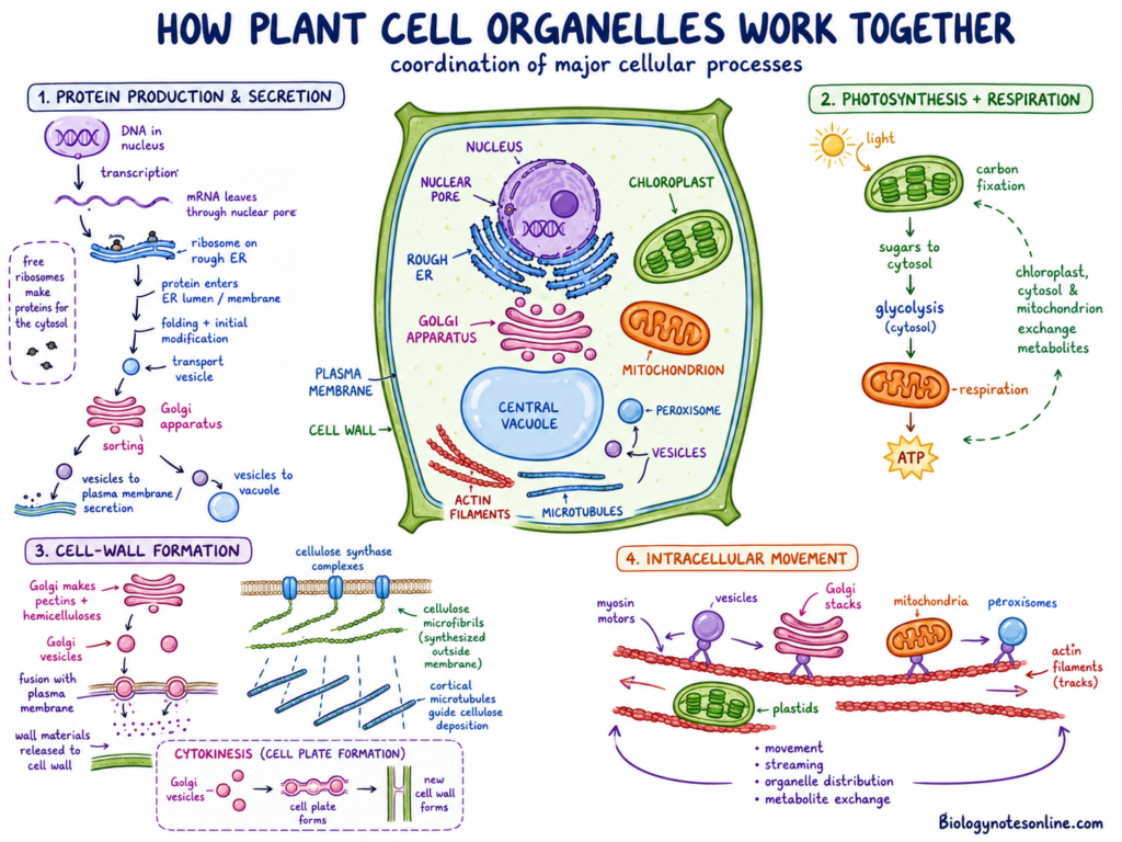
The plant cell organelles perform various specific functions, but these functions are not carried out by one organelle only. During different cellular processes, materials are formed in one organelle and then moved to another. The cytoplasm, membranes and cytoskeleton connect these cellular activities.
Some of the important examples of organelle coordination are as follows-
Protein Production and Secretion
- The genetic information for a protein is present in the DNA within the nucleus. In the first step, the required gene is transcribed and an mRNA molecule is formed.
- The mRNA moves from the nucleus into the cytoplasm through the nuclear pores. It then becomes attached with a ribosome.
- Proteins that are used in the cytosol are generally formed by free ribosomes. But proteins used for secretion, membranes and different endomembrane compartments are produced by ribosomes attached to the rough endoplasmic reticulum (RER).
- During this process, the growing protein enters into the ER lumen or becomes inserted into its membrane. Initial folding and modification of the protein also takes place within the ER.
- The proteins are then packed into transport vesicles. These vesicles move from the ER to the Golgi apparatus.
- In the Golgi bodies, the proteins are further modified and sorted according to their final location. Different proteins do not move towards the same cellular region.
- After sorting, new vesicles are formed from the Golgi. These may carry the proteins towards the plasma membrane or vacuole.
- Vesicles reaching the plasma membrane fuse with it. The protein may become a part of the membrane or it is released outside the cell. Other proteins are transported into the vacuole, where they perform storage or degradative functions.
Photosynthesis and Respiration
- Chloroplasts and mitochondria are the two major energy-related organelles of a plant cell. Their reactions are different, but their metabolic activities are connected.
- In the chloroplast, light energy is used to produce ATP and NADPH. These are then used in carbon fixation, resulting in the formation of energy-rich carbon compounds.
- Some of the carbon fixed during photosynthesis is exported from the chloroplast into the cytosol. It can be used in the formation of sucrose and several other cellular compounds.
- Sugars are partially broken down in the cytosol during glycolysis. The products can then enter into the mitochondria.
- In mitochondria, these respiratory substrates are further oxidized. Energy released during this process is used for the formation of ATP by oxidative phosphorylation.
- Mitochondria are active in photosynthetic cells also. They continue to perform respiration and help in maintaining cellular energy and redox condition during light as well as dark periods.
- Chloroplasts and mitochondria do not exchange all materials directly. Different metabolites are transferred through the cytosol with the help of transport proteins present in their envelope membranes.
- Thus, carbon compounds, organic acids and reducing equivalents move between chloroplasts, cytosol and mitochondria. These exchanges connect photosynthesis with respiration and other metabolic pathways.
Cell-Wall Formation
- The plant cell wall is made up of different polysaccharides. These wall materials are not all formed at the same cellular location.
- Pectins and many hemicelluloses are synthesized within the Golgi apparatus. After their formation, they are packed into Golgi-derived vesicles.
- These vesicles are transported through the cytoplasm and delivered towards the plasma membrane. Vesicle fusion releases the polysaccharides into the wall region.
- Cellulose is formed by a different process. The cellulose synthase complexes are assembled within the endomembrane system and transported to the plasma membrane.
- At the plasma membrane, these complexes synthesize cellulose chains from cytoplasmic sugar nucleotides. The chains are released outside the membrane and together form cellulose microfibrils.
- Cortical microtubules are present near the inner surface of the plasma membrane. These microtubules influence the movement of cellulose synthase complexes and the direction of cellulose deposition.
- Actin filaments and motor proteins are used in the transport of vesicles carrying wall materials and enzymes. In this way, the cytoskeleton helps materials to reach the required wall region.
- During cell division, many Golgi-derived vesicles are delivered to the middle of the cell. Their fusion forms the cell plate, which later develops into the new wall between the daughter cells.
Intracellular Movement
- Organelles are not always present at one fixed position within the plant cell. Many of them continuously move through the cytoplasm.
- Actin filaments form an internal network that extends throughout the plant-cell cytoplasm. These filaments provide tracks for movement of different cellular components.
- Motor proteins called myosins move along the actin filaments. They are associated with organelles, vesicles and other cellular materials.
- The interaction of actin filaments and myosin also produces cytoplasmic streaming. During this process, cytosol flows through different regions of the cell.
- Cytoplasmic streaming is important in large plant cells, where diffusion alone may not distribute materials rapidly over longer cellular distances.
- Golgi stacks, mitochondria and peroxisomes can be moved through the cytoplasm with the actin-based transport system. Plastids also change their location according to the cell type and environmental condition.
- The movement does not occur at an equal speed or in the same direction all the time. Organelles may move, stop, change direction or become attached near a particular cellular region.
- This movement helps in organelle distribution, vesicle transport and exchange of metabolites. It also brings different organelles close to one another when their metabolic reactions need to be connected.
Movement of Water and Solutes in Plant Cells
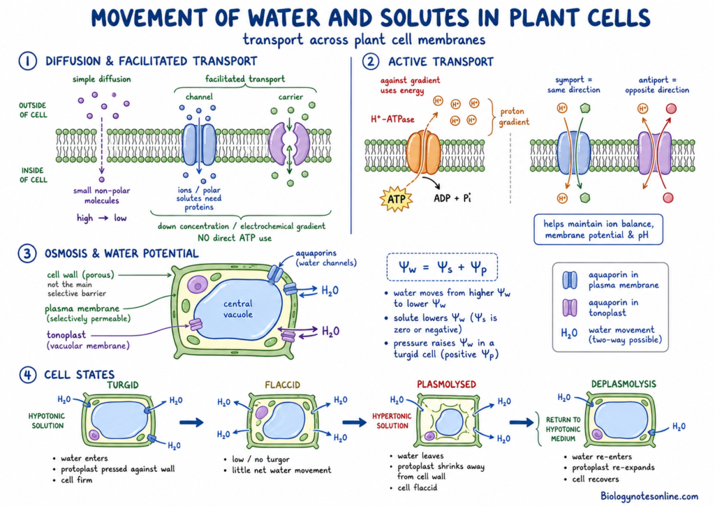
Water, ions and dissolved substances continuously move across the membranes of plant cells. The plasma membrane and tonoplast control this movement. The cell wall is porous, but it does not provide the main selective barrier.
The transport may take place without direct energy use or it may require cellular energy. Different channels, carriers and pumps are involved in this process.
Diffusion and Facilitated Transport
- Diffusion is the movement of molecules from a region of higher concentration towards the region of lower concentration.
- The movement continues down the concentration gradient. It does not directly require metabolic energy.
- Small non-polar molecules can move through the lipid bilayer. Many ions and polar substances cannot pass freely through it.
- During facilitated transport, membrane proteins assist the movement. These proteins include channels and carriers.
- The transported substance still moves down its concentration or electrochemical gradient. ATP is not directly used in this process.
Active Transport
- Active transport moves ions or solutes against their concentration or electrochemical gradient.
- This movement requires energy. The energy may be obtained directly from ATP or indirectly from an ion gradient.
- Proton pumps are important in plant cells. The plasma-membrane H⁺-ATPase uses ATP and transports hydrogen ions outside the cell.
- A proton gradient is produced in this process. Other transporters use this gradient for taking up or removing different ions and solutes.
- Symporters move two substances in the same direction. Antiporters move them in opposite directions.
- Active transport helps in maintaining ion concentration, membrane potential and cellular pH.
Osmosis and Water Potential
- Osmosis is the movement of water across a selectively permeable membrane.
- Water moves from a region of higher water potential towards a region having lower water potential.
- Solutes decrease the water potential. Pressure inside a turgid plant cell increases it.
- In a simple plant-cell system, water potential is mainly written as follows-
Water potential (Ψw) = Solute potential (Ψs) + Pressure potential (Ψp)
- Solute potential is zero or negative. The addition of solutes makes it more negative.
- Pressure potential becomes positive in a turgid living cell. It is produced when the protoplast presses against the cell wall.
- Aquaporins increase the movement of water across membranes. But the direction is still based on the water-potential difference.
Turgid, Flaccid and Plasmolysed Cells
- A turgid cell is formed when a plant cell is placed in a hypotonic condition. The surrounding solution has higher water potential than the cell sap.
- Water enters into the cell by osmosis. The vacuole becomes enlarged and the protoplast presses against the wall.
- The wall prevents excessive expansion. Thus, turgor pressure is developed and the cell becomes firm.
- A flaccid cell has low or no turgor pressure. This condition may occur in an isotonic solution, where no major net entry of water takes place.
- During water loss, the vacuole and protoplast become smaller. The cell becomes less firm, but the plasma membrane may still remain close to the wall.
- A plasmolysed cell is formed in a sufficiently hypertonic solution. Water moves out from the cell due to its lower external water potential.
- The protoplast shrinks during this process. The plasma membrane withdraws from parts of the cell wall.
- The cell wall does not shrink in the same way. Space is now formed between the wall and the retracted protoplast.
- Plasmolysis can be reversed when the cell is returned to a suitable hypotonic medium, if the cell has not been severely damaged. This reverse process is referred to as deplasmolysis.
Major Functions of Plant Cells
Plant cells perform different functions for normal growth and development of a plant. These functions depend on the type and location of the cell. The major functions of plant cells are as follows-
- Photosynthesis- Green plant cells perform photosynthesis within the chloroplasts. During this process, light energy is used to form organic compounds from carbon dioxide. All plant cells do not perform photosynthesis.
- Respiration- Plant cells break down sugars to release energy. Most of the ATP is formed within mitochondria during aerobic respiration. This energy is used in different cellular processes.
- Synthesis- Proteins, lipids, carbohydrates and nucleic acids are formed within plant cells. Different organelles are involved in their formation. Ribosomes are mainly used for protein synthesis.
- Storage- Plant cells store water, ions, sugars and different metabolites. Vacuoles are the major storage region in many cells. Starch, oils and proteins are also stored in specialized cell structures.
- Support- The cell wall gives shape and mechanical support to the cell. It also prevents excess swelling by resisting the internal pressure. Turgor of living cells supports leaves and other soft plant parts.
- Growth- Plant growth occurs by cell division and enlargement of cells. New cells are formed in meristematic regions. These cells later become mature and specialized for a particular function.
- Transport- Water, ions and other substances move across the plasma membrane. Materials are also transported from one organelle to another. Plasmodesmata allow direct transport between neighbouring living cells.
- Communication- Plant cells receive chemical and environmental signals. The signals cause changes in cellular activity and gene expression. Plasmodesmata also help in communication between adjacent cells.
- Homeostasis- The internal condition of cell is maintained by the plasma membrane and vacuole. The vacuole regulates water, ions and cellular pH. Waste substances can also be separated within it.
- Defense- The cell wall provides an initial barrier against injury and pathogens. Plant cells also form different defensive substances. Some of them are stored within the vacuole or cell wall.
Plant Cell vs Animal Cell
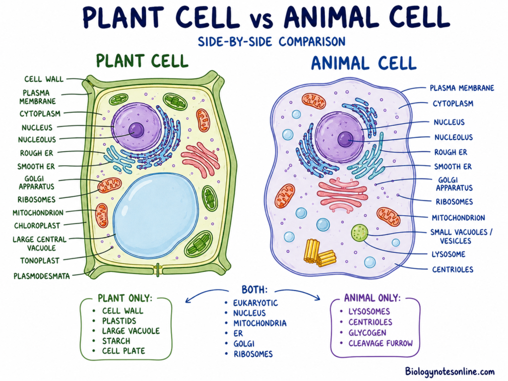
Plant cells and animal cells are eukaryotic cells, which means they have membrane-bound cell organelles. Most of the living differentiated cells contain a nucleus. Both cells have plasma membrane and mitochondria.
Plant cells also have structural components that are not found in typical animal cells, including cell wall and plastids. A large central vacuole is also present in many mature plant cells. Animal cells generally contain centrioles and a typical lysosomal system. The major differences are given below-
| Feature | Plant Cell | Animal Cell |
|---|---|---|
| Cell type | It is a eukaryotic cell. Different membrane-bound organelles are present. | It is also a eukaryotic cell with different membrane-bound organelles. |
| Nucleus | A membrane-bound nucleus is found in most living plant cells. Some specialized cells lose the nucleus during maturation. | The nucleus is present in most differentiated living cells. Some specialized animal cells may lose it. |
| Cell wall | A cell wall is present outside the plasma membrane in most cells. It contains mainly cellulose, hemicelluloses and pectins. | Cell wall is absent. The plasma membrane forms the outer living boundary of cell. |
| Plasma membrane | It is present inside the cell wall. The membrane controls movement of substances into and outside the cell. | It is present as the outer cellular membrane. It also controls selective transport. |
| Plastids | Plastids are present. Chloroplasts, chromoplasts and storage plastids are some of their forms. | Plastids are absent. |
| Chloroplasts | Chloroplasts are present in green and photosynthetic cells. They are not found in every plant cell. | Chloroplasts are absent. Animal cells do not perform chloroplast-based photosynthesis. |
| Vacuole | Many mature cells contain a large and persistent central vacuole. It is used in storage, turgor and degradation. | Small vacuoles, vesicles or related compartments may be present. A large persistent central vacuole is generally absent. |
| Mitochondria | Mitochondria are present in metabolically active cells. They perform cellular respiration and ATP production. | Mitochondria are also present in most metabolically active animal cells. Thus, mitochondria are not restricted to animals. |
| Lysosomal system | Typical animal-type lysosomes are not the main degradative compartment. Lytic vacuoles perform many lysosome-like functions. | Lysosomes are common degradative organelles. They contain enzymes used for breakdown and recycling. |
| Centrioles | Centrioles are absent from most cells of flowering plants and conifers. They occur as basal-body structures in the motile male gametes of some plant groups. | A pair of centrioles is commonly present within the centrosome of animal cells. But all animal cells do not retain centrioles. |
| Cell junctions | Neighbouring living cells are connected by plasmodesmata. The middle lamella also helps in adhesion between their walls. | Different junctions are found based on tissue, such as tight junctions, anchoring junctions and gap junctions. |
| Shape | The shape is commonly fixed, angular or polygonal due to the wall and tissue packing. All plant cells are not rectangular. | Animal cells have more variable shapes because a rigid wall is absent. Their form depends on the cell function and tissue. |
| Energy storage | Carbohydrate is stored mainly in the form of starch. Oils are also important storage materials, especially in seeds. | Carbohydrate is stored mainly in the form of glycogen. Fats are also used as a major energy store. |
| Cytokinesis | In many land-plant cells, cytokinesis takes place by formation of a cell plate. The phragmoplast guides vesicles towards the developing plate. | Cytokinesis commonly takes place by formation of a cleavage furrow. An actomyosin ring constricts the cell. |
| Motile structures | Cilia and flagella are absent from most somatic cells of land plants. Motile sperm are found in bryophytes, ferns, cycads and Ginkgo. | Cilia or flagella are present in some specialized cells. But these structures are not found in every animal cell. |
Important Clarifications
- Both plant cells and animal cells contain a plasma membrane. The plant plasma membrane remains inside the cell wall.
- Both types of cells contain mitochondria. Plant cells do not depend only on chloroplasts for cellular energy.
- A membrane-bound nucleus is present in most differentiated living cells of plants and animals. Some specialized cells are exceptions.
- Animal cells can contain small vacuoles, vesicles and other membrane compartments. They generally do not contain the large persistent central vacuole found in many mature plant cells.
- Plant cells do not completely lack a lysosomal system. The lytic vacuole performs breakdown, recycling and several other lysosome-like functions.
- These differences are not absolute for every cell and organism. Structure changes according to lineage, tissue and stage of differentiation. Flagellated plant sperm and animal cells without centrioles are some examples.
Plant Cells Compared with Other Cell Types
Plant cells are different from bacterial and fungal cells in their internal organization. Cell wall and vacuoles can be present in more than one group. But their composition and cellular arrangement are different.
Plant Cell vs Bacterial Cell
| Feature | Plant Cell | Bacterial Cell |
|---|---|---|
| Cell organization | Plant cell is a eukaryotic cell. It has a complex internal organization. | Bacterial cell is a prokaryotic cell. Its organization is comparatively simpler. |
| Nucleus | A membrane-bound nucleus is generally present. | True nucleus is absent. DNA is present within the nucleoid region. |
| Cell organelles | Mitochondria, endoplasmic reticulum, Golgi bodies and plastids are present. | Typical membrane-bound organelles are absent. Some bacteria have specialized membrane regions. |
| Ribosomes | Cytoplasmic ribosomes are mainly 80S type. Plastids and mitochondria contain smaller bacterial-like ribosomes. | Ribosomes are mainly 70S type. |
| DNA | Nuclear DNA occurs as linear chromosomes. DNA is also present in plastids and mitochondria. | The main chromosome is generally circular. Plasmids may also be present. |
| Cell wall | The wall mainly contains cellulose, hemicelluloses and pectins. | Most bacterial walls contain peptidoglycan. Some bacteria do not have a normal wall. |
| Cell division | Cell division takes place by mitosis. Cytokinesis commonly involves cell-plate formation. | Division generally takes place by binary fission. Mitosis is absent. |
Plant Cell vs Fungal Cell
| Feature | Plant Cell | Fungal Cell |
| Cell organization | It is a eukaryotic cell with a nucleus and membrane-bound organelles. | It is also a eukaryotic cell. Nucleus and membrane-bound organelles are present. |
| Cell wall | The wall is mainly rich in cellulose, hemicelluloses and pectins. | The wall commonly contains chitin and glucans. Its composition varies between fungal groups. |
| Plastids | Plastids are present. Green cells contain chloroplasts for photosynthesis. | Plastids and chloroplasts are absent. |
| Vacuoles | A large central vacuole is common in many mature cells. | Vacuoles are also present. These are used in storage, degradation and ion balance. |
| Storage carbohydrate | Carbohydrate is stored mainly in the form of starch. | Carbohydrate is generally stored as glycogen. Trehalose is also present in many fungi. |
| Nutrition | Green plants are mainly photoautotrophic. Organic compounds are formed through photosynthesis. | Fungi are heterotrophic. Nutrients are absorbed after external digestion. |
Plant Cells Observation Under a Microscope
Plant cells can be observed by using a compound light microscope. A thin plant material is placed on a glass slide and covered with a coverslip. Some structures are visible directly. Other structures need staining for producing better contrast.
The type of structures observed depends on the specimen and microscope used. Onion epidermis and aquatic green leaves are commonly used for this study.
Onion Epidermal Cell
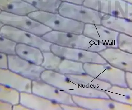
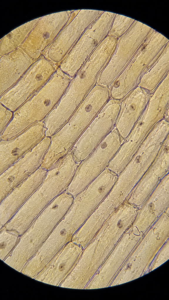
- The inner epidermis of onion bulb (Allium cepa) is commonly used for observing plant cells. It can be removed as a thin and almost transparent layer.
- The cells appear elongated or rectangular in shape. A clear boundary is observed around each cell. This boundary is mainly the cell wall.
- The plasma membrane is present inside the wall. But it is generally not seen separately in a normal water-mounted cell.
- A large vacuole occupies most part of the mature epidermal cell. It usually appears as a clear central region. The cytoplasm remains towards the sides of cell.
- The nucleus is transparent and may not be clearly observed in an unstained preparation. After adding a suitable stain, the nucleus becomes more visible.
- Bulb epidermal cells generally do not contain chloroplasts. The onion bulb is an underground storage organ and its inner epidermal tissues are non-photosynthetic. Green aerial onion tissues have chloroplasts.
Green Leaf Cell
- A thin green leaf is used for observing chloroplasts and the movement of cytoplasm. Aquatic plants such as Elodea canadensis are suitable for this preparation.
- The leaf is placed directly in a drop of water. Its cells are arranged closely and the cell walls can be observed under the light microscope.
- Several green chloroplasts are present near the wall region of each photosynthetic cell. Chlorophyll present within them gives the green colour.
- A large central vacuole pushes the cytoplasm and chloroplasts towards the outer part of cell.
- In a living leaf preparation, chloroplasts may be seen moving around the cell. This movement is used to observe cytoplasmic streaming.
- The movement may become slow or stop when the specimen is damaged, dried or heated. Strong illumination can also change the position and movement of chloroplasts.
Basic Slide Preparation
The following are the basic steps used for preparation of a plant-cell slide-
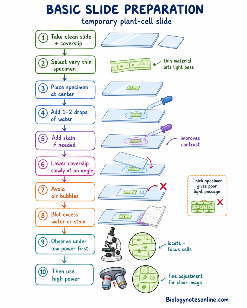
- A clean glass slide and coverslip are taken.
- A very thin specimen is selected. Thick plant material does not allow proper passage of light.
- The specimen is placed at the centre of slide.
- One or two drops of water are added over it. This forms a temporary water mount.
- A stain can be added when required. Staining gives contrast to the nucleus and other transparent cellular materials.
- The coverslip is lowered slowly over the specimen. It can be placed from one side at an angle and then lowered.
- Air bubbles should not remain below the coverslip. These bubbles can block the specimen and disturb the image.
- Excess water or stain is removed from the sides with absorbent paper.
- The slide is first observed under the low-power objective. The required cells are located and focused.
- After this, the high-power objective is used. Fine adjustment is used for obtaining a clear image.
What Can and Cannot Be Seen
A light microscope shows the general organization of plant cells. Very small cell components cannot be separated due to its limited resolution. Electron microscopes are used for studying the fine internal structure.
| Structure or observation | Light microscope | Electron microscope |
|---|---|---|
| Cell shape | General cell shape and arrangement can be observed. | Cell shape can be studied with much greater detail. |
| Cell wall | The wall is clearly visible as the outer cell boundary. Its fine layers are not generally resolved. | Primary wall, secondary wall and some fine wall organization can be studied. |
| Plasma membrane | It is usually not separated clearly from the wall in a normal preparation. It becomes noticeable during plasmolysis. | The plasma membrane and its relation with the wall can be observed. |
| Nucleus | The nucleus can be seen, mainly after staining. Fine nuclear organization is not clear. | Nuclear envelope, pores, nucleolus and chromatin regions can be studied in detail. |
| Large vacuole | It is seen as a large clear region in many mature cells. The tonoplast is difficult to resolve by ordinary brightfield microscopy. | Tonoplast and vacuolar contents can be observed with greater detail. |
| Chloroplasts | Chloroplasts are clearly visible in green cells. Their green colour and movement can be observed. | Thylakoids, grana, stroma and starch grains can be studied. Images do not show natural green colour. |
| Cytoplasmic streaming | It can be observed in living cells. Movement of chloroplasts or cytoplasmic particles indicates the process. | It cannot be followed in a conventional fixed electron-microscope specimen. |
| Mitochondria | Individual mitochondria are generally not clearly seen in an ordinary classroom preparation. Special stains or fluorescence methods are required. | Outer membrane, inner membrane and cristae can be observed. |
| Golgi and ER | These are not resolved clearly by an ordinary light microscope. Fluorescent labeling can show their distribution. | Golgi cisternae and endoplasmic-reticulum membranes can be studied. |
| Ribosomes | Ribosomes are too small to be resolved. | Ribosomes can be observed as small dense particles, mainly by transmission electron microscopy. |
| Plasmodesmata | Individual plasmodesmata are generally below normal light-microscope resolution. | Their channels, wall passage and desmotubule can be studied. |
| Living cell | Living cells can be observed in a water mount. Cell movement and changes can also be followed. | Conventional electron microscopy requires fixation and a vacuum. The specimen is not living. |
| Natural colour | Natural pigments such as chlorophyll can be observed. | Electron images are produced in shades of grey. Colour may be added later for representation. |
The normal light microscope can resolve details approximately 0.2 µm apart. It is suitable for observing cell outlines, nuclei, chloroplasts and large vacuolar regions. Electron microscopy provides much higher resolution. It shows membranes and the fine internal structures of cell organelles.
Common Misconceptions about Plant Cells
There are several misconceptions related to plant cells. Most of them are formed from the generalized plant-cell diagram. The common misconceptions and their correct facts are given below-
| Misconception | Fact check | Correct fact |
|---|---|---|
| All plant cells are rectangular. | False | Plant cells occur in different shapes. They may be polygonal, rounded, elongated, tubular or highly lobed. Cell wall, turgor and packing of cells affect their shape. |
| Every plant cell contains chloroplasts. | False | Chloroplasts are mainly found in green photosynthetic cells. Root cells and many internal tissues do not contain chloroplasts. They contain other types of plastids. |
| The cell wall replaces the plasma membrane. | False | A living plant cell contains both structures. The plasma membrane is present immediately inside the cell wall. It forms the selective living boundary of the protoplast. |
| The cell wall is a membrane-bound organelle. | False | The cell wall is an extracellular matrix formed outside the plasma membrane. It is not a membrane-bound organelle. Its major components include cellulose, hemicelluloses and pectins. |
| Plant cells do not contain mitochondria. | False | Plant cells contain mitochondria along with plastids. Mitochondria perform cellular respiration and provide ATP for different cellular activities. They are present in photosynthetic and non-photosynthetic tissues. |
| Plants perform photosynthesis but not respiration. | False | Living plant cells perform respiration. It occurs in green leaves as well as roots and other non-green tissues. Photosynthesis and mitochondrial respiration can occur within the same photosynthetic cell. |
| The central vacuole is an empty space. | False | The vacuole contains cell sap, ions, metabolites, pigments and different enzymes. It is used in storage, turgor, detoxification, degradation and cellular homeostasis. |
| Every plant cell has one large central vacuole. | False | A large central vacuole is common in many mature plant cells. Young and dividing cells generally have smaller vacuoles. Different vacuole types can also occur according to the cell and tissue. |
| Plant cells have no lysosomal activity. | False | Plants generally use lytic vacuoles for many lysosome-like functions. These vacuoles break down proteins, damaged organelles and other cellular materials. |
| The plant cell wall is completely impermeable. | False | The wall is a hydrated and porous matrix. Water and many dissolved substances can move through it. Selective transport is mainly controlled by the plasma membrane. |
| All mature plant cells are living and contain a nucleus. | False | Some mature cells lose their protoplast. Tracheids and vessel elements are dead at functional maturity. Mature sieve elements remain living, but they lose the nucleus and depend on associated companion cells. |
| All plant cells have the same size and organelles. | False | Plant cells change according to their tissue, age and function. Meristematic cells are small, whereas many mature cells become enlarged by vacuolar expansion. Their plastids and other cellular components are also different. |
| The cell wall is only a dead supporting layer. | False | The wall provides support, but it is also involved in growth, signaling and defense. Its composition changes during development and cell specialization. |
| Chloroplasts provide all the ATP needed by a plant cell. | False | Chloroplasts form ATP during photosynthesis, mainly for reactions inside the chloroplast. Mitochondrial respiration provides ATP for many other cellular processes. |
Plant cell Worksheet
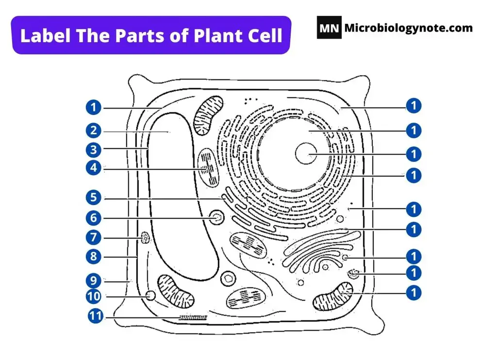
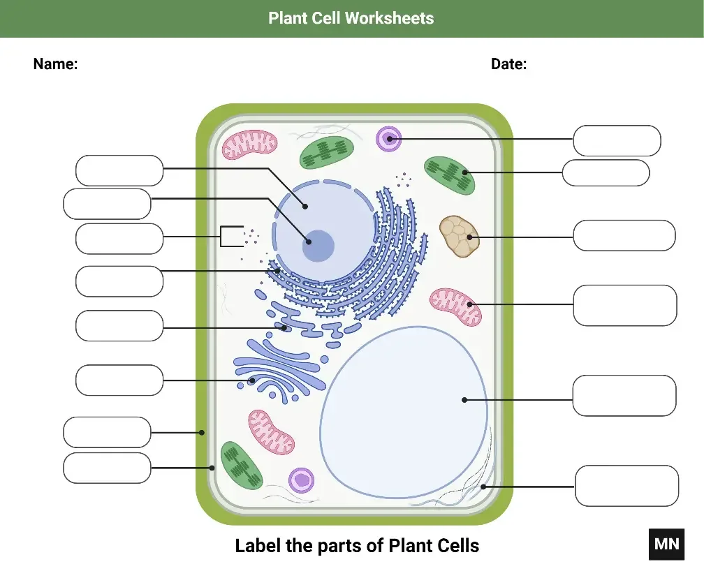
Frequently Asked Questions
What is a plant cell?
A plant cell is the smallest living structural and functional unit of plant tissues. It is a eukaryotic cell with a membrane-bound nucleus.
What are the main parts of a plant cell?
The major parts include cell wall, plasma membrane, cytoplasm, nucleus, plastids, vacuole, mitochondria, ER, Golgi bodies and ribosomes.
Which structures distinguish plant cells from animal cells?
The main structures are cell wall, plastids, large central vacuole and plasmodesmata. Cell-plate formation is also characteristic during cytokinesis.
Do all plant cells have chloroplasts?
No. Chloroplasts are mainly present in green photosynthetic cells. Root cells and many storage tissues contain other plastid types.
Do plant cells have mitochondria?
Yes. Plant cells contain mitochondria and perform cellular respiration. They are present in green and non-green living cells.
Why do plant cells have a cell wall?
The cell wall gives shape, protection and mechanical support. It also resists the internal turgor pressure of cell.
Why are plant cells usually shown as rectangular?
The wall, turgor pressure and close tissue packing produce angular forms. The diagram is also a simplified two-dimensional structure.
What is the function of the central vacuole?
The central vacuole stores water, ions, pigments and metabolites. It maintains turgor and also performs degradative functions.
What is the difference between the cell wall and plasma membrane?
The cell wall is an extracellular and porous structure. The plasma membrane is a living selectively permeable membrane present inside the wall.
What are plasmodesmata?
Plasmodesmata are small membrane-lined channels between neighbouring plant cells. They allow direct transport and cellular communication.
How do plant cells divide?
The nucleus divides by mitosis. During cytokinesis, vesicles gather near the middle region and form a cell plate.
What is the function of the cell plate?
The cell plate separates the two daughter cells. It later forms the new plasma membranes and wall region between them.
Do plant cells have lysosomes?
Plant cells generally use lytic vacuoles for many lysosome-like functions. These include breakdown and recycling of cellular materials.
What happens to a plant cell in a concentrated salt solution?
Water moves outside the cell by osmosis. The protoplast shrinks and the plasma membrane withdraws from the wall. This is called plasmolysis.
Which plant-cell structures can be seen under a light microscope?
The cell wall, general cell outline, nucleus after staining, chloroplasts and large vacuolar region can be observed. Ribosomes and fine membranes cannot be resolved.
What are the main types of specialized plant cells?
The major types include meristematic, parenchyma, collenchyma, sclerenchyma, xylem, phloem, guard and root-hair cells.
Which plant cells are dead at maturity?
Tracheids and vessel elements are dead at functional maturity. Most sclerenchyma fibres and sclereids are also dead when mature.
How are plant and fungal cells different?
Plant walls are mainly cellulose-rich, while fungal walls commonly contain chitin and glucans. Fungi also lack plastids and chloroplasts.
What is the typical size of a plant cell?
Plant-cell size varies considerably. Meristematic cells are generally small, while mature vacuolated cells can become much larger. One size range is not universal.
Why do onion epidermal cells lack chloroplasts?
The bulb epidermis of Allium cepa is an underground non-photosynthetic tissue. Therefore, its cells generally contain colourless plastids instead of chloroplasts.
References
- Alberts, B., Johnson, A., Lewis, J., Raff, M., Roberts, K., & Walter, P. (2002). Molecular biology of the cell (4th ed.). Garland Science.
- Baron, S. (Ed.). (1996). Medical microbiology (4th ed.). University of Texas Medical Branch at Galveston.
- Clark, M. A., Douglas, M., & Choi, J. (2018). Biology 2e. OpenStax.
- Cooper, G. M. (2000). The cell: A molecular approach (2nd ed.). Sinauer Associates.
- Fowler, S., Roush, R., & Wise, J. (2013). Concepts of biology. OpenStax.
- Parker, N., Schneegurt, M., Thi Tu, A.-H., Lister, P., & Forster, B. M. (2016). Microbiology. OpenStax.
- Pyke, K. A. (2009). Plastid biology. Cambridge University Press.
- Rudall, P. J. (2007). Anatomy of flowering plants: An introduction to structure and development (3rd ed.). Cambridge University Press. https://doi.org/10.1017/CBO9780511801709
- Rudall, P. J. (2020). Anatomy of flowering plants: An introduction to plant structure and development (4th ed.). Cambridge University Press. https://doi.org/10.1017/9781108782104
- Varki, A., Cummings, R. D., Esko, J. D., Stanley, P., Hart, G. W., Aebi, M., Mohnen, D., Kinoshita, T., Packer, N. H., Prestegard, J. H., Schnaar, R. L., & Seeberger, P. H. (Eds.). (2022). Essentials of glycobiology (4th ed.). Cold Spring Harbor Laboratory Press. https://doi.org/10.1101/glycobiology.4e
- Assmann, S. M., & Shimazaki, K. (1999). The multisensory guard cell: Stomatal responses to blue light and abscisic acid. Plant Physiology, 119(3), 809–816. https://doi.org/10.1104/pp.119.3.809
- Bayer, E. M., & Benitez-Alfonso, Y. (2024). Plasmodesmata: Channels under pressure. Annual Review of Plant Biology, 75, 291–317. https://doi.org/10.1146/annurev-arplant-070623-093110
- Beauzamy, L., Nakayama, N., & Boudaoud, A. (2014). Flowers under pressure: Ins and outs of turgor regulation in development. Annals of Botany, 114(7), 1517–1533. https://doi.org/10.1093/aob/mcu187
- Bhawana, Miller, J. L., & Cahoon, A. B. (2014). 3D plant cell architecture of Arabidopsis thaliana (Brassicaceae) using focused ion beam–scanning electron microscopy. Applications in Plant Sciences, 2(6), Article 1300090. https://doi.org/10.3732/apps.1300090
- Bollhöner, B., Prestele, J., & Tuominen, H. (2012). Xylem cell death: Emerging understanding of regulation and function. Journal of Experimental Botany, 63(3), 1081–1094. https://doi.org/10.1093/jxb/err438
- Cheng, X., Lang, I., Adeniji, O. S., & Griffing, L. (2017). Plasmolysis-deplasmolysis causes changes in endoplasmic reticulum form, movement, flow, and cytoskeletal association. Journal of Experimental Botany, 68(15), 4075–4087. https://doi.org/10.1093/jxb/erx243
- Codjoe, J. M., Miller, K., & Haswell, E. S. (2022). Plant cell mechanobiology: Greater than the sum of its parts. The Plant Cell, 34(1), 129–145. https://doi.org/10.1093/plcell/koab230
- Delmer, D., Dixon, R. A., Keegstra, K., & Mohnen, D. (2024). The plant cell wall—dynamic, strong, and adaptable—is a natural shapeshifter. The Plant Cell, 36(5), 1257–1311. https://doi.org/10.1093/plcell/koad325
- Farquharson, K. L. (2018). A lipid droplet-associated degradation system in plants. The Plant Cell, 30(9), 1952–1953. https://doi.org/10.1105/tpc.18.00610
- Fernández-de-Uña, L. (2026). Don’t add pressure to stress: Measured water potentials differ by method under severe drought. Plant Physiology, 200(1), Article kiaf634. https://doi.org/10.1093/plphys/kiaf634
- Grierson, C., Nielsen, E., Ketelaar, T., & Schiefelbein, J. (2014). Root hairs. The Arabidopsis Book, 12, e0172. https://doi.org/10.1199/tab.0172
- Gu, Y., & Rasmussen, C. G. (2022). Cell biology of primary cell wall synthesis in plants. The Plant Cell, 34(1), 103–128. https://doi.org/10.1093/plcell/koab249
- Guimil, S., & Dunand, C. (2007). Cell growth and differentiation in Arabidopsis epidermal cells. Journal of Experimental Botany, 58(14), 3829–3840. https://doi.org/10.1093/jxb/erm253
- Haywood, V., Kragler, F., & Lucas, W. J. (2002). Plasmodesmata: Pathways for protein and ribonucleoprotein signaling. The Plant Cell, 14(Suppl. 1), S303–S325. https://doi.org/10.1105/tpc.000778
- Hepler, P. K., Vidali, L., & Cheung, A. Y. (2001). Polarized cell growth in higher plants. Annual Review of Cell and Developmental Biology, 17, 159–187. https://doi.org/10.1146/annurev.cellbio.17.1.159
- Hernández-Verdeja, T., & Strand, Å. (2018). Retrograde signals navigate the path to chloroplast development. Plant Physiology, 176(2), 967–976. https://doi.org/10.1104/pp.17.01299
- Huang, A. H. C. (2018). Plant lipid droplets and their associated proteins: Potential for rapid advances. Plant Physiology, 176(3), 1894–1918. https://doi.org/10.1104/pp.17.01677
- Kang, B.-H., Anderson, C. T., Arimura, S.-I., Bayer, E. M., Bezanilla, M., Botella, M. A., Brandizzi, F., Burch-Smith, T. M., Chapman, K. D., Dünser, K., Gu, Y., Jaillais, Y., Kirchhoff, H., Otegui, M. S., Rosado, A., Tang, Y., Kleine-Vehn, J., Wang, P., & Zolman, B. K. (2022). A glossary of plant cell structures: Current insights and future questions. The Plant Cell, 34(1), 10–52. https://doi.org/10.1093/plcell/koab247
- Kao, Y.-T., Gonzalez, K. L., & Bartel, B. (2018). Peroxisome function, biogenesis, and dynamics in plants. Plant Physiology, 176(1), 162–177. https://doi.org/10.1104/pp.17.01050
- Khoulali, C., Pastor, J. M., Galeano, J., Vissenberg, K., & Miedes, E. (2025). Cell wall–based machine learning models to predict plant growth using onion epidermis. International Journal of Molecular Sciences, 26(7), Article 2946. https://doi.org/10.3390/ijms26072946
- Koenig, A. M., Liu, B., & Hu, J. (2023). Visualizing the dynamics of plant energy organelles. Biochemical Society Transactions, 51(6), 2029–2040. https://doi.org/10.1042/BST20221093
- Liu, S., Jobert, F., Rahneshan, Z., Doyle, S. M., & Robert, S. (2021). Solving the puzzle of shape regulation in plant epidermal pavement cells. Annual Review of Plant Biology, 72, 525–550. https://doi.org/10.1146/annurev-arplant-080720-081920
- Liu, Y.-L., Guo, Y.-H., Song, X.-Q., Hu, M.-X., & Zhao, S.-T. (2023). A method for analyzing programmed cell death in xylem development by flow cytometry. Frontiers in Plant Science, 14, Article 1196618. https://doi.org/10.3389/fpls.2023.1196618
- Marty, F. (1999). Plant vacuoles. The Plant Cell, 11(4), 587–600. https://doi.org/10.1105/tpc.11.4.587
- Millar, A. H., Whelan, J., Soole, K. L., & Day, D. A. (2011). Organization and regulation of mitochondrial respiration in plants. Annual Review of Plant Biology, 62, 79–104. https://doi.org/10.1146/annurev-arplant-042110-103857
- Murata, Y., Mori, I. C., & Munemasa, S. (2015). Diverse stomatal signaling and the signal integration mechanism. Annual Review of Plant Biology, 66, 369–392. https://doi.org/10.1146/annurev-arplant-043014-114707
- Nebenführ, A., & Dixit, R. (2018). Kinesins and myosins: Molecular motors that coordinate cellular functions in plants. Annual Review of Plant Biology, 69, 329–361. https://doi.org/10.1146/annurev-arplant-042817-040024
- Ostermeier, M., Garibay-Hernández, A., Holzer, V. J. C., Schroda, M., & Nickelsen, J. (2024). Structure, biogenesis, and evolution of thylakoid membranes. The Plant Cell, 36(10), 4014–4035. https://doi.org/10.1093/plcell/koae102
- Pierre-Jerome, E., Drapek, C., & Benfey, P. N. (2018). Regulation of division and differentiation of plant stem cells. Annual Review of Cell and Developmental Biology, 34, 289–310. https://doi.org/10.1146/annurev-cellbio-100617-062459
- Quan, S., Yang, P., Cassin-Ross, G., Kaur, N., Switzenberg, R., Aung, K., Li, J., & Hu, J. (2013). Proteome analysis of peroxisomes from etiolated Arabidopsis seedlings identifies a peroxisomal protease involved in β-oxidation and development. Plant Physiology, 163(4), 1518–1538. https://doi.org/10.1104/pp.113.223453
- Renzaglia, K. S., Lopez, R. A., Henry, J. S., Flowers, N. D., & Vaughn, K. C. (2017). Transmission electron microscopy of centrioles, basal bodies and flagella in motile male gametes of land plants. Bio-protocol, 7(19), e2448. https://doi.org/10.21769/BioProtoc.2448
- Roeder, A. H. K., Otegui, M. S., Dixit, R., Anderson, C. T., Faulkner, C., Zhang, Y., Harrison, M. J., Kirchhelle, C., Goshima, G., Coate, J. E., Doyle, J. J., Hamant, O., Sugimoto, K., Dolan, L., Meyer, H., Ehrhardt, D. W., Boudaoud, A., & Messina, C. (2022). Fifteen compelling open questions in plant cell biology. The Plant Cell, 34(1), 72–102. https://doi.org/10.1093/plcell/koab225
- Schmidt, U. G., Endler, A., Schelbert, S., Brunner, A., Schnell, M., Neuhaus, H. E., Marty-Mazars, D., Marty, F., Baginsky, S., & Martinoia, E. (2007). Novel tonoplast transporters identified using a proteomic approach with vacuoles isolated from cauliflower buds. Plant Physiology, 145(1), 216–229. https://doi.org/10.1104/pp.107.096917
- Schmidt-Marcec, S., Parish, A., Smertenko, T., Hickey, M., Piette, B. M. A. G., & Smertenko, A. (2024). The microtubule-nucleating factor MACERATOR tethers AUGMIN7 to microtubules and governs phragmoplast architecture. The Plant Cell, 36(4), 1072–1097. https://doi.org/10.1093/plcell/koad304
- Schramma, N., Israëls, C. P., & Jalaal, M. (2023). Chloroplasts in plant cells show active glassy behavior under low-light conditions. Proceedings of the National Academy of Sciences of the United States of America, 120(3), Article e2216497120. https://doi.org/10.1073/pnas.2216497120
- Schuster, M. (2025). From the archives: Plasmodesmata—regulation of function, targeting by pathogenic bacteria, and plasmodesmal-associated proteins. The Plant Cell, 37(3), Article koaf040. https://doi.org/10.1093/plcell/koaf040
- Spicer, R. (2014). Symplasmic networks in secondary vascular tissues: Parenchyma distribution and activity supporting long-distance transport. Journal of Experimental Botany, 65(7), 1829–1848. https://doi.org/10.1093/jxb/ert459
- Sweetlove, L. J., & Fernie, A. R. (2013). The spatial organization of metabolism within the plant cell. Annual Review of Plant Biology, 64, 723–746. https://doi.org/10.1146/annurev-arplant-050312-120233
- Szymanski, D. B., & Staiger, C. J. (2018). The actin cytoskeleton: Functional arrays for cytoplasmic organization and cell shape control. Plant Physiology, 176(1), 106–118. https://doi.org/10.1104/pp.17.01519
- van Spoordonk, R., Schneider, R., & Sampathkumar, A. (2023). Mechano-chemical regulation of complex cell shape formation: Epidermal pavement cells—A case study. Quantitative Plant Biology, 4, e5. https://doi.org/10.1017/qpb.2023.4
- Verma, D. P. S. (2001). Cytokinesis and building of the cell plate in plants. Annual Review of Plant Physiology and Plant Molecular Biology, 52, 751–784. https://doi.org/10.1146/annurev.arplant.52.1.751
- Wolf, S. (2022). Cell wall signaling in plant development and defense. Annual Review of Plant Biology, 73, 323–353. https://doi.org/10.1146/annurev-arplant-102820-095312
- Wurtzel, E. T. (2019). Changing form and function through carotenoids and synthetic biology. Plant Physiology, 179(3), 830–843. https://doi.org/10.1104/pp.18.01122
- Zwieniecki, M. A., & Holbrook, N. M. (2000). Bordered pit structure and vessel wall surface properties: Implications for embolism repair. Plant Physiology, 123(3), 1015–1020. https://doi.org/10.1104/pp.123.3.1015