

Storage granules are the intracellular inclusions present inside many microorganisms, especially bacterial cells. These are reserve materials that are stored inside the cell when nutrients or energy sources are available in excess. The stored materials can be used when nutrient becomes limited. Some of the important storage granules include glycogen granules, polyhydroxyalkanoate (PHA) granules such as poly-β-hydroxybutyrate (PHB), polyphosphate granules, sulfur granules and cyanophycin. Glycogen and PHB are used as carbon and energy reserve. Polyphosphate granules store inorganic phosphate, whereas sulfur granules contain elemental sulfur which is used during metabolism. Cyanophycin is a nitrogen-rich storage granule. These substances are generally stored in concentrated or polymerized form inside the cell. This also reduces the osmotic effect which can occur if large amount of soluble nutrients remain freely in the cytoplasm.
General Characteristics of Storage Granules
- Storage granules are intracellular inclusion bodies present in the cytoplasm of many prokaryotic cells. Their number may differ in different organisms and growth conditions.
- These granules are mainly used for the storage of reserve materials. The materials are accumulated when they are present in excess and used when the particular nutrient becomes limited.
- Different types of substances can be stored in these granules. These include glycogen, polyhydroxyalkanoates (PHA), inorganic phosphate and elemental sulfur.
- Storage of nutrients in polymerized form reduces the presence of large amount of soluble substances freely in the cytoplasm. It also helps in reducing the osmotic effect inside the cell.
- Storage granules are not always surrounded by a typical membrane. Some granules such as polyhydroxybutyrate (PHB) granules have phospholipids and proteins associated with their surface.
- The size, number and composition of storage granules are not same in every cell. These can change according to the microbial species and its growth condition.
- The stored material can be mobilized during nutrient-limiting or unfavorable conditions. It then acts as an internal reserve for cellular metabolism.
General Structure of Storage Granules
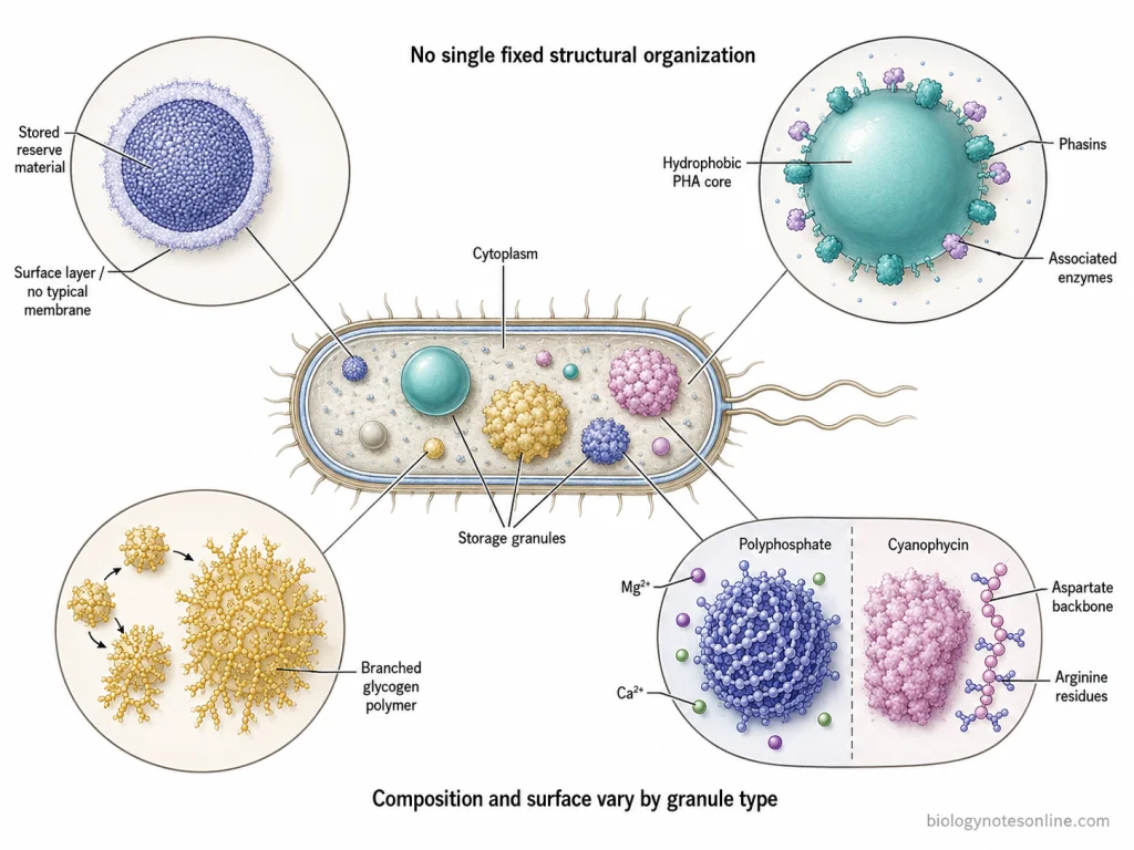
- Storage granules are generally present as distinct inclusion bodies within the cytoplasm. Their shape and size are not same in all microorganisms and these may occur as small particles or comparatively large granules inside the cell.
- The inner part of a storage granule is mainly formed by the accumulated reserve material. These substances are generally present in a concentrated or polymerized form rather than remaining freely dissolved in the cytoplasm.
- A typical membrane is not present around all the storage granules. Some of them are membraneless inclusions, while certain granules have proteins and lipid components associated with their outer surface.
- Polyhydroxyalkanoate (PHA) granules have a hydrophobic polymer core. The surface is covered mainly by proteins called phasins, together with enzymes involved in synthesis and degradation of PHA. Thus, the granule remains separated from the surrounding aqueous cytoplasm.
- Glycogen granules are made up of highly branched glucose polymers. Glycogen molecules can occur as smaller particles and may also associate to form larger particle structures.
- The structure of polyphosphate granules is different. They contain long chains of inorganic phosphate and are commonly associated with divalent cations such as Mg²⁺ and Ca²⁺. These granules generally appear as dense intracellular bodies during electron microscopic examination.
- Cyanophycin granules are generally membraneless granules. The stored polymer consists of an aspartic acid backbone having arginine residues attached to it and is deposited as an insoluble material in the cytoplasm.
- Therefore, storage granules do not have one fixed structural organization. Their internal material and the covering present around them depend on the type of substance that is stored.
Why Do Bacteria Store Nutrients in Granules?
Bacteria store excess nutrients in the form of storage granules inside the cell. These materials are generally accumulated when they are present in sufficient amount in the surrounding. The stored nutrients are then utilized during nutrient limiting conditions. The following are some of the important reasons for storing nutrients in granules-
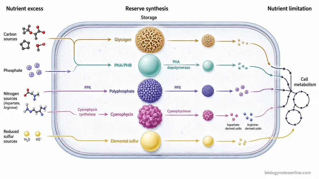
- Nutrient reserve- The major function of storage granules is the storage of reserve materials. When the nutrient becomes less available in the environment, the stored material can be utilized by the bacterial cell.
- Carbon and energy reserve- Glycogen and polyhydroxyalkanoates (PHA) are important carbon reserve materials. These are accumulated when carbon source is available in excess. During shortage of carbon, the compounds are broken down and used for energy and other cellular activities.
- Survival during starvation- Nutrients are not continuously available to bacteria in their natural environment. During such condition, the reserve present inside the granules can be used. This helps the bacterial cells to survive for a longer period under starvation condition.
- Reduction of osmotic stress- If large amount of nutrients remain freely dissolved inside the cytoplasm, it can increase the osmotic pressure of the cell. Therefore, many reserve substances are stored in polymerized or insoluble form. In this form, a large quantity of material can be accumulated without greatly increasing the concentration of soluble substances.
- Phosphate reserve- Excess phosphate is stored in some bacteria in the form of polyphosphate granules. The stored phosphate is used when phosphate becomes limited. It is required for different cellular components such as nucleic acids and phospholipids and in some bacteria, polyphosphate can also act as an energy reserve.
- Sulfur reserve- In some sulfur bacteria, elemental sulfur is accumulated as sulfur granules. This stored sulfur can later be used during metabolism and serves as an energy source under suitable conditions.
- Nitrogen reserve- Some bacteria and cyanobacteria contain cyanophycin granules as nitrogen-rich reserve material. Cyanophycin is accumulated when sufficient nitrogen is present. During nitrogen limiting condition, it is degraded and the stored nitrogen can be used by the cell.
Major Types of Storage Granules in Bacteria
Different bacteria accumulate different reserve materials inside their cells. The type of granule formed mainly depends upon the organism and the material which is available for storage. Some of the important types of storage granules found in bacteria are-
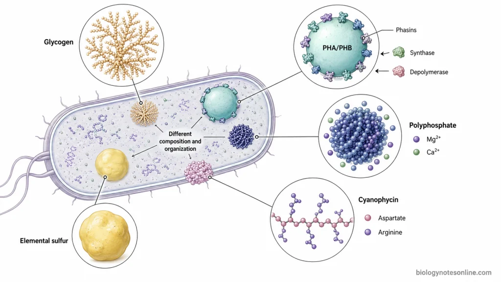
- Glycogen granules- Glycogen is a highly branched polymer of glucose and is stored as carbon reserve in many bacteria. It is generally accumulated when carbon source is available in excess. The stored glycogen can again be utilized as a source of carbon and energy during its shortage.
- Polyhydroxyalkanoate (PHA) granules- These are carbon and energy storing granules found in different groups of bacteria. Poly-β-hydroxybutyrate (PHB) is one of the best-known form of PHA. It is accumulated inside the cell as water-insoluble granules. Large amount of this reserve may be formed particularly when carbon is present in excess but another nutrient required for growth becomes limited.
- Polyphosphate granules- These granules contain long chains of inorganic phosphate and act mainly as a phosphate reserve. They are also referred to as volutin granules or metachromatic granules in bacteria. When phosphate is required, the stored polyphosphate can be degraded and used by the cell.
- Sulfur granules- Some sulfur-oxidizing bacteria accumulate elemental sulfur as intracellular granules during oxidation of reduced sulfur compounds such as hydrogen sulfide. The sulfur remains temporarily stored and is further oxidized when the external reduced sulfur source becomes limited. Such granules are common in several sulfur bacteria.
- Cyanophycin granules- Cyanophycin is a nitrogen-rich reserve polymer, particularly common in cyanobacteria and also present in some other bacteria. It consists mainly of aspartate and arginine and accumulates as granules inside the cell. It acts as a reserve of nitrogen and carbon which can be used during suitable metabolic conditions.
How Are Storage Granules Formed and Mobilized?
The formation and mobilization of storage granules mainly depends on the availability of nutrients and metabolic condition of the bacterial cell. When a particular material is available in excess, it can be changed into a reserve form and accumulated inside the cell. During shortage, the stored material is again degraded and used. The process is as follows-
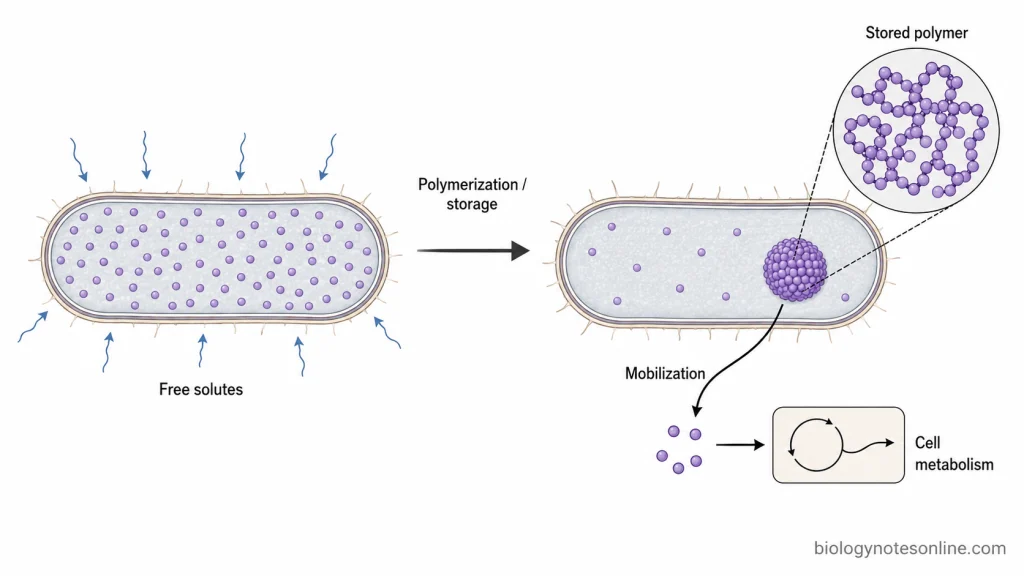
- Accumulation of nutrients- In the first step, nutrients are taken up from the surrounding environment. When their supply is greater than the immediate requirement of cell, some of these materials are directed towards storage instead of being completely used for growth.
- Formation of reserve material- The accumulated nutrients are converted into different storage compounds by specific enzymes. Carbon can be stored as glycogen or polyhydroxyalkanoates (PHA), whereas phosphate is polymerized to form polyphosphate. Cyanophycin is formed from aspartate and arginine with the help of cyanophycin synthetase. Thus, the material stored is not same in all bacteria.
- Granule formation- The reserve compounds now become accumulated as visible intracellular granules. In PHA-producing bacteria, newly synthesized PHA forms hydrophobic granules inside the cytoplasm and different proteins remain associated with its surface. The number and size of these granules can change with the organism and growth condition.
- Storage inside the cell- After formation, the granules remain as a reserve until the stored material is required. Glycogen serves as carbon and energy storage, polyphosphate stores phosphate, and cyanophycin is mainly a nitrogen-rich reserve. Some sulfur-oxidizing bacteria also accumulate elemental sulfur in sulfur granules.
- Mobilization of granules- When the external nutrient becomes limited, degradation of stored material is initiated. During this process, specific enzymes break the reserve polymers into smaller compounds which can again be used in cellular metabolism. PHA, for example, is degraded by PHA depolymerase enzymes.
- Release of stored phosphate- Polyphosphate granules are also mobilized enzymatically. Polyphosphate can be synthesized by polyphosphate kinase (PPK), whereas its degradation is carried out by enzymes such as exopolyphosphatase (PPX), releasing phosphate for cellular use.
- Mobilization of nitrogen reserve- In cyanophycin-containing cells, the stored polymer is broken down by cyanophycinase. The products are further converted into aspartate and arginine which can be reused by the cell. This is particularly useful when available nitrogen becomes limited.
- Use of stored material- The released carbon, phosphate, nitrogen or sulfur compounds now enter the respective metabolic pathways. In this way, formation of granules takes place mainly during excess availability, while their mobilization provides stored material during its requirement.
How Are Storage Granules Detected and Visualized?
Different staining and microscopic methods are used for the detection of storage granules present inside bacterial cells. The method used is different according to the type of stored material. Some granules can be detected with specific dyes, while electron microscopy is used for studying their size, location and internal structure. The following are the common methods used-
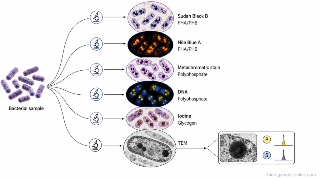
- Light microscopy- Storage granules can be observed after staining the bacterial cells with suitable stains. The granules take up the stain differently from the remaining portion of the cell and thus become visible under the microscope.
- Sudan Black B staining- It is used for the detection of polyhydroxyalkanoate (PHA) granules, especially poly-β-hydroxybutyrate (PHB). After staining, PHB is seen as dark or blue-black granules inside the bacterial cells.
- Nile Blue A staining- Nile Blue A is a fluorescent stain used for visualization of PHA and PHB granules. The stained granules show a bright orange fluorescence when examined under fluorescence microscope. It is commonly used for further detection of PHB-producing bacteria after initial screening with Sudan Black B.
- Metachromatic staining- Polyphosphate granules can be detected by their metachromatic staining property. Dyes such as toluidine blue produce a colour in the granules different from the normal colour of the dye. These granules were therefore also called metachromatic or volutin granules.
- DAPI staining- DAPI is also used to detect polyphosphate inside bacterial cells. At suitable higher concentration, polyphosphate granules produce yellow fluorescence, whereas DAPI bound with DNA generally gives blue fluorescence. However, the method is not completely specific because DAPI can also interact with some other cellular materials.
- Iodine staining- Bacterial glycogen can be detected with iodine. Iodine-stained glycogen has also been visualized as intracellular spots using confocal fluorescence microscopy.
- Electron microscopy- Transmission electron microscopy (TEM) is used to study storage granules at much higher resolution. It can show their number, position, size and appearance within the bacterial cell. Sulfur granules, glycogen deposits and polyphosphate bodies have been studied by this method.
- Elemental analysis- Electron microscopy can also be combined with energy-dispersive X-ray spectroscopy (EDX/EDS). In this method, the elemental composition of a granule can be determined, such as phosphorus in polyphosphate bodies or sulfur in sulfur granules.
Significance of Storage Granules
Storage granules have an important role in bacterial cells because different nutrients can be stored in a reserve form. These materials are used according to the requirement of cell and the surrounding conditions. The following are some of the important significance of storage granules–
- Carbon and energy reserve- Storage compounds such as glycogen and polyhydroxyalkanoates (PHA) act as reserve source of carbon and energy. When the external carbon source is not sufficient, these compounds are degraded and used by the bacterial cell.
- Survival during starvation- Bacteria commonly face periods where sufficient nutrients are not available. During this condition, stored materials can provide the compounds required for maintenance. Thus, storage granules help the cells to survive under nutrient-limiting condition.
- Adaptation to changing nutrients- Nutrient availability in natural environments may change frequently. Glycogen can be stored and mobilized rapidly during such changes, which allows bacteria to respond when carbon becomes unavailable and again resume growth when suitable nutrient is obtained.
- Phosphate reserve- Polyphosphate granules provide a stored source of phosphate inside the bacterial cell. Polyphosphate is also associated with survival during nutritional stress and different stress responses.
- Nitrogen reserve- Cyanophycin is an important nitrogen-rich storage material of many cyanobacteria and some other bacteria. It is accumulated when nitrogen is available and can be broken down during nitrogen shortage. In some cyanobacteria, it also helps in the transfer and temporary storage of newly fixed nitrogen.
- Sulfur storage- Some sulfur-oxidizing bacteria store elemental sulfur in sulfur granules. The stored sulfur can be further used in redox reactions when required and therefore takes part in energy metabolism of these organisms.
- Reduced osmotic effect- Storage in a polymeric form allows large quantity of reserve material to remain inside the cell without producing the same osmotic effect as many freely dissolved molecules. Glycogen is one important example, which has little effect on the internal osmotic pressure of the cell.
- Stress resistance- Storage granules are not only useful during nutrient shortage. PHA accumulation has also been associated with increased resistance against different environmental stresses, while polyphosphate contributes to bacterial stress survival.
References
- Achbergerová, L., & Nahálka, J. (2011). Polyphosphate—An ancient energy source and active metabolic regulator. Microbial Cell Factories, 10, 63. https://doi.org/10.1186/1475-2859-10-63
- Bowlin, M. Q., & Gray, M. J. (2021). Inorganic polyphosphate in host and microbe biology. Trends in Microbiology, 29(11), 1013–1023. https://doi.org/10.1016/j.tim.2021.02.002
- Cleveland, J. L., Binks, A. P., & LeClair, R. J. (2025). Microbiology, pharmacology, and immunology for pre-clinical students. Virginia Tech Carilion School of Medicine, Open Education Initiative, and Virginia Tech Publishing. https://doi.org/10.21061/micropharmimmuno
- Docampo, R. (2024). Advances in the cellular biology, biochemistry, and molecular biology of acidocalcisomes. Microbiology and Molecular Biology Reviews, 88(1), e00042-23. https://doi.org/10.1128/mmbr.00042-23
- Esteban-Torres, M., Ruiz, L., Rossini, V., Nally, K., & van Sinderen, D. (2023). Intracellular glycogen accumulation by human gut commensals as a niche adaptation trait. Gut Microbes, 15(1), 2235067. https://doi.org/10.1080/19490976.2023.2235067
- Fukala, I., & Kučera, I. (2024). Natural polyhydroxyalkanoates—An overview of bacterial production methods. Molecules, 29(10), 2293. https://doi.org/10.3390/molecules29102293
- George, G. N., Gnida, M., Bazylinski, D. A., Prince, R. C., & Pickering, I. J. (2008). X-ray absorption spectroscopy as a probe of microbial sulfur biochemistry: The nature of bacterial sulfur globules revisited. Journal of Bacteriology, 190(19), 6376–6383. https://doi.org/10.1128/JB.00539-08
- Grage, K., Jahns, A. C., Parlane, N., Palanisamy, R., Rasiah, I. A., Atwood, J. A., & Rehm, B. H. A. (2009). Bacterial polyhydroxyalkanoate granules: Biogenesis, structure, and potential use as nano-/micro-beads in biotechnological and biomedical applications. Biomacromolecules, 10(4), 660–669. https://doi.org/10.1021/bm801394s
- Guerrero, R., Haselton, A., Solé, M., Wier, A., & Margulis, L. (1999). Titanospirillum velox: A huge, speedy, sulfur-storing spirillum from Ebro Delta microbial mats. Proceedings of the National Academy of Sciences of the United States of America, 96(20), 11584–11588. https://doi.org/10.1073/pnas.96.20.11584
- Jurečková, Ľ., Chmelová, D., Ondrejovič, M., & Miertuš, S. (2026). Isolation and purification of bacterially produced polyhydroxyalkanoates: Mechanisms, limitations, and current advances. Life, 16(2), 269. https://doi.org/10.3390/life16020269
- Kwiatos, N., & Steinbüchel, A. (2021). Cyanophycin modifications—Widening the application potential. Frontiers in Bioengineering and Biotechnology, 9, 763804. https://doi.org/10.3389/fbioe.2021.763804
- Legat, A., Gruber, C., Zangger, K., Wanner, G., & Stan-Lotter, H. (2010). Identification of polyhydroxyalkanoates in Halococcus and other haloarchaeal species. Applied Microbiology and Biotechnology, 87(3), 1119–1127. https://doi.org/10.1007/s00253-010-2611-6
- Liu, Q.-H., Tang, J.-W., Wen, P.-B., Wang, M.-M., Zhang, X., & Wang, L. (2021). From prokaryotes to eukaryotes: Insights into the molecular structure of glycogen particles. Frontiers in Molecular Biosciences, 8, 673315. https://doi.org/10.3389/fmolb.2021.673315
- Madigan, M. T., Bender, K. S., Buckley, D. H., Sattley, W. M., & Stahl, D. A. (2018). Brock biology of microorganisms (15th ed., Global ed.). Pearson. https://www.pearson.com/en-gb/subject-catalog/p/brock-biology-of-microorganisms-global-edition/P200000008849/9781292235196
- Madison, L. L., & Huisman, G. W. (1999). Metabolic engineering of poly(3-hydroxyalkanoates): From DNA to plastic. Microbiology and Molecular Biology Reviews, 63(1), 21–53. https://doi.org/10.1128/MMBR.63.1.21-53.1999
- Maki, J. S. (2013). Bacterial intracellular sulfur globules: Structure and function. Journal of Molecular Microbiology and Biotechnology, 23(4–5), 270–280. https://doi.org/10.1159/000351335
- Mandala, V. S., Loh, D. M., Shepard, S. M., Geeson, M. B., Sergeyev, I. V., Nocera, D. G., Cummins, C. C., & Hong, M. (2020). Bacterial phosphate granules contain cyclic polyphosphates: Evidence from 31P solid-state NMR. Journal of the American Chemical Society, 142(43), 18407–18421. https://doi.org/10.1021/jacs.0c06335
- Obruca, S., Sedlacek, P., Slaninova, E., Fritz, I., Daffert, C., Meixner, K., Sedrlova, Z., & Koller, M. (2020). Novel unexpected functions of PHA granules. Applied Microbiology and Biotechnology, 104(11), 4795–4810. https://doi.org/10.1007/s00253-020-10568-1
- Ovecka, M., Bahaji, A., Muñoz, F. J., Almagro, G., Ezquer, I., Baroja-Fernández, E., Li, J., & Pozueta-Romero, J. (2012). A sensitive method for confocal fluorescence microscopic visualization of starch granules in iodine stained samples. Plant Signaling & Behavior, 7(9), 1146–1150. https://doi.org/10.4161/psb.21370
- Parker, N., Schneegurt, M., Tu, A.-H. T., Forster, B. M., & Lister, P. (2016). Microbiology. OpenStax. https://openstax.org/books/microbiology/pages/1-introduction
- Ruiz-Haddad, L., Ali, M., Pronk, M., van Loosdrecht, M. C. M., & Saikaly, P. E. (2024). Demystifying polyphosphate-accumulating organisms relevant to wastewater treatment: A review of their phylogeny, metabolism, and detection. Environmental Science and Ecotechnology, 21, 100387. https://doi.org/10.1016/j.ese.2024.100387
- Saier, M. H., Jr. (2013). Microcompartments and protein machines in prokaryotes. Journal of Molecular Microbiology and Biotechnology, 23(4–5), 243–269. https://doi.org/10.1159/000351625
- Salton, M. R. J., & Kim, K.-S. (1996). Structure. In S. Baron (Ed.), Medical microbiology (4th ed., Chapter 2). University of Texas Medical Branch at Galveston. https://www.ncbi.nlm.nih.gov/books/NBK8477/
- San Miguel-González, G. de J., Alemán-Huerta, M. E., Martínez-Herrera, R. E., Quintero-Zapata, I., de la Torre-Zavala, S., Avilés-Arnaut, H., Gandarilla-Pacheco, F. L., & de Luna-Santillana, E. de J. (2024). Alkaline-tolerant Bacillus cereus 12GS: A promising polyhydroxybutyrate (PHB) producer isolated from the north of Mexico. Microorganisms, 12(5), 863. https://doi.org/10.3390/microorganisms12050863
- Sasazawa, M., Tomares, D. T., Childers, W. S., & Saurabh, S. (2024). Biomolecular condensates as stress sensors and modulators of bacterial signaling. PLOS Pathogens, 20(8), e1012413. https://doi.org/10.1371/journal.ppat.1012413
- Sekar, K., Linker, S. M., Nguyen, J., Grünhagen, A., Stocker, R., & Sauer, U. (2020). Bacterial glycogen provides short-term benefits in changing environments. Applied and Environmental Microbiology, 86(9), e00049-20. https://doi.org/10.1128/AEM.00049-20
- Sharon, I., & Schmeing, T. M. (2023). Bioinformatics of cyanophycin metabolism genes and characterization of promiscuous isoaspartyl dipeptidases that catalyze the final step of cyanophycin degradation. Scientific Reports, 13, 8314. https://doi.org/10.1038/s41598-023-34587-w
- Tarayre, C., Nguyen, H.-T., Brognaux, A., Delepierre, A., De Clercq, L., Charlier, R., Michels, E., Meers, E., & Delvigne, F. (2016). Characterisation of phosphate accumulating organisms and techniques for polyphosphate detection: A review. Sensors, 16(6), 797. https://doi.org/10.3390/s16060797
- Tocheva, E. I., Dekas, A. E., McGlynn, S. E., Morris, D., Orphan, V. J., & Jensen, G. J. (2013). Polyphosphate storage during sporulation in the gram-negative bacterium Acetonema longum. Journal of Bacteriology, 195(17), 3940–3946. https://doi.org/10.1128/JB.00712-13
- Watzer, B., & Forchhammer, K. (2018). Cyanophycin synthesis optimizes nitrogen utilization in the unicellular cyanobacterium Synechocystis sp. strain PCC 6803. Applied and Environmental Microbiology, 84(20), e01298-18. https://doi.org/10.1128/AEM.01298-18
- Willey, J. M., Sandman, K. M., & Wood, D. H. (2020). Prescott’s microbiology (11th ed.). McGraw-Hill Education. https://www.mheducation.com/unitas/highered/sample-chapters/9781260211887.pdf
- Wilson, W. A., Roach, P. J., Montero, M., Baroja-Fernández, E., Muñoz, F. J., Eydallin, G., Viale, A. M., & Pozueta-Romero, J. (2010). Regulation of glycogen metabolism in yeast and bacteria. FEMS Microbiology Reviews, 34(6), 952–985. https://doi.org/10.1111/j.1574-6976.2010.00220.x