

Cilia are membrane-covered, microtubule-based projections that extend from the surface of many eukaryotic cells. Motile cilia generate movement of cells, fluids, or particles, whereas most primary cilia function as specialized sensory and signaling compartments. Their structure centers on an axoneme anchored to the cell by a basal body.
What Are Cilia?
Cilia are short hair-like structures present on the surface of many eukaryotic cells and are covered by the cell membrane. These structures contain a microtubule core called the axoneme. It develops from a basal body present at the base of the cilium. Cilia are mainly of two types, motile cilia and primary cilia. Motile cilia generally have a 9+2 arrangement of microtubules and show repeated beating movement. This movement is used to move fluid or other materials over the surface of cells, such as the movement of mucus in the respiratory tract. Primary cilia, on the other hand, are usually present as a single non-motile structure and commonly have a 9+0 arrangement of microtubules. It mainly functions in sensing and signalling. These cilia help the cell to detect different signals present outside the cell and respond to them.
Characteristics of Cilia
The following are some of the important characteristics of cilia-
- Cilia are short hair-like projections present on the surface of many eukaryotic cells. They are covered by the plasma membrane of the cell.
- Axoneme– The inner core of a cilium is formed of microtubules and is called the axoneme. It forms the main structural framework of cilia.
- Each cilium develops from a basal body which is present at its base. The basal body is a modified centriole from where the microtubules of axoneme are extended.
- Based on their structure and movement, cilia are mainly of two types, motile cilia and primary cilia. Motile cilia generally contain nine outer microtubule doublets and two microtubules at the centre. This is referred to as 9+2 arrangement. In primary cilia, the central pair is usually absent and hence they have 9+0 arrangement. Some motile cilia also possess the 9+0 arrangement.
- Motile cilia are commonly present in large numbers on the surface of cell. These cilia show repeated beating movement. The movement is used to move fluids or other materials over the cells, such as movement of mucus in the respiratory tract.
- Primary cilia– These are generally present as a single cilium on a cell and most of them are non-motile. It mainly performs sensory and signalling functions. Different extracellular signals can be detected with the help of these cilia.
- The microtubules in motile cilia are associated with axonemal dynein proteins. These proteins act as molecular motors which use ATP and produce the force required for movement of cilia.
- The membrane of cilia forms a specialized region of the cell membrane. Different receptors and signalling proteins can be concentrated in this region. This is particularly important in primary cilia for carrying out its sensory function.
Structure of a Cilium
The structure of a cilium consists of a membrane covering, basal region and the internal microtubular framework. The following are the major structural parts of a cilium-
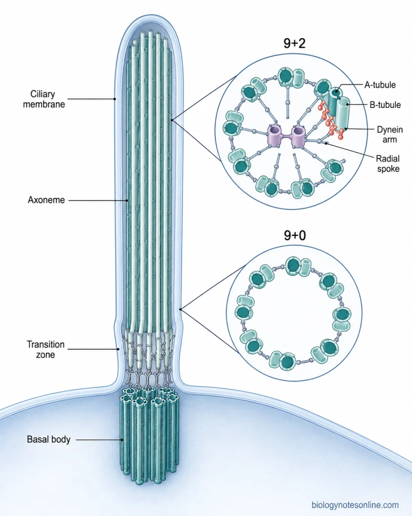
- Ciliary membrane– The entire cilium is surrounded by a ciliary membrane which is continuous with the plasma membrane of the cell. It forms a specialized membrane region and contains different ciliary proteins and receptors.
- Basal body– It is present at the base of cilium and anchors the cilium to the cell. The basal body is formed from a modified centriole. It contains nine sets of microtubule triplets, from which the microtubules of cilium are developed.
- Transition zone– Between the basal body and the ciliary axoneme is a specialized region called the transition zone. In this region, the C-tubules of the basal body terminate and the nine microtubule doublets continue into the cilium. This region also forms an important boundary between cilium and rest of the cell.
- Axoneme– The major internal structural framework of a cilium is called the axoneme. It is made up mainly of nine peripheral microtubule doublets arranged in a circular pattern.
- A-tubule and B-tubule– Each outer microtubule doublet consists of an A-tubule and a B-tubule. The A-tubule is a complete microtubule whereas the B-tubule is an incomplete microtubule attached to it.
- Microtubule arrangement– In most motile cilia, nine outer microtubule doublets surround two single microtubules present at the centre. This is referred to as the 9+2 arrangement. Primary cilia usually do not contain the central pair and hence show a 9+0 arrangement. Some motile cilia, such as nodal cilia, also possess 9+0 structure.
- Dynein arms– Motile cilia contain inner and outer dynein arms associated with the outer microtubule doublets. These are motor proteins which are important for producing ciliary movement.
- Radial spokes and nexin links– These are also present in the typical motile axoneme. Radial spokes extend from the outer doublets towards the central pair, while adjacent outer doublets are connected by structures associated with the dynein regulatory complex (nexin). These structures help in organization of the axoneme during ciliary movement.
Types of Cilia
Cilia are generally divided into two major types depending upon their movement and function. These are motile cilia and primary or non-motile cilia. A specialized type of motile cilia called nodal cilia is also present during embryonic development.
1. Motile cilia
Motile cilia are the cilia which show regular beating movement. These are generally present in large numbers on the surface of a cell and commonly possess the 9+2 arrangement of microtubules. Dynein arms are present which help in producing the movement of cilia. The major function of these cilia is movement of fluids or materials over the cell surface. They are present in the respiratory epithelium, ependymal cells of brain and the oviduct, where their coordinated beating helps in movement of mucus, cerebrospinal fluid or other materials.
2. Primary cilia (Non-motile cilia)
Primary cilia are usually present as a single cilium on the cell. Most of them are non-motile and contain nine peripheral microtubule doublets without the central pair, which is referred to as the 9+0 arrangement. It mainly acts as a sensory structure rather than producing movement. Different receptors and signalling proteins are present in the ciliary membrane which allows the cell to detect chemical and mechanical signals from its surrounding environment.
3. Nodal cilia
These are a specialized type of motile cilia found in the embryonic node during early development. Unlike the usual motile cilia, nodal cilia have 9+0 arrangement, but they contain dynein motors and hence are capable of movement. They show a rotational or rotary movement instead of the typical beating movement of 9+2 cilia. During this process, a directional fluid flow is produced across the embryonic node which is important for establishment of left-right arrangement of organs in the developing body.
How Do Motile Cilia Move?
The movement of motile cilia is based on the sliding of microtubule doublets present in the axoneme. However, these microtubules cannot slide freely and the sliding force is changed into bending of the cilium. The process takes place in the following steps-
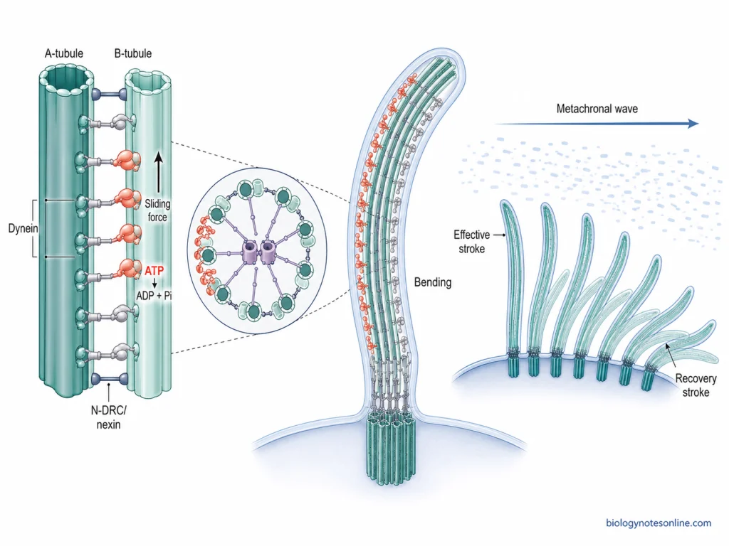
Step 1- Arrangement of axoneme
A typical motile cilium contains nine peripheral microtubule doublets surrounding two central microtubules, forming the 9+2 arrangement. Each outer doublet contains an A-tubule and B-tubule. Dynein arms are attached to the A-tubule, while nexin-dynein regulatory complex and radial spokes are also associated with the axoneme.
Step 2- Activation of dynein
The dynein arms act as motor proteins. In this step, ATP is hydrolyzed by dynein and the energy produced causes a conformational change in the dynein protein. The dynein attached to one A-tubule interacts with the B-tubule of the neighbouring microtubule doublet.
Step 3- Sliding of microtubules
After activation, dynein moves along the B-tubule of the adjacent doublet and a sliding force is produced between the two microtubule doublets. Thus, one doublet tends to slide with respect to another. If these doublets were free, the result would mainly be sliding rather than bending.
Step 4- Conversion of sliding into bending
The microtubule doublets are not free to move apart. They are connected by the nexin-dynein regulatory complex (N-DRC) and the axoneme is also anchored at its basal region. These restrictions limit the amount of sliding between neighbouring doublets. As a result, the sliding force produced by dynein is converted into bending of the entire axoneme. This is the major reason why a cilium bends instead of simply allowing its microtubules to slide past one another.
Step 5- Regulation of the bend
Dynein activity does not occur equally on all sides of the axoneme at the same time. Dyneins on one side become more active and produce bending in one direction, while activity then changes to the opposite side. The central pair, radial spokes and N-DRC take part in controlling the activity of axonemal dyneins. In this way, bending can occur repeatedly rather than remaining fixed in one direction.
Step 6- Formation of ciliary beat
Repeated bending is finally changed into the characteristic beating movement of motile cilia. In many epithelial cilia, the movement consists of an effective stroke, in which the cilium moves forward and pushes the material, followed by a recovery stroke that brings it back towards its original position. Repetition of these strokes produces continuous ciliary movement and nearby cilia can act together for movement of fluid or material over the cell surface.
How Do Primary Cilia Function as Signaling Compartments?
Primary cilia do not only act as projections from the cell surface. They form a specialized signaling compartment where different receptors, signalling proteins and other regulatory molecules are concentrated. The ciliary membrane is continuous with the plasma membrane, but its protein composition is maintained differently from rest of the cell membrane.
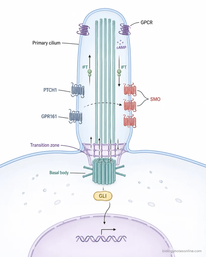
Ciliary compartment formation
At the base of primary cilium is a specialized region called the transition zone. It acts as a selective barrier and controls movement of many membrane and soluble proteins between the cilium and rest of the cell. Thus, proteins required for ciliary signalling can be maintained within a small specialized region instead of being distributed throughout the plasma membrane.
Concentration of receptors
Different receptors are specifically transported and concentrated in the ciliary membrane. Many G-protein coupled receptors (GPCRs) and their signalling components are found in primary cilia. This allows an extracellular signal to be received in a restricted region and the downstream reaction can be controlled from this compartment.
Transport of signalling proteins
Proteins do not remain permanently fixed inside the cilium. They are moved into, along and out of the cilium by controlled trafficking systems. Intraflagellar transport (IFT) moves different cargo along the axonemal microtubules, while other ciliary trafficking proteins also help in selection of membrane proteins. In this way, the composition of primary cilium can change depending upon the signalling condition.
Hedgehog signalling
One of the best established examples is the Hedgehog (Hh) pathway. In the absence of Hedgehog signal, Patched1 (PTCH1) and GPR161 are associated with the ciliary compartment and the pathway remains in its inhibited state. When Hedgehog ligand is received, the distribution of these proteins changes and Smoothened (SMO) becomes concentrated in the cilium. This eventually changes the activity of GLI transcription factors which regulate Hedgehog-responsive genes. Thus, entry and removal of signalling proteins from cilium is an important part of the reaction.
GPCR signalling
Primary cilia also contain several GPCRs which can regulate intracellular second messengers such as cyclic AMP (cAMP). The signalling molecules present inside the small ciliary space can therefore be regulated separately to some extent from those present in the main cell body. Different cell types contain different combinations of these ciliary receptors.
Signal organization
Bringing receptors, transport proteins and downstream signalling components into one small compartment helps in organization of a signalling pathway. It can also control when a protein enters or leaves the cilium. The primary cilium therefore acts as a signalling hub rather than simply acting as a passive surface projection.
Mechanical signal sensing
Primary cilia have also been associated with sensing mechanical conditions such as fluid flow, particularly in tissues such as kidney. Earlier models proposed that bending of the cilium activates polycystin proteins and directly produces a ciliary Ca²⁺ signal. However, this mechanism is not completely settled. Some experimental studies did not detect a flow-induced ciliary Ca²⁺ influx and the exact relationship between ciliary bending, polycystins and calcium signalling is still being studied. Thus, a sensory role of primary cilia is well supported, but all proposed mechanisms of direct ciliary mechanosensation should not be considered identical or completely established.
How Are Cilia Built and Maintained?
Formation of a cilium is a controlled process which begins from the mother centriole of the cell. The centriole changes into a basal body, the axoneme starts to grow and ciliary membrane is formed around it. Further growth and maintenance mainly depends upon a transport process called intraflagellar transport (IFT).
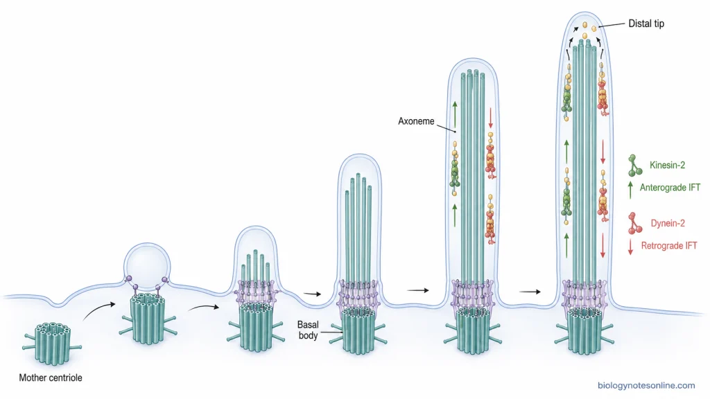
Formation of the basal body
In the initial stage, the mother centriole becomes associated with the cell membrane or with a ciliary vesicle and is converted into the basal body. It becomes attached near the cell surface by its distal appendages. The basal body now acts as the site from which the microtubules of cilium are formed.
Growth of the axoneme
After basal body formation, nine microtubule doublets extend from it and form the axoneme. New axonemal components are mainly added at the distal tip of the growing cilium. At the same time, membrane is supplied around the developing axoneme and the cilium gradually extends outward from the cell surface.
Why is IFT required?
Cilia do not contain their own machinery for protein synthesis. The proteins required for building the axoneme are therefore produced in the cell body, while the site of their addition is present at the distant ciliary tip. These materials must be carried from the base to the tip. This is carried out by intraflagellar transport (IFT).
Anterograde transport
During anterograde IFT, IFT particles containing ciliary proteins and building materials move from the base towards the tip of cilium. This movement is mainly carried out by kinesin-2 motor proteins along the axonemal microtubules. Tubulin and other components delivered to the tip can then be used for further growth and assembly of the cilium.
Retrograde transport
Materials do not move only towards the tip. After delivery of cargo, IFT components and other materials are transported back from the tip towards the cell body. This is referred to as retrograde IFT and is mainly carried out by dynein-2. It helps in recycling transport machinery and removal of materials from the cilium.
Maintenance of cilia
A mature cilium is not a permanently fixed structure. Its proteins and microtubule components undergo continuous turnover, with new materials being supplied and other materials returned towards the cell body. Thus, IFT is required not only during formation of cilia but also for maintaining its normal structure and length. Disturbance of this transport can therefore result in shortened, abnormal or loss of cilia.
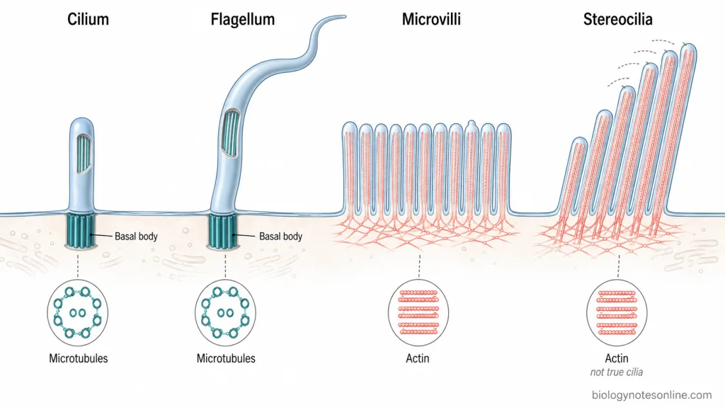
Cilia vs Flagella, Microvilli and Stereocilia
Cilia, flagella, microvilli and stereocilia are projections extending from the surface of cells. However, they differ in their internal structure, movement and major functions. The important differences are as follows-
| Characteristics | Cilia | Flagella | Microvilli | Stereocilia |
|---|---|---|---|---|
| Basic structure | Cilia are membrane-covered cell projections having an internal axoneme. | Eukaryotic flagella have almost the same basic structural organization as cilia. The terms cilia and flagella are therefore sometimes used interchangeably. | These are small finger-like projections of the plasma membrane. | Stereocilia are long specialized projections similar to microvilli and are not true cilia. |
| Cytoskeleton | Their internal core is made up of microtubules. Motile cilia commonly possess 9+2 arrangement, whereas primary cilia usually have 9+0 arrangement. | The core is also a microtubule-based axoneme. In motile eukaryotic flagella, the typical arrangement is 9+2. | Microvilli contain a central bundle of parallel actin filaments. | They contain closely packed bundles of actin filaments rather than microtubules. |
| Basal attachment | Each cilium develops from a basal body, which is a modified centriole. | A flagellum is also anchored by a basal body. | They do not arise from basal bodies. Their actin bundles are attached to the cortical cytoskeleton of the cell. | No basal body is present for each stereocilium. Their actin bundles extend into rootlets that help in anchoring them at the apical region of the cell. |
| Number and length | Motile cilia are commonly short and present in large numbers, while a primary cilium is generally single. | Flagella are generally longer and occur singly or in small numbers on the cell. | They are short and usually occur in very large numbers, forming structures such as the brush border. | These are much longer than ordinary microvilli and are arranged together in bundles of different heights in sensory hair cells. |
| Movement | Motile cilia show repeated beating movement. Primary cilia are generally non-motile. | Motile flagella produce bending or wave-like movement which can propel the entire cell. | Microvilli do not show cilia-like active beating movement. | Stereocilia are non-motile in the ciliary sense, but their bundles can be mechanically deflected. |
| Major function | Cilia are used for movement of fluids and materials over cell surfaces. Primary cilia mainly perform sensory and signalling functions. | Their major function is locomotion of the cell, such as movement of sperm. | The major function is to increase the surface area of cells, especially for absorption and other interactions with luminal contents. | In inner ear hair cells, stereocilia are used for mechanosensation and take part in detection of sound and body movement. |
| Examples | Respiratory epithelial cilia, oviduct cilia and primary cilia of many vertebrate cells. | Sperm flagellum and flagella of different unicellular eukaryotes. | Microvilli of intestinal enterocytes and kidney epithelial cells. | Stereocilia of cochlear and vestibular hair cells. |
What Happens When Cilia Do Not Function Properly?
Defects in cilia can affect their normal movement as well as signalling functions. Such disorders caused by abnormal formation or functioning of cilia are referred to as ciliopathies. The following are some of the important effects of ciliary defects-
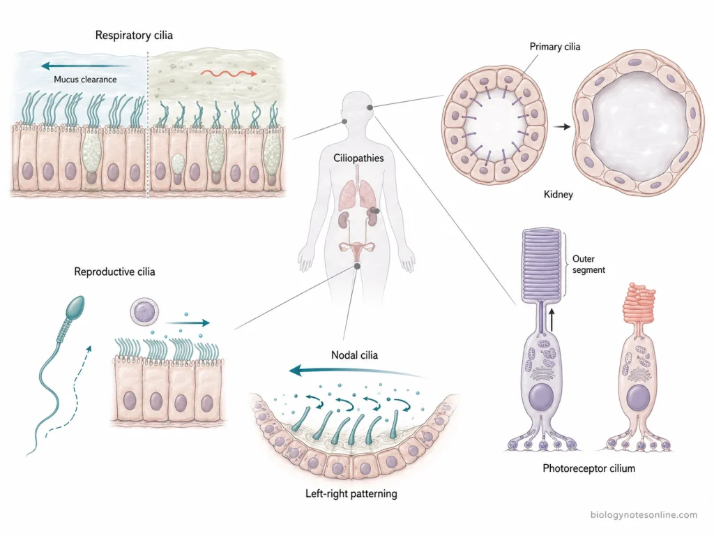
- Respiratory tract defects– Motile cilia of the respiratory epithelium are used to remove mucus and trapped particles from the airways. If their beating becomes abnormal, mucus starts accumulating and microorganisms are not cleared properly. This can result in repeated respiratory infections, sinusitis and middle ear infections. In severe conditions, bronchiectasis can also develop. These defects are commonly seen in Primary Ciliary Dyskinesia (PCD).
- Infertility– The reproductive system is also affected by defective cilia. In males, abnormal movement of sperm flagellum reduces sperm motility and may result in infertility. In females, cilia of the fallopian tube are involved in transport of the oocyte. Improper functioning of these cilia can reduce fertility and the chance of ectopic pregnancy can also increase.
- Abnormal arrangement of organs– During embryonic development, nodal cilia produce directional fluid movement which is important for normal left-right body pattern. When these cilia fail to function, normal position of internal organs may be disturbed. This can result in situs inversus. Other laterality defects such as heterotaxy can also occur.
- Kidney disorders– Primary cilia are present on the cells of kidney tubules and take part in normal signalling and maintenance of tubular structure. Defects in these cilia can result in abnormal growth of tubules and formation of cysts. Polycystic kidney disease (PKD) and nephronophthisis (NPHP) are some of the important examples associated with ciliary defects.
- Vision problems– Photoreceptor cells of retina contain a specialized ciliary region. It is necessary for transport of different proteins to the outer segment of photoreceptor. If this transport becomes disturbed, photoreceptor cells gradually lose their normal function. Retinal degeneration and progressive loss of vision can occur.
- Developmental defects– Primary cilia are involved in different signalling pathways required during growth and development. Disturbance of these signalling processes can affect more than one organ system. The brain, skeleton, eyes, kidneys, liver and other tissues may show abnormalities depending upon the type of ciliopathy.
Functions of Cilia
Cilia perform different functions depending upon their type and location in the cell. The following are some of the important functions of cilia-
- Mucociliary clearance– Motile cilia present in the respiratory tract show coordinated beating movement. This movement is used to move mucus containing dust particles, microorganisms and other inhaled materials towards the upper respiratory tract for their removal.
- Movement of cerebrospinal fluid– Cilia present on the ependymal cells of brain ventricles help in movement of cerebrospinal fluid (CSF). Their repeated beating produces fluid flow along the ventricular surfaces.
- Transport in reproductive tract– In females, motile cilia of the oviduct are involved in collection and movement of the ovulated oocyte and also help in transport towards the uterus. Cilia are also present in parts of male reproductive tract where the movement produced by them supports sperm transport and prevents their aggregation.
- Sensory function– Primary cilia mainly act as sensory structures of the cell. They can detect different chemical and mechanical signals present outside the cell and transfer these information into cellular responses. Thus, primary cilium is often involved in sensing the surrounding environment of a cell.
- Cell signalling– Different receptors and signalling proteins are concentrated in the membrane of primary cilia. It is involved in several signalling pathways, including Hedgehog signalling, which is important during development and normal tissue functions.
- Left-right body patterning– During early embryonic development, specialized motile nodal cilia produce a leftward fluid flow at the embryonic node. This flow is important for establishment of the normal left-right arrangement of internal organs. Sensory cilia present in this region also take part in detecting the flow-associated signal.
- Locomotion and food movement– In many unicellular eukaryotes, cilia are used for movement of the entire cell through surrounding fluid. They can also take part in movement of food particles towards the region of food uptake.
Examples of Cilia
Cilia are present in different cells of animals as well as many unicellular eukaryotes. The following are some of the important examples of cilia-
- Respiratory tract cilia– Motile cilia are present in large numbers on the epithelial cells lining the respiratory tract. Their coordinated beating is used for movement of mucus and trapped particles towards the upper respiratory tract.
- Oviduct cilia– The epithelial lining of the oviduct or fallopian tube contains numerous motile cilia. These cilia help in movement of the ovulated oocyte through the oviduct and towards the uterus.
- Ependymal cilia– These are found on ependymal cells lining the ventricles of brain. They are motile cilia and their beating helps in movement of cerebrospinal fluid (CSF) along the ventricular surface.
- Kidney primary cilia– Primary cilia are present on epithelial cells of different regions of kidney tubules and collecting ducts. These are generally single and non-motile structures. It mainly performs sensory and signalling functions in these cells.
- Olfactory cilia– Olfactory sensory neurons contain specialized cilia extending from their dendritic ends. Different proteins necessary for detection of odorants are concentrated in these cilia and hence they play an important role in the sense of smell.
- Photoreceptor cilia– Rod and cone photoreceptor cells of retina contain a highly modified sensory cilium. The light-sensitive outer segment is associated with this ciliary structure and is connected to the inner segment through the connecting cilium.
- Nodal cilia– These are single motile cilia present temporarily in the embryonic node during early development. Unlike most motile cilia, they have a 9+0 arrangement and show rotational movement.
- Cilia of Paramecium– Paramecium is covered by thousands of motile cilia arranged over its cell surface. The cilia beat in coordinated waves which are used for swimming and also assist in feeding.
References
- Anvarian, Z., Mykytyn, K., Mukhopadhyay, S., Pedersen, L. B., & Christensen, S. T. (2019). Cellular signalling by primary cilia in development, organ function and disease. Nature Reviews Nephrology, 15(4), 199–219. https://doi.org/10.1038/s41581-019-0116-9
- Antony, D., Brunner, H. G., & Schmidts, M. (2021). Ciliary dyneins and dynein related ciliopathies. Cells, 10(8), 1885. https://doi.org/10.3390/cells10081885
- Bai, Y., Wei, C., Li, P., Sun, X., Cai, G., Chen, X., & Hong, Q. (2022). Primary cilium in kidney development, function and disease. Frontiers in Endocrinology, 13, 952055. https://doi.org/10.3389/fendo.2022.952055
- Bouhouche, K., Valentine, M. S., Le Borgne, P., Lemullois, M., Yano, J., Lodh, S., Nabi, A., Tassin, A. M., & Van Houten, J. L. (2022). Paramecium, a model to study ciliary beating and ciliogenesis: Insights from cutting-edge approaches. Frontiers in Cell and Developmental Biology, 10, 847908. https://doi.org/10.3389/fcell.2022.847908
- Brooks, E. R., & Wallingford, J. B. (2014). Multiciliated cells: A review. Current Biology, 24(19), R973–R982. https://doi.org/10.1016/j.cub.2014.08.047
- Brown, J. M., & Witman, G. B. (2014). Cilia and diseases. BioScience, 64(12), 1126–1137. https://doi.org/10.1093/biosci/biu174
- Collison, R., Hyatali, S. A., Kamenova, A., Rashed, A., Riley, D., Kumar, K., Stowell, J. M., & Loebinger, M. R. (2025). Primary ciliary dyskinesia: Aetiology, diagnosis and clinical management. Clinical Medicine, 25(3), 100319. https://doi.org/10.1016/j.clinme.2025.100319
- Czarnecki, P. G., & Shah, J. V. (2012). The ciliary transition zone: From morphology and molecules to medicine. Trends in Cell Biology, 22(4), 201–210. https://doi.org/10.1016/j.tcb.2012.02.001
- Ezzati, M., Djahanbakhch, O., Arian, S., & Carr, B. R. (2014). Tubal transport of gametes and embryos: A review of physiology and pathophysiology. Journal of Assisted Reproduction and Genetics, 31(10), 1337–1347. https://doi.org/10.1007/s10815-014-0309-x
- Fry, A. M., Leaper, M. J., & Bayliss, R. (2014). The primary cilium: Guardian of organ development and homeostasis. Organogenesis, 10(1), 62–68. https://doi.org/10.4161/org.28910
- Ghanaeian, A., Majhi, S., McCafferty, C. L., Nami, B., Black, C. S., Yang, S. K., Legal, T., Papoulas, O., Janowska, M., Valente-Paterno, M., Marcotte, E. M., Wloga, D., & Bui, K. H. (2023). Integrated modeling of the Nexin-dynein regulatory complex reveals its regulatory mechanism. Nature Communications, 14(1), 5741. https://doi.org/10.1038/s41467-023-41480-7
- Gudis, D., Zhao, K.-Q., & Cohen, N. A. (2012). Acquired cilia dysfunction in chronic rhinosinusitis. American Journal of Rhinology & Allergy, 26(1), 1–6. https://doi.org/10.2500/ajra.2012.26.3716
- Hilgendorf, K. I., Johnson, C. T., & Jackson, P. K. (2016). The primary cilium as a cellular receiver: Organizing ciliary GPCR signaling. Current Opinion in Cell Biology, 39, 84–92. https://doi.org/10.1016/j.ceb.2016.02.008
- Ho, E. K., & Stearns, T. (2021). Hedgehog signaling and the primary cilium: Implications for spatial and temporal constraints on signaling. Development, 148(9), dev195552. https://doi.org/10.1242/dev.195552
- Horani, A., Ferkol, T. W., Dutcher, S. K., & Brody, S. L. (2016). Genetics and biology of primary ciliary dyskinesia. Paediatric Respiratory Reviews, 18, 18–24. https://doi.org/10.1016/j.prrv.2015.09.001
- Hsiao, Y.-C., Tuz, K., & Ferland, R. J. (2012). Trafficking in and to the primary cilium. Cilia, 1(1), 4. https://doi.org/10.1186/2046-2530-1-4
- Hu, Q., & Nelson, W. J. (2011). Ciliary diffusion barrier: The gatekeeper for the primary cilium compartment. Cytoskeleton, 68(6), 313–324. https://doi.org/10.1002/cm.20514
- Ishikawa, T. (2017). Axoneme structure from motile cilia. Cold Spring Harbor Perspectives in Biology, 9(1), a028076. https://doi.org/10.1101/cshperspect.a028076
- Jenkins, P. M., McEwen, D. P., & Martens, J. R. (2009). Olfactory cilia: Linking sensory cilia function and human disease. Chemical Senses, 34(5), 451–464. https://doi.org/10.1093/chemse/bjp020
- Katoh, T. A. (2024). Function of nodal cilia in left-right determination: Mechanical regulation in initiation of symmetry breaking. Biophysics and Physicobiology, 21(3), e210018. https://doi.org/10.2142/biophysico.bppb-v21.0018
- Khanna, H. (2015). Photoreceptor sensory cilium: Traversing the ciliary gate. Cells, 4(4), 674–686. https://doi.org/10.3390/cells4040674
- Kim, S., & Dynlacht, B. D. (2013). Assembling a primary cilium. Current Opinion in Cell Biology, 25(4), 506–511. https://doi.org/10.1016/j.ceb.2013.04.011
- Kumar, V., Umair, Z., Kumar, S., Goutam, R. S., Park, S., & Kim, J. (2021). The regulatory roles of motile cilia in CSF circulation and hydrocephalus. Fluids and Barriers of the CNS, 18, 31. https://doi.org/10.1186/s12987-021-00265-0
- Leigh, M. W., Horani, A., Kinghorn, B., O’Connor, M. G., Zariwala, M. A., & Knowles, M. R. (2019). Primary ciliary dyskinesia (PCD): A genetic disorder of motile cilia. Translational Science of Rare Diseases, 4(1–2), 51–75. https://doi.org/10.3233/TRD-190036
- Long, H., Wang, Q., & Huang, K. (2015). Ciliary/flagellar protein ubiquitination. Cells, 4(3), 474–482. https://doi.org/10.3390/cells4030474
- McPherson, D. R. (2018). Sensory hair cells: An introduction to structure and physiology. Integrative and Comparative Biology, 58(2), 282–300. https://doi.org/10.1093/icb/icy064
- Mirvis, M., Stearns, T., & Nelson, W. J. (2018). Cilium structure, assembly, and disassembly regulated by the cytoskeleton. Biochemical Journal, 475(14), 2329–2353. https://doi.org/10.1042/BCJ20170453
- Morales, E. A., Gaeta, I., & Tyska, M. J. (2023). Building the brush border, one microvillus at a time. Current Opinion in Cell Biology, 80, 102153. https://doi.org/10.1016/j.ceb.2023.102153
- Mukhopadhyay, S., & Rohatgi, R. (2014). G-protein-coupled receptors, Hedgehog signaling and primary cilia. Seminars in Cell & Developmental Biology, 33, 63–72. https://doi.org/10.1016/j.semcdb.2014.05.002
- Nachury, M. V. (2014). How do cilia organize signalling cascades? Philosophical Transactions of the Royal Society B: Biological Sciences, 369(1650), 20130465. https://doi.org/10.1098/rstb.2013.0465
- Olm, M. A. K., Caldini, E. G., & Mauad, T. (2015). Diagnosis of primary ciliary dyskinesia. Jornal Brasileiro de Pneumologia, 41(3), 251–263. https://doi.org/10.1590/S1806-37132015000004447
- Ostrowski, L. E., Dutcher, S. K., & Lo, C. W. (2011). Cilia and models for studying structure and function. Proceedings of the American Thoracic Society, 8(5), 423–429. https://doi.org/10.1513/pats.201103-027SD
- Park, J., & Bird, J. E. (2023). The actin cytoskeleton in hair bundle development and hearing loss. Hearing Research, 436, 108817. https://doi.org/10.1016/j.heares.2023.108817
- Park, K., & Leroux, M. R. (2022). Composition, organization and mechanisms of the transition zone, a gate for the cilium. EMBO Reports, 23(12), e55420. https://doi.org/10.15252/embr.202255420
- Pazour, G. J. (2024). Cilia structure and function in human disease. Current Opinion in Endocrine and Metabolic Research, 34, 100509. https://doi.org/10.1016/j.coemr.2024.100509
- Prasad, R. M., Jin, X., & Nauli, S. M. (2014). Sensing a sensor: Identifying the mechanosensory function of primary cilia. Biosensors, 4(1), 47–62. https://doi.org/10.3390/bios4010047
- Saternos, H., Ley, S., & AbouAlaiwi, W. (2020). Primary cilia and calcium signaling interactions. International Journal of Molecular Sciences, 21(19), 7109. https://doi.org/10.3390/ijms21197109
- Satir, P., & Christensen, S. T. (2008). Structure and function of mammalian cilia. Histochemistry and Cell Biology, 129(6), 687–693. https://doi.org/10.1007/s00418-008-0416-9
- Satir, P., Pedersen, L. B., & Christensen, S. T. (2010). The primary cilium at a glance. Journal of Cell Science, 123(4), 499–503. https://doi.org/10.1242/jcs.050377
- Shapiro, A. J., Zariwala, M. A., Ferkol, T., Davis, S. D., Sagel, S. D., Dell, S. D., Rosenfeld, M., Olivier, K. N., Milla, C., Daniel, S. J., Kimple, A. J., Manion, M., Knowles, M. R., Leigh, M. W., & Genetic Disorders of Mucociliary Clearance Consortium. (2016). Diagnosis, monitoring, and treatment of primary ciliary dyskinesia: PCD Foundation consensus recommendations based on state of the art review. Pediatric Pulmonology, 51(2), 115–132. https://doi.org/10.1002/ppul.23304
- Sun, Z. (2020). Regulation and function of calcium in the cilium. Current Opinion in Physiology, 17, 278–283. https://doi.org/10.1016/j.cophys.2020.08.019
- Taschner, M., & Lorentzen, E. (2016). The intraflagellar transport machinery. Cold Spring Harbor Perspectives in Biology, 8(10), a028092. https://doi.org/10.1101/cshperspect.a028092
- Veland, I. R., Awan, A., Pedersen, L. B., Yoder, B. K., & Christensen, S. T. (2009). Primary cilia and signaling pathways in mammalian development, health and disease. Nephron Physiology, 111(3), p39–p53. https://doi.org/10.1159/000208212
- Wheway, G., Parry, D. A., & Johnson, C. A. (2014). The role of primary cilia in the development and disease of the retina. Organogenesis, 10(1), 69–85. https://doi.org/10.4161/org.26710
- Wloga, D., Joachimiak, E., Osinka, A., Ahmadi, S., & Majhi, S. (2024). Motile cilia in female and male reproductive tracts and fertility. Cells, 13(23), 1974. https://doi.org/10.3390/cells13231974
ډیر ښه
له تاسو مننه