

Cell proliferation is the biological process that increases the number of cells through repeated cell division, usually coordinated with cell growth. It is essential for development, tissue renewal, and repair, but must be tightly regulated because abnormal or uncontrolled proliferation contributes to diseases such as cancer.
What Is Cell Proliferation?
Cell proliferation is a process by which cells increase in number through cell growth and cell division. In eukaryotic cells, this process takes place by progression of the cells through the cell cycle. During this process, the cell grows, duplicates its DNA and finally divides to produce daughter cells. The process is controlled by internal checkpoints and external signals such as growth factors. Cell proliferation is necessary for the growth and development of multicellular organisms. It is also required for replacement of old or damaged cells in many tissues. The rate of cell proliferation is different in different cells and tissues, depending upon their function and requirement. When the normal control of this process is lost, the cells may continue to divide abnormally and this is an important characteristic of cancer.
Cell Proliferation vs Cell Growth and Cell Division
| Features | Cell Proliferation | Cell Growth | Cell Division |
|---|---|---|---|
| Meaning | Cell proliferation is a process by which number of cells is increased through cell division. | Cell growth refers to increase in size or mass of an individual cell. | Cell division is the process in which a parent cell divides to form daughter cells. |
| Main change | Here, the cell number increases. | In this process, cellular mass and size increases. Cell number does not necessarily increase. | One parent cell is separated into daughter cells, therefore producing new cells. |
| Relation with cell cycle | It involves repeated progression of cells through the cell cycle and division. | Cell growth takes place during the cell cycle and is coordinated with cell division. | It occurs after the necessary cell-cycle events including DNA replication and chromosome distribution. |
| Relationship | Cell proliferation depends on cell division for increasing the cell population. | Growth alone is not referred to as cell proliferation because it can increase cell size without increasing cell number. | Cell division is the direct process by which proliferation increases the number of cells. |
Cell Proliferation vs Tissue Growth
| Features | Cell Proliferation | Tissue Growth |
|---|---|---|
| Definition | Cell proliferation refers to an increase in number of cells by cell division. | Tissue growth is an increase in the size or mass of a tissue. It can occur by increase in cell number, cell size or both. |
| Major change | Here, mainly the cell number is increased. | The overall tissue mass or volume increases. |
| How it occurs | It takes place when cells progress through the cell cycle and divide to form more cells. | It involves cell proliferation together with cell growth, and may also be affected by extracellular matrix and other tissue components. |
| Relationship | Proliferation can contribute to tissue growth by increasing number of cells. It does not itself mean increase in total tissue mass. | Tissue growth is a broader process. It may occur due to more cells or enlargement of the existing cells. |
| Measurement | It is generally considered in terms of cell number over a period of time. | It is determined from change in tissue size, volume or mass. |
What is Normal Cell Proliferation?
Normal cell proliferation is a controlled process by which cells divide and increase their number according to the requirement of the body. It takes place during growth, development, tissue renewal and repair. The rate of proliferation is not same in all tissues, and some cells divide continuously while others divide only after receiving suitable signals. The following are some of the important features of normal cell proliferation-
- Controlled cell division- Cells progress through the cell cycle in a regulated manner. Cell division takes place only when required.
- Growth signals- Normal cells generally require extracellular signals such as mitogens for proliferation. In absence of these signals, many cells stop division and may enter G0 phase.
- Tissue renewal- Cells of skin, intestinal epithelium and blood are continuously replaced by proliferation of stem or progenitor cells.
- Different proliferation rate- The rate differs among different tissues. Some cells divide frequently, whereas cells such as liver cells normally divide rarely but can proliferate after tissue loss.
- Limited proliferation- Normal cells stop proliferating when sufficient cell number is obtained or stimulatory signals are removed. This helps in maintaining normal tissue homeostasis.
- Repair and regeneration- After tissue injury, proliferation of remaining cells is increased to replace the cells which are lost or damaged.
What is Abnormal Cell Proliferation?
Abnormal cell proliferation occurs when the normal control of cell division is disturbed. The cells may continue to divide even when new cells are not required and normal signals controlling the cell cycle are not followed properly. This causes abnormal accumulation of cells and can affect the normal tissue. Some of the important features are-
- Loss of growth control- Normal control over cell division becomes ineffective and the cells continue proliferation. This is an important feature of cancer cells.
- Persistent proliferation- Abnormal cells do not always divide very rapidly. Continued division for a long period can also result in accumulation of large number of cells.
- Hyperplasia- An increase in number of cells within a tissue is called hyperplasia. The cells may still appear normal and hyperplasia itself is not cancer.
- Tumor formation- Uncontrolled proliferation can produce an abnormal mass of cells called a tumor or neoplasm. It may be benign or malignant.
- Abnormal cell-cycle regulation- Defects in proteins and signaling pathways controlling the cell cycle can result in inappropriate proliferation.
- Invasion in malignant cells- Malignant cells not only proliferate abnormally but can also invade the surrounding normal tissues and spread to other sites.
Cell Proliferation and the Cell Cycle
Cell proliferation is closely associated with the cell cycle, which is an ordered series of events by which a cell grows, duplicates its DNA and finally divides into daughter cells. Repeated completion of this cycle results in increase in cell number.
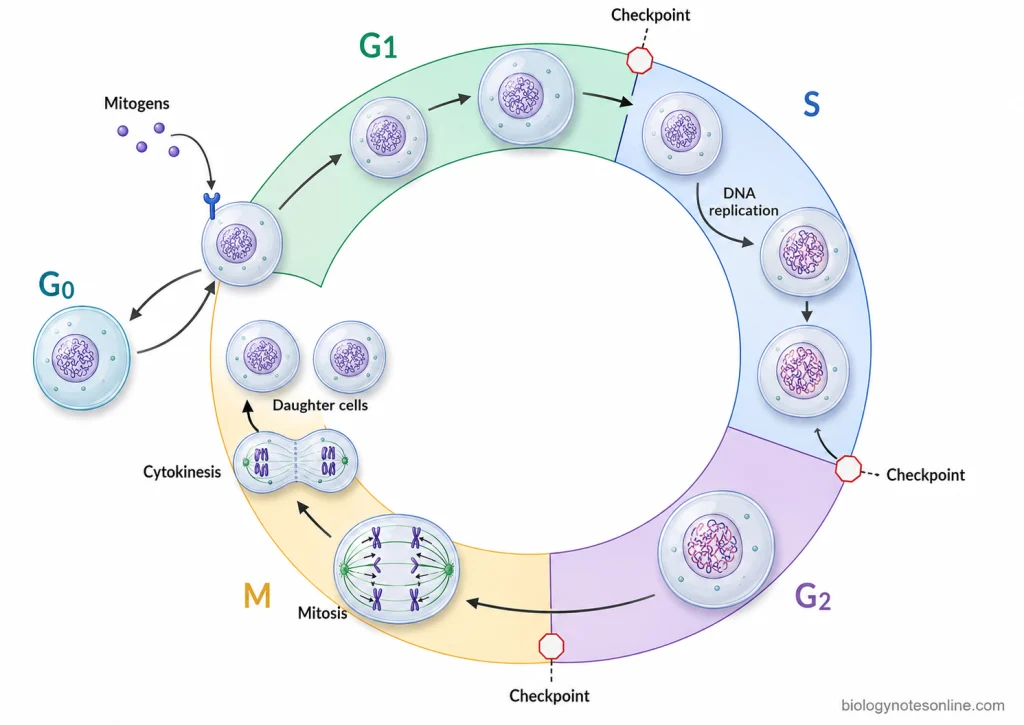
- G1 phase- It is the first gap phase after cell division. During this phase, the cell grows and synthesizes cellular components required for the next phase. Progression through G1 is also affected by extracellular signals and condition of the cell.
- S phase- In this phase, DNA replication takes place. The DNA content is duplicated so that complete genetic material can be supplied to the daughter cells during division.
- G2 phase- It occurs after completion of DNA synthesis. Cell growth continues and different proteins required for mitosis are synthesized. It prepares the cell for the M phase.
- M phase- This is the phase of cell division. The replicated chromosomes are separated during mitosis, which is usually followed by cytokinesis to form two daughter cells. Thus, completion of M phase directly increases the number of cells.
- G0 phase- Some cells can leave the active cell cycle from G1 and enter a resting or quiescent state called G0. These cells are not actively proliferating, although some of them can again enter the cell cycle after receiving suitable signals.
The progression of cells through different phases is regulated by cyclins, cyclin-dependent kinases (CDKs) and cell-cycle checkpoints. Checkpoints can stop further progression when conditions are not suitable or when DNA is damaged. In this way, regulation of the cell cycle controls cell proliferation.
How Is Cell Proliferation Regulated?
Cell proliferation is a regulated process in which cells divide according to the requirement of the tissue. The regulation is carried out by different extracellular and intracellular signals which control the progression of cell cycle. Depending upon these signals, the cell may continue the cycle, stop at a particular checkpoint or remain in non-dividing condition. Some of the important mechanisms regulating cell proliferation are-
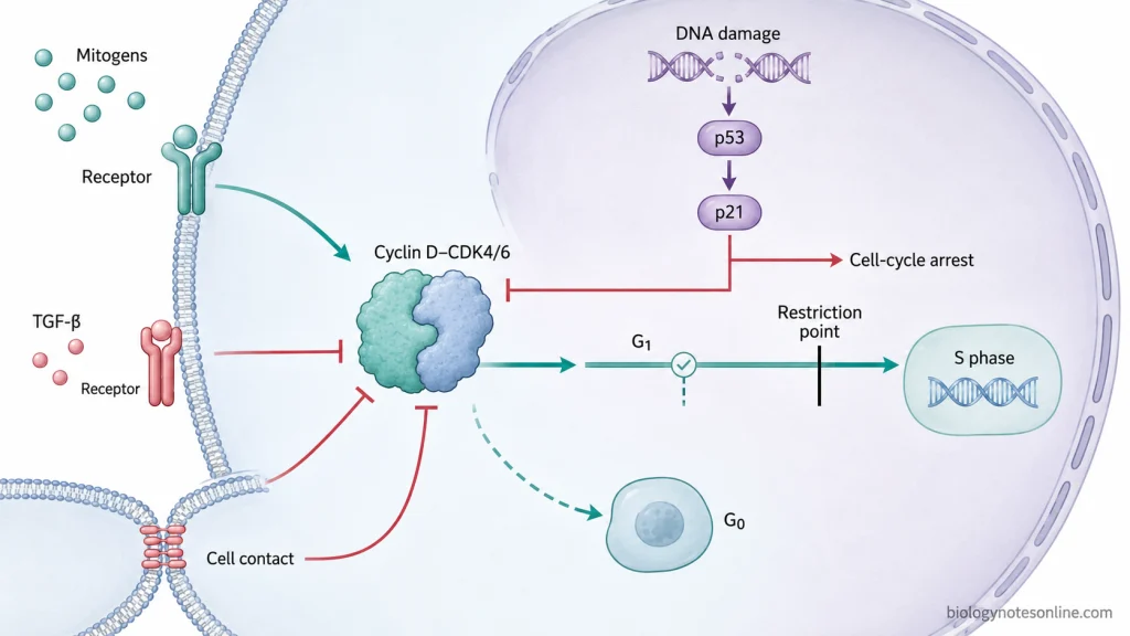
- Growth factors and mitogens- Growth factors and mitogens are extracellular signals that stimulate the cell to enter into the cell cycle and continue its progression. They bind with specific receptors present on the cell surface and activate intracellular signaling pathways. Their effect is mainly important during G1 phase. In this phase, these signals help the cell to pass the restriction point and proceed towards DNA synthesis.
- Cyclins and Cyclin-dependent kinases (CDKs)- The different stages of cell cycle are controlled by cyclins and CDKs. Cyclin concentration changes during different phases of the cycle and their binding activates specific CDKs. Cyclin D-CDK4/6, for example, has an important role in progression of G1 phase.
- Restriction point- It is an important regulatory point present during late G1 phase. Before passing this point, the cell requires extracellular proliferative signals. When suitable signals are not available, proliferation may stop and the cell can enter into G0 phase. After crossing the restriction point, the cell becomes committed to complete the remaining cell cycle.
- Cell-cycle checkpoints- These checkpoints monitor different events taking place during cell cycle. DNA damage, incomplete replication of DNA or improper attachment of chromosomes can stop further progression. Thus, cells which are damaged or not properly prepared are prevented from continuing normal cell division.
- p53 and CDK inhibitors- p53 is a tumor suppressor protein which can become activated when DNA is damaged. It induces the CDK inhibitor p21. p21 inhibits cyclin-CDK complexes and cell-cycle arrest takes place. This gives time for repair of the damaged DNA, while severe or unrepaired damage can also result in cell death.
- Inhibitory extracellular signals and cell contact- Cell proliferation is also controlled by signals which inhibit division. TGF-β can inhibit progression of the cell cycle, whereas increased contact between cells can suppress proliferation in many cell types. Therefore, the continuation of cell proliferation depends upon the balance between stimulatory and inhibitory signals.
Proliferation, Quiescence, Differentiation, Senescence, and Cell Death
Proliferation vs Quiescence
| Features | Proliferation | Quiescence |
|---|---|---|
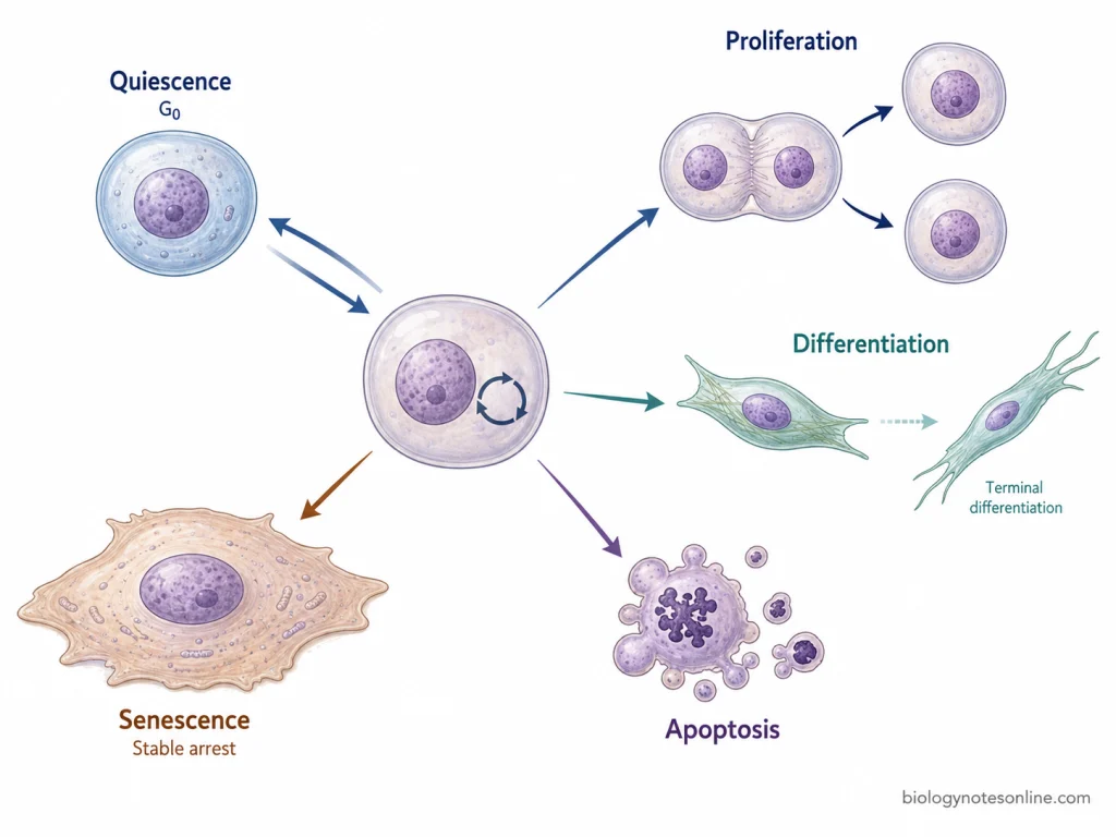
| Cell condition | In proliferation, cells are actively progressing through the cell cycle and new cells are produced by division. | Quiescent cells are alive and metabolically active, but they are not dividing. They generally remain outside the active cell cycle in G0 phase. |
| Cell fate | The cell continues through growth, DNA replication and division when suitable proliferative signals are present. | It is a temporary non-proliferating condition. The cell is maintained without being committed to cell death or permanent arrest. |
| Reversibility | Proliferation can be stopped when the required signals are removed or inhibitory signals are received. | Quiescence is reversible. After receiving suitable growth or mitogenic signals, these cells can again enter into the cell cycle and start proliferation. |
| Major difference | Cell number is being increased. | Cell division is stopped for a period, but the capacity to proliferate is retained. |
Proliferation vs Differentiation
| Features | Proliferation | Differentiation |
|---|---|---|
| Cell condition | Proliferating cells continue division and increase the cell population. | Differentiation is a process in which cells acquire specialized structural and functional characteristics. |
| Cell fate | The major fate is production of more cells of the proliferating population. | Here, the cell moves towards a specialized cell state. Differentiation and cell-cycle exit are commonly associated during terminal differentiation. |
| Reversibility | A proliferating cell can leave the cycle and undergo differentiation depending upon the cell type and signals. | Terminal differentiation is generally associated with permanent or very stable exit from cell cycle. However, reversibility is not identical in every differentiated cell type. |
| Major difference | It is mainly concerned with increasing cell number. | It is mainly associated with development of specialized cell function and identity, and not simply increase in cell number. |
Proliferation vs Senescence
| Features | Proliferation | Senescence |
|---|---|---|
| Cell condition | Cells remain capable of progressing through cell cycle and division takes place. | Cellular senescence is a stable cell-cycle arrest. The cells remain alive and metabolically active but normally do not continue division. |
| Cell fate | Daughter cells are produced and the cell population increases. | The cell remains present in the tissue but stays in a non-proliferating state. It is not the same as cell death. |
| Reversibility | Proliferative activity can change according to cell-cycle regulatory signals. | Senescence is generally considered a long-lasting or largely irreversible arrest under normal physiological conditions. Unlike quiescent cells, senescent cells do not readily start proliferation after normal mitogenic stimulation. |
| Major difference | Cell division continues. | Division is stably stopped, although the cell remains viable. |
Proliferation vs Apoptosis
| Features | Proliferation | Apoptosis |
|---|---|---|
| Cell condition | During proliferation, viable cells progress through the cell cycle and divide. | Apoptosis is a regulated form of programmed cell death in which an intracellular death mechanism is activated. |
| Cell fate | One cell gives rise to daughter cells and therefore contributes to increase in cell number. | The affected cell is eliminated. Caspases participate in carrying out the apoptotic process by cleavage of cellular proteins. |
| Reversibility | A cell can stop proliferation and enter another cell state without necessarily dying. | Apoptosis represents commitment towards cell death. Once the death program reaches its execution stage, the normal proliferating state is not restored. |
| Major difference | It adds cells to a cell population through division. | It removes cells from the population by programmed cell death. Thus, proliferation and apoptosis together have an important role in controlling cell number in multicellular tissues. |
Factors Affecting Cell Proliferation
Cell proliferation is affected by many internal and external factors. Some factors increase cell division, whereas others can slow or stop the progression of cells through the cell cycle. Some of the important factors affecting cell proliferation are-
- Growth factors and mitogens- These are extracellular signals which stimulate the cells to enter into cell cycle and continue cell division.
- Nutrients and energy- Nutrients and sufficient energy are required for normal growth and proliferation of cells. Deficiency of nutrients can decrease the rate of proliferation.
- Cell density and contact- When the number of cells increases, contact between neighbouring cells can inhibit further proliferation in many normal cells. This is referred to as density-dependent inhibition.
- Extracellular matrix and cell attachment- Many normal cells require attachment to extracellular matrix for their growth and proliferation. This attachment is mainly carried out through integrins.
- DNA damage- Damage in DNA can activate cell-cycle checkpoints and further division is stopped. p53 and p21 are some of the important proteins involved in this process.
- Hormones- Hormones can increase or decrease proliferation depending upon the type of cell and tissue. Steroid hormones are one of the important examples.
- Inhibitory signals- Some extracellular signals inhibit cell-cycle progression. TGF-β can suppress proliferation in several types of cells.
Biological Functions of Cell Proliferation
Cell proliferation is necessary for growth, development and maintenance of tissues. It increases the number of cells and also replaces the cells which are lost or damaged. Some of the important functions of cell proliferation are-
- Growth and development- During development, repeated cell division increases the total number of cells. These cells later form different tissues and organs.
- Tissue maintenance- Old and damaged cells are continuously replaced by newly formed cells. This maintains the normal cell population of tissues.
- Wound healing- After injury, different cells start proliferating at the damaged region. It helps in repair and replacement of lost tissue.
- Regeneration- Some tissues can restore the lost cells by proliferation of remaining cells or stem cells. Liver is an important example of this process.
- Blood cell formation- Hematopoietic stem cells proliferate in bone marrow and produce different blood cells. These cells replace the blood cells lost during normal life span.
- Immune response- Activated lymphocytes undergo repeated cell division after recognition of specific antigen. This produces a large number of antigen-specific cells, called clonal expansion.
What Happens When Cell Proliferation Becomes Abnormal?
Abnormal cell proliferation occurs when the normal balance between production and loss of cells is disturbed. The cells may divide excessively or normal cell replacement can become reduced. This affects tissue growth and its normal maintenance. Some of the important effects are-
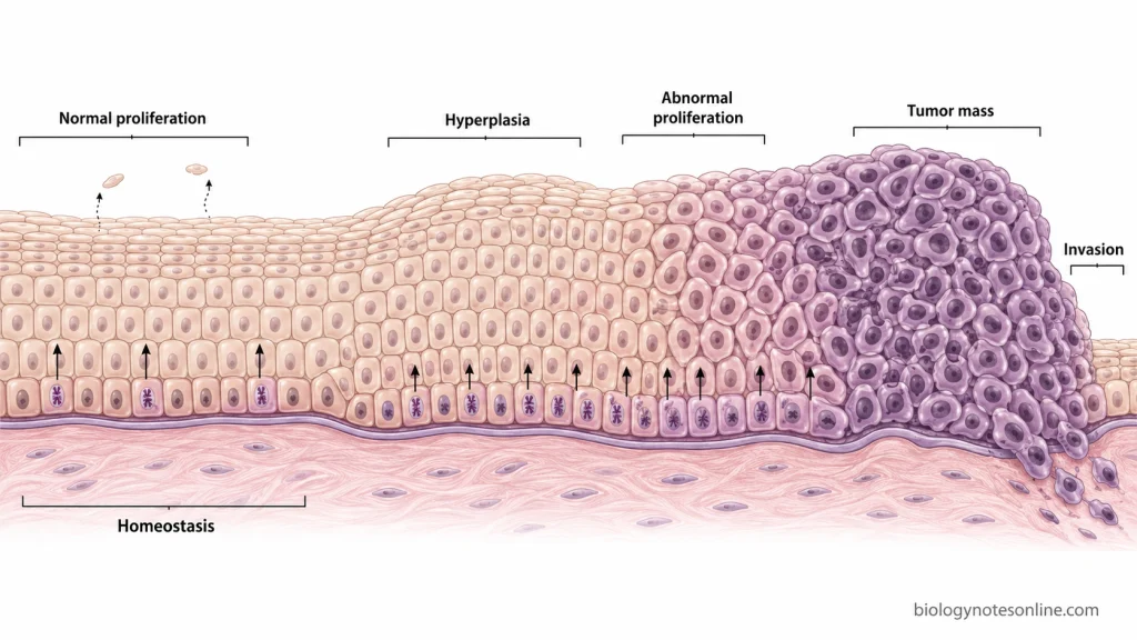
- Hyperplasia- Excess proliferation can increase the number of cells within a tissue. This increase in cell number is called hyperplasia.
- Tumor formation- When cells continue to proliferate without normal growth control, an abnormal mass of cells can be formed. Such uncontrolled proliferation is an important feature of cancer.
- Loss of tissue organization- Abnormally proliferating cells can disturb the normal arrangement and function of surrounding tissue. Malignant cells may also invade the nearby tissues.
- Reduced tissue renewal- If cell proliferation becomes too low, lost cells are not replaced properly. This can result in reduction of tissue mass or tissue atrophy.
- Improper tissue repair- Reduced or disturbed proliferation can affect replacement of cells after tissue injury. Normal proliferation is required during tissue maintenance and wound repair.
- Cell-cycle arrest or cell death- Abnormal proliferative signals do not always cause more cell division. In some normal cells, excessive growth signals can activate checkpoints resulting in cell-cycle arrest or apoptosis.
How Is Cell Proliferation Measured?
Cell proliferation can be measured by determining increase in cell number, DNA synthesis or the presence of proliferation-related markers. Different methods are used depending upon the type of cells and experimental condition. Some of the common methods are-
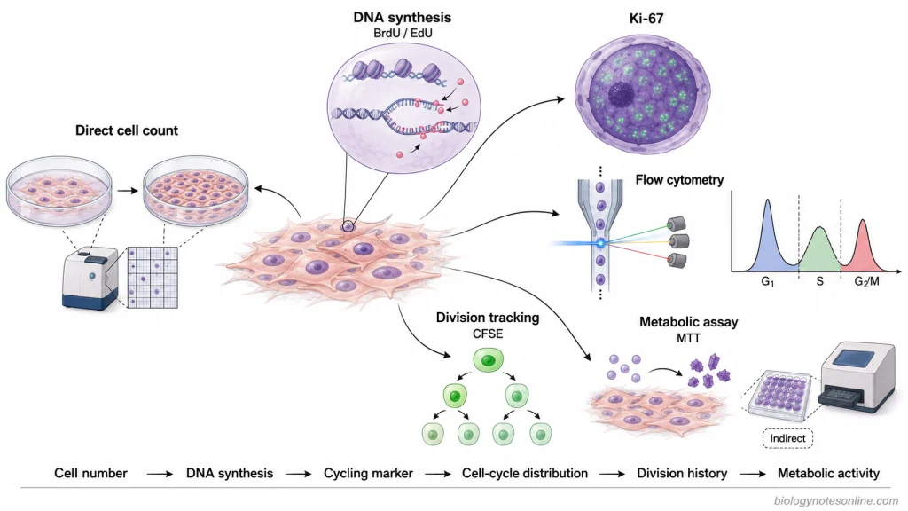
- Direct cell counting- Cells are counted at different time intervals and the increase in cell number is determined. It is one of the direct methods for measuring proliferation.
- BrdU and EdU assay- BrdU or EdU is incorporated into newly synthesized DNA during S phase. The labelled cells are then detected to determine the cells undergoing DNA synthesis.
- Ki-67 staining- Ki-67 is a proliferation-associated nuclear protein present in actively cycling cells and absent from resting G0 cells. The percentage of Ki-67 positive cells is used as a proliferation index.
- Flow cytometry– It is used to determine the distribution of cells in different phases of the cell cycle based on their DNA content. Proliferation markers can also be measured along with it.
- Cell-tracking dyes- Dyes such as CFSE are distributed between daughter cells after every division. Reduction of dye intensity is therefore used to follow successive cell divisions.
- Metabolic assays- Assays such as MTT estimate proliferation indirectly from cellular metabolic activity. These assays measure the whole cell population and do not directly count cell divisions.
Diseases Related to Cell Proliferation
Abnormal cell proliferation is associated with different diseases. In some diseases, excessive proliferation increases the number of cells, whereas reduced proliferation can affect normal cell replacement. Some of the important diseases related to cell proliferation are-
- Cancer- It is the major disease associated with uncontrolled and persistent cell proliferation. The normal control of cell cycle is lost and tumor cells continue to divide.
- Psoriasis- It is characterized by abnormal proliferation of epidermal keratinocytes. This increased proliferation contributes to thickening of the epidermis and formation of psoriatic lesions.
- Benign Prostatic Hyperplasia (BPH)- BPH is a non-malignant proliferative disease of prostate. Proliferation of stromal and epithelial cells causes enlargement of the prostate tissue.
- Atherosclerosis- Proliferation of vascular smooth muscle cells (VSMCs) occurs during development of atherosclerotic lesions, particularly during early atherogenesis and vascular injury. These cells contribute to changes in the vessel wall.
- Aplastic anemia- In this disease, hematopoietic stem and progenitor cells are greatly reduced and normal production of blood cells is affected. Defective survival and proliferative capacity of these cells can also occur in some forms of the disease.
References
- Alberts, B., Johnson, A., Lewis, J., Raff, M., Roberts, K., & Walter, P. (2002). Molecular biology of the cell (4th ed.). Garland Science. https://www.ncbi.nlm.nih.gov/books/NBK21054/
- Andreeff, M., Goodrich, D. W., & Pardee, A. B. (2003). Proliferation. In D. W. Kufe, R. E. Pollock, R. R. Weichselbaum, R. C. Bast Jr., T. S. Gansler, J. F. Holland, & E. Frei III (Eds.), Holland-Frei cancer medicine (6th ed.). BC Decker. https://www.ncbi.nlm.nih.gov/books/NBK13035/
- Bennett, M. R., Sinha, S., & Owens, G. K. (2016). Vascular smooth muscle cells in atherosclerosis. Circulation Research, 118(4), 692–702. https://doi.org/10.1161/CIRCRESAHA.115.306361
- Blagosklonny, M. V., & Pardee, A. B. (2000–2013). The restriction point of the cell cycle. In Madame Curie Bioscience Database. Landes Bioscience. https://www.ncbi.nlm.nih.gov/books/NBK6318/
- Buttitta, L. A., & Edgar, B. A. (2007). Mechanisms controlling cell cycle exit upon terminal differentiation. Current Opinion in Cell Biology, 19(6), 697–704. https://doi.org/10.1016/j.ceb.2007.10.004
- Cooper, G. M. (2000). The cell: A molecular approach (2nd ed.). Sinauer Associates. https://www.ncbi.nlm.nih.gov/books/NBK9839/
- Cossarizza, A., Chang, H.-D., Radbruch, A., Akdis, M., Andrä, I., Annunziato, F., Bacher, P., Barnaba, V., Battistini, L., Bauer, W. M., Baumgart, S., Becher, B., Beisker, W., Berek, C., Blanco, A., Borsellino, G., Boulais, P. E., Brinkman, R. R., Büscher, M., . . . Zimmermann, J. (2017). Guidelines for the use of flow cytometry and cell sorting in immunological studies. European Journal of Immunology, 47(10), 1584–1797. https://doi.org/10.1002/eji.201646632
- Dang, C., Gilewski, T. A., Surbone, A., & Norton, L. (2003). Cell proliferation. In D. W. Kufe, R. E. Pollock, R. R. Weichselbaum, R. C. Bast Jr., T. S. Gansler, J. F. Holland, & E. Frei III (Eds.), Holland-Frei cancer medicine (6th ed.). BC Decker. https://www.ncbi.nlm.nih.gov/books/NBK12640/
- Ge, Y., & Fuchs, E. (2018). Stretching the limits: From homeostasis to stem cell plasticity in wound healing and cancer. Nature Reviews Genetics, 19(5), 311–325. https://doi.org/10.1038/nrg.2018.9
- Hata, J., Harigane, Y., Matsuoka, K., Akaihata, H., Yaginuma, K., Meguro, S., Hoshi, S., Sato, Y., Ogawa, S., Uemura, M., & Kojima, Y. (2023). Mechanism of androgen-independent stromal proliferation in benign prostatic hyperplasia. International Journal of Molecular Sciences, 24(14), 11634. https://doi.org/10.3390/ijms241411634
- Janeway, C. A., Jr., Travers, P., Walport, M., & Shlomchik, M. J. (2001). Immunobiology: The immune system in health and disease (5th ed.). Garland Science. https://www.ncbi.nlm.nih.gov/books/NBK10757/
- Johnson, M. S., & Cook, J. G. (2023). Cell cycle exits and U-turns: Quiescence as multiple reversible forms of arrest. Faculty Reviews, 12, 5. https://doi.org/10.12703/r/12-5
- Kaldis, P. (2016). Quo vadis cell growth and division? Frontiers in Cell and Developmental Biology, 4, 95. https://doi.org/10.3389/fcell.2016.00095
- Kim, K. H., & Sederstrom, J. M. (2015). Assaying cell cycle status using flow cytometry. Current Protocols in Molecular Biology, 111(1), 28.6.1–28.6.11. https://doi.org/10.1002/0471142727.mb2806s111
- Kumari, R., & Jat, P. (2021). Mechanisms of cellular senescence: Cell cycle arrest and senescence associated secretory phenotype. Frontiers in Cell and Developmental Biology, 9, 645593. https://doi.org/10.3389/fcell.2021.645593
- Liang, N., & Zhang, K. (2026). Factors influencing the proliferation of keratinocytes in psoriasis. iScience, 29(2), 114745. https://doi.org/10.1016/j.isci.2026.114745
- Martin, G. M. (2007). The genetics and epigenetics of altered proliferative homeostasis in ageing and cancer. Mechanisms of Ageing and Development, 128(1), 9–12. https://doi.org/10.1016/j.mad.2006.11.003
- McKinnon, K. M. (2018). Flow cytometry: An overview. Current Protocols in Immunology, 120, 5.1.1–5.1.11. https://doi.org/10.1002/cpim.40
- Mead, T. J., & Lefebvre, V. (2014). Proliferation assays (BrdU and EdU) on skeletal tissue sections. In M. J. Hilton (Ed.), Skeletal development and repair: Methods and protocols (pp. 233–243). Humana Press. https://doi.org/10.1007/978-1-62703-989-5_17
- Musgrove, E. A., & Sutherland, R. L. (1994). Cell cycle control by steroid hormones. Seminars in Cancer Biology, 5(5), 381–389. https://pubmed.ncbi.nlm.nih.gov/7849266/
- Ruijtenberg, S., & van den Heuvel, S. (2016). Coordinating cell proliferation and differentiation: Antagonism between cell cycle regulators and cell type-specific gene expression. Cell Cycle, 15(2), 196–212. https://doi.org/10.1080/15384101.2015.1120925
- Salama, R., Sadaie, M., Hoare, M., & Narita, M. (2014). Cellular senescence and its effector programs. Genes & Development, 28(2), 99–114. https://doi.org/10.1101/gad.235184.113
- Su, T. T., & O’Farrell, P. H. (1998). Size control: Cell proliferation does not equal growth. Current Biology, 8(19), R687–R689. https://doi.org/10.1016/S0960-9822(98)70436-1
- Yang, X., & Xu, T. (2011). Molecular mechanism of size control in development and human diseases. Cell Research, 21(5), 715–729. https://doi.org/10.1038/cr.2011.63
- Zeng, Y., & Katsanis, E. (2015). The complex pathophysiology of acquired aplastic anaemia. Clinical & Experimental Immunology, 180(3), 361–370. https://doi.org/10.1111/cei.12605