Microvilli are tiny, finger-like projections found on the surface of epithelial cells throughout the body, particularly in the small intestine. These structures significantly increase the surface area of the cells, enhancing their ability to absorb nutrients and other substances. The primary functions of microvilli include facilitating nutrient absorption, aiding in the process of fertilization, supporting immune cell interactions, and playing roles in sensory detection and secretion. Located on the inner surface of the small intestine, microvilli are crucial for efficient digestion and nutrient uptake. They also help anchor sperm cells during fertilization and contribute to various physiological processes. Understanding what microvilli are and their functions is essential for grasping their importance in maintaining overall health and bodily functions.
What are microvilli?
- Microvilli are minute, finger-like extensions of the plasma membrane present on the surface of various epithelial cells. Typically measuring around 0.1 micrometers in diameter and up to 2 micrometers in length, microvilli are narrower than cilia but thicker than them. They play a critical role in enhancing the surface area of cells, particularly in tissues involved in absorption and secretion.
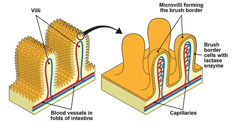
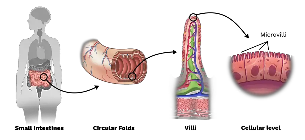
- These structures are most notably found in the mucosa of the small intestine, where they form what is known as the brush border. This term describes the densely packed array of microvilli that significantly increases the absorptive surface area of the intestinal epithelium. By doing so, microvilli facilitate the efficient uptake of nutrients from digested food.
- Besides the intestines, microvilli are also present in various other locations throughout the body, including egg cells, white blood cells, and the proximal convoluted tubules of the kidneys. In the liver, microvilli are found on both the absorptive surfaces, such as the space of Disse, and the secretory poles, such as the bile canaliculus of hepatocytes. This dual presence underscores their functional versatility.
- The primary function of microvilli is to maximize the surface area of the cell membrane for absorption or secretion. For instance, in the small intestine, microvilli work in conjunction with villi to further amplify the surface area available for nutrient absorption, enhancing the digestive efficiency of the gut.
- Although microvilli extend from the cell surface, they lack the complex organelles found in the cell body, which distinguishes them from other cellular extensions. Variations in the length and surface coating of microvilli can occur depending on their location and function. For example, microvilli in the small intestine may differ in length and surface properties compared to those in the large intestine or other tissues.
- In addition to their role in absorption, microvilli are also prevalent on lymphocytes within the immune system, highlighting their importance across different biological contexts. Overall, microvilli are essential for increasing cellular surface area, thereby enhancing the cell’s ability to interact with its environment and perform specific physiological functions.
Microvilli Definition
Microvilli are tiny, finger-like projections on the surface of epithelial cells that increase the cell’s surface area to enhance absorption and secretion.
Locations of Microvilli
- Small Intestine:
- Microvilli are densely packed on the apical surface of epithelial cells lining the small intestine, forming the brush border.
- This structure significantly increases the absorptive surface area, enhancing nutrient uptake during digestion.
- Egg Cells:
- Microvilli are present on the plasma membrane of egg cells.
- They facilitate the adherence and stabilization of sperm cells that have penetrated the egg’s extracellular matrix, promoting successful fertilization.
- White Blood Cells:
- On the surface of white blood cells, microvilli contribute to cellular movement and interaction with other cells or pathogens.
- Their presence aids in various immune responses and cell migration.
- Other Locations:
- Microvilli can also be found on cells in various other tissues, such as the proximal convoluted tubules of the kidneys and certain secretory surfaces in the liver.
- They play a role in enhancing the surface area for absorption or secretion in these locations.
Structure of Microvilli
- Plasma Membrane:
- Microvilli are encased by a plasma membrane, which forms their outer boundary. This membrane provides a protective layer and facilitates interactions with the extracellular environment.
- Actin Filaments:
- Internally, microvilli are supported by tightly packed bundles of actin filaments, which serve as their structural core. These filaments are closely arranged, typically comprising 20-30 filaments per bundle.
- Actin filaments are anchored at the tip of each microvillus and extend into the dense core, providing rigidity and support. This arrangement is crucial for maintaining the shape and function of the microvilli.
- Actin-Bundling Proteins:
- Cross-bridges of actin-bundling proteins, such as fascin and fimbrin, link the actin filaments together. These proteins stabilize the filaments and ensure their proper alignment.
- Additionally, myosin I and calmodulin form another bridge, connecting the actin bundles to the plasma membrane. Myosin I has a dual function, with a binding site for filamentous actin and a lipid-binding domain for interaction with the membrane.
- Terminal Web:
- At the base of the microvilli, the actin filaments are embedded in the apical cytoplasm and integrated into a network known as the terminal web. This web consists of transversely running actin filaments stabilized by spectrin and is underlain by keratin intermediate filaments.
- The terminal web is anchored to tight junctions and zonula adherens at the apical epithelial junctional complex, providing structural stability to the microvilli.
- Contractile Proteins:
- The terminal web contains tropomyosin and myosin II, which contribute to its contractile properties. These proteins facilitate changes in the intermicrovillous space, which expands and contracts in response to cellular activity.
- Glycocalyx:
- Specialized Forms:
- Filopodia: Irregular microvilli found on certain cells, such as macrophages and fibroblasts, are associated with phagocytosis and cell motility.
- Stereocilia: Long, branching microvilli, more accurately termed stereovilli, are found on cochlear and vestibular receptor cells as well as the epididymis. Despite their appearance, they are non-motile and lack microtubules, serving primarily as sensory transducers.

Functions of microvilli
- Nutrient Absorption:
- Microvilli on the surface of epithelial cells, particularly in the small intestine, dramatically increase the cell’s surface area.
- This enhanced surface area facilitates the efficient absorption of nutrients, such as proteins, carbohydrates, and lipids, as well as water molecules from ingested food.
- Fertilization:
- In egg cells, microvilli play a crucial role in the fertilization process.
- They anchor sperm cells to the egg’s surface, aiding in the successful attachment and subsequent fusion necessary for fertilization.
- Immune Function:
- Microvilli on white blood cells serve as anchoring points, contributing to cell movement and interaction with other immune cells or pathogens.
- Enzyme Function:
- The membrane of microvilli contains various enzymes essential for digestion.
- For instance, glycosidases present on enterocyte microvilli are crucial for carbohydrate digestion, breaking down complex sugars into simpler forms that can be absorbed more easily.
- Sensory Detection:
- Microvilli also have a role in sensory functions, particularly in the auditory system.
- In the inner ear, they are involved in detecting sound vibrations, which are converted into electrical signals sent to the brain.
- Secretion:
- On hepatocytes, microvilli contribute to the secretion processes.
- They assist in the release of substances into the bile canaliculus, supporting liver function.
- Clinical Relevance:
- The absence or malfunction of microvilli can lead to serious conditions.
- For example, congenital microvillous atrophy, a rare but fatal condition, is characterized by the loss of microvilli in the intestinal tract, leading to severe nutrient malabsorption in newborns.

FAQ
What are microvilli?
Microvilli are small, finger-like projections that extend from the surface of cells in various organs of the body. They are typically 0.5 to 1 micrometer in diameter and several micrometers in length.
Where are microvilli found in the body?
Microvilli are found in many different tissues and organs, but they are most abundant in the small intestine, where they help with nutrient absorption.
What is the function of microvilli?
Microvilli serve several important functions, including increasing the surface area of cells, facilitating the transport of molecules, sensing stimuli, and aiding in cell adhesion.
How do microvilli increase the surface area of cells?
Microvilli extend from the surface of cells, creating a brush-like structure that increases the surface area available for absorption and secretion of nutrients and fluids.
What is the structure of microvilli?
Microvilli consist of a core of actin filaments surrounded by a plasma membrane. They may also contain various proteins, such as transporters, channels, and receptors.
Can microvilli regenerate?
Yes, microvilli have a high turnover rate and can regenerate quickly after being damaged or lost.
What happens if microvilli are damaged or lost?
Damage or loss of microvilli can lead to a variety of health problems, such as malabsorption of nutrients, diarrhea, and hearing loss.
What is microvillus inclusion disease?
Microvillus inclusion disease is a rare genetic disorder that affects the microvilli in the small intestine. It leads to severe diarrhea and malabsorption of nutrients, and is typically diagnosed in infancy.
Can microvilli be seen under a microscope?
Yes, microvilli can be visualized using an electron microscope, which can magnify them to a size that is visible to the human eye.
Are microvilli unique to humans?
No, microvilli are found in many different animal species, and are thought to have evolved as a way to increase the surface area of cells in order to improve nutrient absorption and other cellular functions.
References
- https://www.biologydiscussion.com/cell/cell-surface/3-types-of-cell-surface-projection-cell-biology/26375
- https://unacademy.com/content/neet-ug/study-material/biology/microvilli/
- http://histologyguide.com/EM-view/EM-093-microvilli/02-photo-1.html
- https://biology.kenyon.edu/edwards/project/greg/pd.htm
- https://study.com/academy/lesson/microvilli-definition-function.html
- http://hyperphysics.phy-astr.gsu.edu/hbase/Biology/micvil.html
- https://www.sciencedirect.com/topics/agricultural-and-biological-sciences/microvillus
- https://www.sciencedirect.com/topics/biochemistry-genetics-and-molecular-biology/microvillus
- https://www.biologyonline.com/dictionary/microvillus
- https://rupress.org/jcb/article-pdf/12/3/623/1064187/623.pdf
- https://www.easybiologyclass.com/difference-between-cilia-and-microvilli-comparison-table/