

Chlamydomonas is a unicellular green algae, which belongs to the class Chlorophyceae. It is microscopic and mostly found in fresh water, stagnant pools, moist soil, sea water and snow also. Some species are present in mountain snow and they give red colour to snow, which is called watermelon snow.
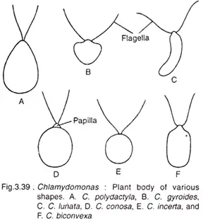
It is a simple motile algae. The body is single celled and oval or pear shaped. The cell is surrounded by a firm cell wall made up of glycoprotein. At the anterior side, two equal flagella are present, which help in movement of the cell in water.
The cell contains a large cup-shaped chloroplast. This chloroplast helps in photosynthesis and food preparation. Inside the chloroplast, a pyrenoid is present, which helps in formation and storage of starch. A red coloured eyespot or stigma is also present, which is sensitive to light.
Chlamydomonas is generally photoautotrophic in nature. It prepares its own food by the help of sunlight, carbon dioxide and water. It acts as a primary producer in aquatic ecosystem. Some species can also grow in darkness by using organic carbon source like acetate. This condition is called facultative heterotrophic nature.
Two contractile vacuoles are present near the base of flagella. These vacuoles remove excess water from the cell. They also maintain osmotic balance of the cell. The nucleus is present in cytoplasm and other cell organelles are also present.
Reproduction in Chlamydomonas takes place by asexual and sexual methods. During favourable condition, asexual reproduction occurs by the formation of zoospores. The protoplast divides inside the parent cell and forms 2 to 16 daughter cells. These daughter cells are flagellated and come out from the parent wall.
During unfavourable condition, sexual reproduction takes place. The haploid cells form gametes. These gametes fuse and form diploid zygospore. The zygospore has thick wall and it can survive in dry condition, nutrient deficiency and other unfavourable condition. When favourable condition comes, it undergoes meiosis and produces new haploid cells.
Chlamydomonas reinhardtii is used as an important model organism in biological studies. It is easy to culture and has simple life cycle. It is used to study photosynthesis, genetics, cell division, flagella, cell motility and ciliary diseases. Its flagella are similar to human cilia, so it is useful in medical and cell biological research.
Chlamydomonas Classification
| Taxonomic Rank | Classification |
|---|---|
| Kingdom | Plantae |
| Division | Chlorophyta |
| Class | Chlorophyceae |
| Order | Chlamydomonadales (frequently treated as synonymous with Volvocales) |
| Family | Chlamydomonadaceae |
| Genus | Chlamydomonas |
Habitat of Chlamydomonas
- Chlamydomonas is widely distributed in fresh water habitat. It is commonly found in ponds, pools, lakes, ditches and other stagnant water.
- It is also found in marine water. Some species live in sea water, saline water and oceanic water.
- Some species of Chlamydomonas are present in terrestrial habitat. They grow on damp soil, moist soil, wet rock surface and along the bank of ponds and lakes.
- Chlamydomonas can also grow in cold habitat. Some species are found in mountain snow, ice, melt water and polar or alpine region.
- Chlamydomonas nivalis is found in snow. It produces red colouration of snow, which is called red snow or watermelon snow.
- Some species can live in acidic habitat also. Chlamydomonas acidophila can survive in very low pH condition.
- It generally prefers nutrient rich water. It grows well in water containing organic matter, nitrogenous matter and ammonium salts.
- Thus, Chlamydomonas is found in fresh water, marine water, damp soil and extreme habitat also. It shows wide distribution due to its simple body and adaptable nature.
Nutrition of Chlamydomonas
- Chlamydomonas is mostly photoautotrophic algae which prepare food by photosynthesis in presence of light.
- The large cup-shaped chloroplast is the main site of food preparation. It contains chlorophyll which absorb light energy.
- Water, dissolved CO₂ and inorganic salts enter into the cell from surrounding water by general body surface.
- There is no root like organ for absorption. The whole cell surface takes part in absorption of raw materials.
- In photosynthesis, CO₂ and water are converted into carbohydrate. The process occurs inside the chloroplast.
- The carbohydrate produced is used in respiration, growth and other metabolic activities of the cell.
- The reserve food of Chlamydomonas is starch. It is deposited as starch grains around the pyrenoid.
- Pyrenoid is a proteinaceous body present inside the chloroplast and it is associated with starch formation.
- Some species are not fully dependent on light for nutrition. They may grow in dark also when organic carbon is present.
- This type of nutrition is called facultative heterotrophic nutrition.
- Chlamydomonas reinhardtii and Chlamydomonas dysostosis can absorb acetate from the medium.
- Acetate acts as an organic carbon source and supports growth in absence of light.
Characteristics of Chlamydomonas
- Chlamydomonas is a unicellular green algae which is motile in nature and freely moves in water by flagella.
- The cell is generally oval, spherical, ellipsoidal or pear-shaped, with a slightly pointed anterior end called papilla.
- Two equal whiplash type flagella are present at the anterior end, which help in locomotion of the cell in water.
- The cell is surrounded by a thin and firm cell wall, which is mainly made up of glycoprotein and non-cellulosic polysaccharides.
- A large cup-shaped or bowl-shaped chloroplast is present in the cell, occupying greater part of posterior cytoplasm and it is concerned with photosynthesis.
- One or more pyrenoids are present inside the chloroplast, which are associated with formation and storage of starch.
- A reddish or orange coloured eyespot or stigma is present in anterior part of chloroplast, which is sensitive to light and helps the cell to move towards light.
- Two contractile vacuoles are present near the base of flagella, which remove excess water from the cell and helps in osmoregulation and excretion.
- A single nucleus is present inside the cavity of chloroplast, and other eukaryotic organelles like mitochondria, ribosomes and endoplasmic reticulum are also present.
- The flagella arise from basal granules, which are connected with each other by paradesmos and connected with nucleus by rhizoplast, forming the neuromotor apparatus.
Structure of Chlamydomonas/Thallus Structure of Chlamydomonas
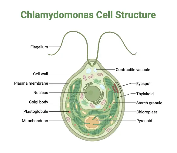
- The thallus of Chlamydomonas is unicellular and motile. It is generally pear-shaped, oval, spherical or ellipsoidal, with narrow anterior end and broad posterior end.
- The cell is surrounded by a thin and firm cell wall, which gives definite shape to the cell. The wall is covered externally by mucilaginous envelope.
- The cell wall is not truly cellulosic in nature. It is mainly composed of hydroxyproline rich glycoproteins and non-cellulosic polysaccharides.
- At the anterior end, the cell wall forms a slightly pointed or rounded projection called papilla. It is present between the two flagella.
- Two equal whiplash type flagella arise from the anterior end near the papilla. These flagella help in swimming movement of the cell.
- The flagella arise from two basal granules or basal bodies present inside the anterior region of the cell.
- The two basal granules are connected with each other by a fine thread like structure called paradesmos. They are also connected with the nucleus by rhizoplast and this forms the neuromotor apparatus.
- Two contractile vacuoles are present at the anterior end, just below the base of flagella. They expand and contract regularly and remove excess water from the cell.
- The contractile vacuoles help in osmoregulation and also help in excretion of waste materials.
- A large cup-shaped or bowl-shaped chloroplast is present in the posterior part of the cell. It occupies greater part of cytoplasm and it is the main site of photosynthesis.
- In some species, the chloroplast may be different in shape, such as H-shaped, stellate or reticulate type.
- One or more pyrenoids are present inside the chloroplast. They are dense proteinaceous bodies and concerned with starch formation.
- The pyrenoid is generally surrounded by starch plates. Starch is deposited around it as reserve food material.
- A small reddish or orange coloured eyespot or stigma is present in the anterior lateral part of chloroplast.
- The eyespot contains carotenoid rich lipid droplets and acts as a photoreceptive organ. It helps the cell to detect light direction and intensity.
- A single nucleus is present in the central region of the cell. It usually lies inside the cavity formed by the cup-shaped chloroplast.
- The cytoplasm is present between the cell wall and chloroplast. It contains mitochondria, ribosomes, dictyosomes, endoplasmic reticulum and other eukaryotic organelles.
Life Cycle of Chlamydomonas
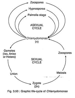
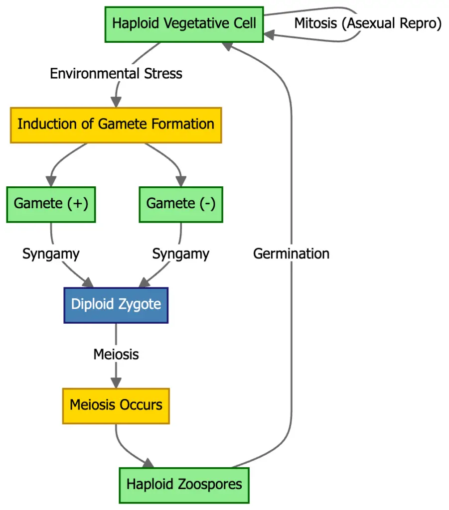
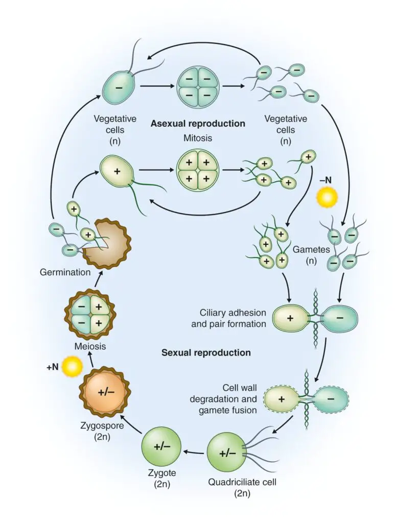
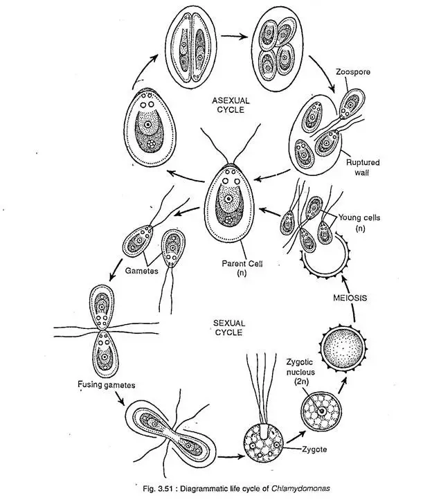
The life cycle of Chlamydomonas is mostly haplontic type. The vegetative cell is haploid and it passes through asexual and sexual reproduction. The diploid condition is found only in zygospore stage.
Asexual Reproduction
Asexual reproduction takes place generally during favourable condition. In this method, the parent cell becomes non-motile and the flagella are withdrawn. The protoplast then divides inside the old cell wall and forms new daughter cells.
Zoospore Formation
In zoospore formation, the nucleus and cytoplasm divide mitotically. The division may form 2, 4, 8 or 16 daughter protoplasts. Each daughter protoplast develops cell wall and two flagella and becomes a motile zoospore.
The parent cell wall breaks and the zoospores come outside. These zoospores swim freely in water and each of them grow into a new Chlamydomonas cell.
Aplanospores
During unfavourable condition, the parent cell loses its flagella and divides inside the cell wall. The daughter cells formed are non-motile and they develop thin wall around them. These non-motile spores are called aplanospores.
The aplanospores remain in resting condition for some time. When favourable condition comes, they develop into new vegetative cells.
Hypnospores
In very severe unfavourable condition, hypnospores are formed. They are thick walled resting spores. They contain reserve food material and can withstand dry condition and other environmental stress.
Palmella Stage
During adverse or dry condition, the protoplast divides repeatedly but the daughter cells do not form flagella. The daughter cells remain together inside a common mucilaginous envelope. This stage is called palmella stage.
In this stage, the cells remain protected inside gelatinous covering. When favourable condition returns, the mucilage dissolves. The cells form flagella and come out as motile zoospores.
Sexual Reproduction
Sexual reproduction occurs mostly during unfavourable condition, such as nitrogen deficiency, drought or other stress condition. The haploid vegetative cells form gametes or sometimes they themselves behave as gametes.
Gamete Formation
The gametes may be similar or dissimilar according to species. Sexual reproduction may be isogamous, anisogamous or oogamous type. In isogamy, similar gametes fuse. In anisogamy, unequal gametes fuse. In oogamy, small motile male gamete fuses with large non-motile female gamete.
Fusion
The gametes of opposite mating type come close to each other. They are commonly called plus and minus strains. The gametes pair and then their cytoplasm and nuclei fuse together.
Zygospore Formation
After fusion, diploid zygote is formed. The zygote loses flagella and secretes a thick resistant wall around it. This thick walled resting structure is known as zygospore.
The zygospore is a resistant stage of life cycle. It can survive drought, freezing temperature and other unfavourable condition.
Meiosis and Germination
When favourable condition returns, the zygospore germinates. The diploid nucleus undergoes meiosis and four haploid nuclei are formed. After this, four haploid zoospores are liberated by breaking of zygospore wall.
These zoospores swim in water and develop into new haploid vegetative cells of Chlamydomonas.
Genome Structure of Chlamydomonas
- Chlamydomonas has three different genomes in its cell, such as nuclear genome, chloroplast genome and mitochondrial genome. These genomes are separately present and their inheritance pattern also not same.
- The nuclear genome is compact type genome of about 120 megabases. It contains the main genetic material of Chlamydomonas and controls most of the cellular characters.
- The genetic material of nucleus is arranged in 17 chromosomes. These chromosomes are related with the 17 linkage groups which are described in Chlamydomonas.
- The vegetative cell is normally haploid. The diploid condition is very short and it is found only in the zygote or zygospore stage.
- In the nuclear genome, duplicated genes are very few. For this reason single gene function and mutation effect can be studied easily in this algae.
- The reference genome was sequenced from CC-503 cw92 mt+ strain. It is a cell wall deficient mutant strain and it was used for isolation of good quality high molecular weight DNA.
- The chloroplast genome is present inside the chloroplast. It is formed by circular double-stranded DNA molecules and present in many copies.
- The chloroplast contains more than 50 copies of chloroplast DNA. These copies remain as circular DNA inside the chloroplast region.
- The chloroplast genome shows uniparental inheritance. It is transmitted through mt+ or plus mating type only.
- The mitochondrial genome is present inside the mitochondria. It contains fewer genes in comparison to chloroplast genome.
- The mitochondrial genome also shows uniparental inheritance. But in this case inheritance takes place through mt- or minus mating type.
Examples of Chlamydomonas
- Chlamydomonas reinhardtii is the most commonly studied species of Chlamydomonas. It is widely used as model organism in genetics, molecular biology, photosynthesis study and biofuel production.
- Chlamydomonas nivalis is a cold adapted species and it can grow in snow and ice region. It produces red pigment astaxanthin, due to which it causes red snow or watermelon snow.
- Chlamydomonas acidophila is a specialized extremophilic species. It grows in very low pH condition and found in highly acidic environment.
- Chlamydomonas yellowstonensis is an alpine species of Chlamydomonas. It gives yellowish-green colour to snow covered mountain region.
- Chlamydomonas hydrae is an epizoic species. It lives on the outer surface of freshwater polyps like Hydra in symbiotic condition.
- Chlamydomonas debaryana is an extremophilic species and can survive in harsh environmental condition. It shows isogamous type of sexual reproduction.
- Chlamydomonas moewusii is a common species used for study of mating behaviour and flagellar function. It is also important in study of motility.
- Chlamydomonas braunii is a species which shows anisogamous sexual reproduction. In this type, gametes are different in their size.
- Chlamydomonas coccifera and Chlamydomonas ooganum are species showing oogamous sexual reproduction. In this condition, large non-motile female gamete and small motile male gametes are formed.
- Chlamydomonas bicilliata, Chlamydomonas arachne and Chlamydomonas reticulata are species having different chloroplast shapes. Their chloroplasts are H-shaped, stellate and reticulate type respectively.
- Chlamydomonas alpina, Chlamydomonas stenii and Chlamydomonas eradians are species with special chloroplast forms. They have discoid, ridged and axile chloroplast respectively.
- Other reported species are Chlamydomonas caudata, Chlamydomonas ehrenbergii, Chlamydomonas elegans, Chlamydomonas muriella, Chlamydomonas ovoidae, Chlamydomonas priscuii, Chlamydomonas smithii, Chlamydomonas grandistigma, Chlamydomonas monadina, Chlamydomonas subcaudata and Chlamydomonas iyengarii.
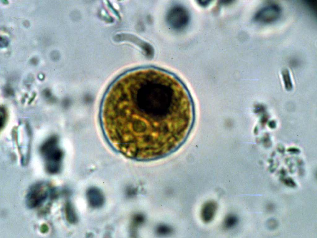
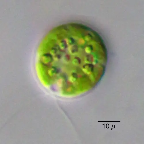
Chlamydomonas under microscope
References
- AlgaeBase. (n.d.). Chlamydomonas nivalis (F.A.Bauer) Wille.
- ALLEN Career Institute. (n.d.). Chlamydomonas: Classification, structure, life cycle and reproduction.
- Balasubramanian, J. (2013). A review on unicellular green algae – Chlamydomonas. Species, 7(8), 7-9.
- Barone, T. (n.d.). Chlamydomonas reinhardtii (green algae). LEVÄT.
- bioRxiv. (n.d.). Chlamylipo, a Chlamydomonas-in-liposome microswimmer: self-propelled swimming and associated lipid membrane flow.
- Briggs, G. M. (2021). Chlamydomonas, a small unicellular green alga. In Inanimate Life. Milne Publishing.
- BYJU’S. (n.d.). Chlamydomonas.
- Chlamydomonas Resource Center. (n.d.). About Chlamydomonas.
- Daggett, S. S. (2024). Chlamydomonas reinhardtii. Health and Medicine | Research Starters | EBSCO Research.
- Egra SSB College. (n.d.). CHLAMYDOMONAS CLASSIFICATI0N HABIT AND HABITAT STRUCTURE.
- GeeksforGeeks. (2025, July 23). Chlamydomonas.
- Kemm, F. (n.d.). What is Chlamydomonas? Singer Instruments.
- Marshall, W. F. (2024). Chlamydomonas as a model system to study cilia and flagella using genetics, biochemistry, and microscopy. Frontiers in Cell and Developmental Biology, 12, 1412641. https://doi.org/10.3389/fcell.2024.1412641
- NCBI. (n.d.). Taxonomy browser (Lobochlamys segnis).
- Nikulin, V. Y., Nikulin, A. Y., Gontcharov, A. A., Bagmet, V. B., & Abdullin, S. R. (2023). Oogamochlamys kurilensis sp. nov. (Chlorophyta, Volvocales) from the Soils of Iturup Island (Sakhalin Region, Russia). Plants, 12(19), 3350. https://doi.org/10.3390/plants12193350
- PMC. (n.d.). Computer-assisted image analysis of human cilia and Chlamydomonas flagella reveals both similarities and differences in axoneme structure.
- PMC. (n.d.). The Chlamydomonas zygospore: mutant strains of Chlamydomonas monoica blocked in zygospore morphogenesis comprise 46 complementation groups.
- PMC. (n.d.). When unity is strength: The strategies used by Chlamydomonas to survive environmental stresses.
- Quarmby, L. M. (1994). Signal transduction in the sexual life of Chlamydomonas. Plant Molecular Biology, 26(5), 1271-1287. https://doi.org/10.1007/BF00016474
- ResearchGate. (n.d.). Molecular phylogeny and taxonomic revision of Chlamydomonas (Chlorophyta). I. Emendation of Chlamydomonas ehrenberg and Chloromonas gobi, and description of Oogamochlamys gen. nov. and Lobochlamys gen. nov.
- ResearchGate. (n.d.). The Chlamydomonas flagellum as a model for human ciliary disease.
- Saraswat, K. (2025, May 30). Chlamydomonas, diagram, structure, reproduction and life cycle. Physics Wallah.
- Taylor & Francis. (n.d.). Full article: Generic and species concepts in Microglena (previously the Chlamydomonas monadina group) revised using an integrative approach.
- UNH Center for Freshwater Biology. (n.d.). Phycokey – Chlamydomonas.
- Unknown Author. (n.d.). Chlamydomonas [PDF]. Googleapis.com.
- Unknown Author. (n.d.). Systematic position, ultrastructural anatomy, and evolutionary reclassification of the genus Chlamydomonas.
- Vedantu. (n.d.). Isogamy anisogamy and oogamy types of sexual reproduction class 12 biology CBSE.
- Wikipedia. (2025, December 2). Chlamydomonas.
- Wikipedia. (2026, May 6). Chlamydomonas nivalis.
- Wingfield, J. L., & Lechtreck, K.-F. (2018). Chlamydomonas basal bodies as flagella organizing centers. Cells, 7(7), 79. https://doi.org/10.3390/cells7070079
- Wirschell, M., Lee, L., & Liu, A. (2025). Editorial: Advances in cilia and flagella research. Frontiers in Cell and Developmental Biology, 13, 1739712. https://doi.org/10.3389/fcell.2025.1739712