

The cell coat, or glycocalyx, is a carbohydrate-rich layer on the outer surface of the plasma membrane, especially prominent in animal cells. It is formed mainly by the carbohydrate portions of glycoproteins, glycolipids and proteoglycans, together with associated extracellular molecules, and contributes to cell protection, recognition, adhesion and interactions with the surrounding environment.
What Is the Cell Coat or Glycocalyx?
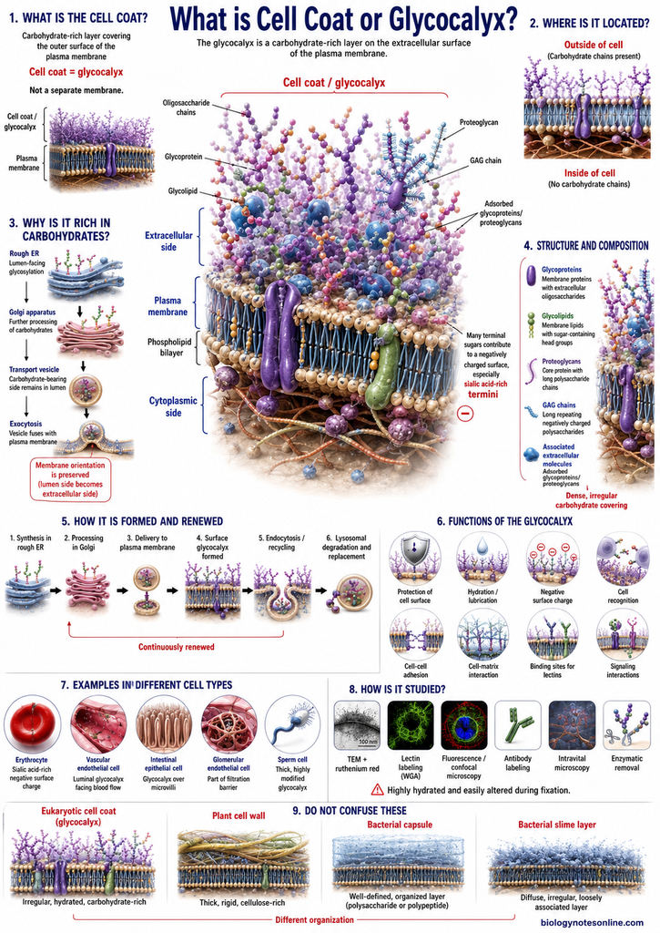
The cell coat or glycocalyx is a carbohydrate-rich layer that covers the outer surface of the plasma membrane. Cell coat and glycocalyx are commonly used synonymously in cell biology. It is mainly formed of oligosaccharide chains which are attached with the membrane glycoproteins and glycolipids, forming a carbohydrate covering towards outside of the cell. Proteoglycans are also present. Their polysaccharide chains contribute to this coat, while some glycoproteins and proteoglycans secreted outside the cell may remain adsorbed on the cell surface and become the part of glycocalyx.
Location of the Cell Coat on the Plasma Membrane
The cell coat is present on the extracellular side of plasma membrane. It covers the outer face. The carbohydrate portions of glycolipids are directed towards outside of the cell, and carbohydrate chains present on the membrane proteins also project into the extracellular region. Thus, these carbohydrate components are present only towards the external surface of the membrane. The glycocalyx is therefore outside the lipid bilayer, not towards its cytoplasmic surface.
Why the Cell Coat Is Rich in Carbohydrates
The cell coat contains a large amount of carbohydrates. This is because many of the proteins and lipids present in plasma membrane contain attached sugar chains. Most of their glycosylation takes place in the lumen of endoplasmic reticulum (ER) and Golgi apparatus.
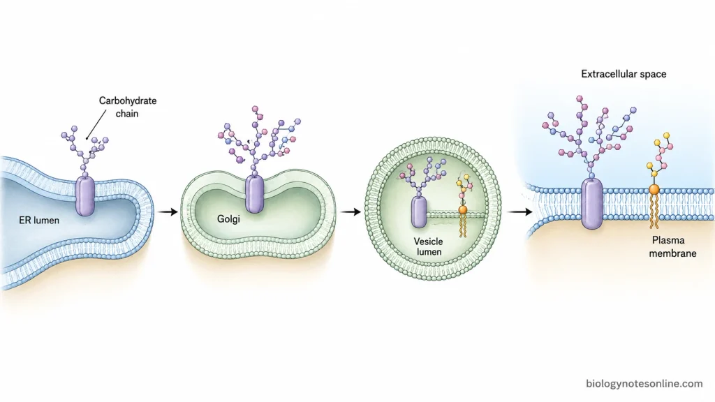
During this process, the carbohydrate chains occur on those portions of membrane molecules which face the lumen. Later these membrane-containing vesicles reach the plasma membrane and fuse with it. The lumen-facing portions now become exposed towards the external side of cell, carrying their carbohydrate chains with them. As a result, carbohydrates become concentrated over the outer membrane surface. This forms the characteristic carbohydrate-rich glycocalyx.
Structure and Composition of the Cell Coat
The cell coat is not a separate membrane structure. Its major part is formed from carbohydrates attached with proteins and lipids of the plasma membrane. Some extracellular macromolecules are also associated with it. The following are the major structural components of cell coat-
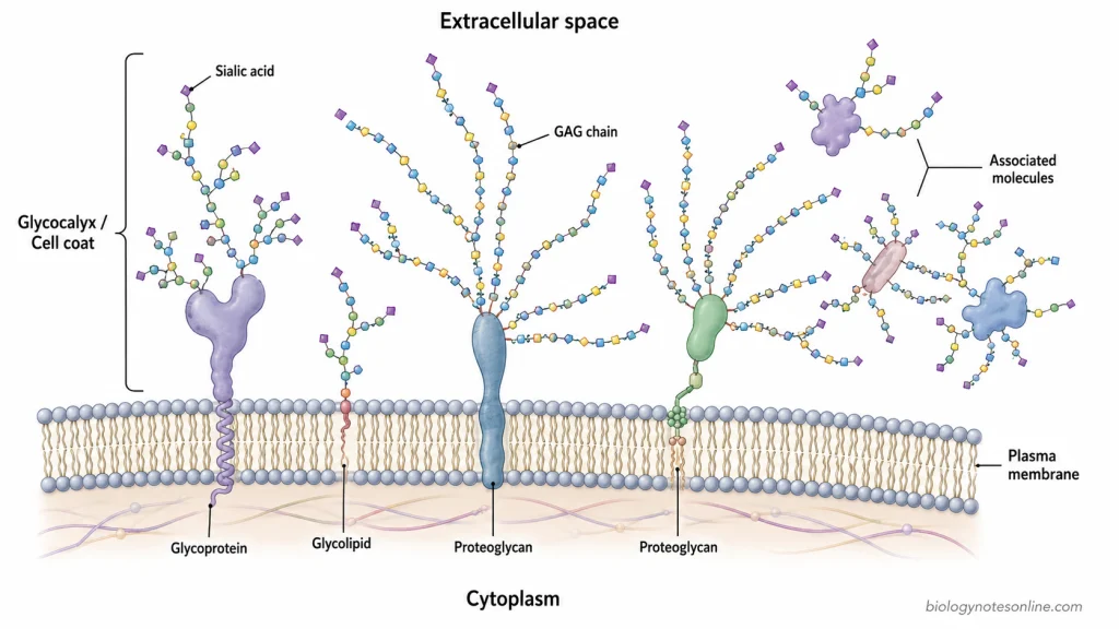
- Glycoproteins- These are membrane proteins having oligosaccharide chains covalently attached with them. The carbohydrate chains are present on the extracellular portions. They project towards outside of the cell and form an important part of the glycocalyx.
- Glycolipids- Glycolipids are membrane lipids containing sugar residue or an oligosaccharide attached to their polar head group. They occur in the non-cytosolic layer of plasma membrane, hence the carbohydrate portion is exposed on the outer cell surface.
- Proteoglycans- It consists of a protein core with long polysaccharide chains attached to it. Some proteoglycans are integral membrane molecules. The protein core may pass through the lipid bilayer, while some are attached with membrane by a glycosylphosphatidylinositol (GPI) anchor. Their long carbohydrate chains extend into the extracellular region and become a part of cell coat.
- Glycosaminoglycan (GAG) chains- These are long polysaccharide chains associated with proteoglycans. They are generally made of repeating disaccharide units. Some GAGs are sulfated and carry considerable negative charge.
- Oligosaccharide chains- The oligosaccharides present on glycoproteins and glycolipids are not arranged in one uniform manner. They are commonly branched. Different sugars and different types of linkages are present, which results in a highly varied carbohydrate surface from one molecule to another.
- Adsorbed glycoproteins and proteoglycans- All the material of glycocalyx is not directly anchored with plasma membrane. Some glycoproteins and proteoglycans are secreted into the extracellular space and later become adsorbed on the cell surface. These molecules are also included as a part of cell coat.
- Sialic acid and other sugars- Different monosaccharides occur in the glycans of cell surface. In vertebrate cells, sialic acids are abundant on many glycoproteins and glycolipids and because of their charge they contribute to the negatively charged nature of the surface coat.
- Overall arrangement- The carbohydrate chains of these molecules extend away from the plasma membrane. Together they form a dense, irregular carbohydrate-rich covering over the cell surface. Thus, the glycocalyx is formed of membrane-bound glycoconjugates along with some material associated from the extracellular side, rather than being a uniform independent layer.
How Is the Cell Coat Formed and Renewed?
The cell coat is not formed as a separate layer at one time. Its components are prepared along with membrane proteins and lipids and finally become exposed on the outer surface of plasma membrane. The major steps are as follows-
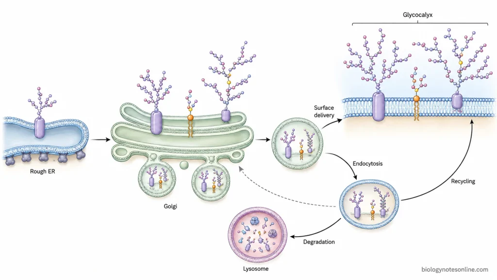
- Formation in Endoplasmic Reticulum (ER)- The membrane proteins are first synthesized in the rough endoplasmic reticulum. In many of these proteins, N-linked oligosaccharides are also added while the protein is present in ER. The carbohydrate-bearing portion remains towards the lumen of ER.
- Processing in Golgi apparatus- These proteins are transported to the Golgi apparatus, where their carbohydrate chains undergo further modification. O-linked sugars are mainly added here. The Golgi also adds glycosaminoglycan chains to the protein core of proteoglycans and is involved in formation of many glycolipids. Thus, different major carbohydrate components of the glycocalyx are processed in this organelle.
- Transport towards the cell surface- After processing, glycoproteins and glycolipids are carried from Golgi in transport vesicles. These vesicles then reach the plasma membrane and fuse with it. The side which was facing the lumen of ER and Golgi now faces outside of the cell. As a result, their carbohydrate chains become exposed at cell surface.
- Formation of the carbohydrate coat- The exposed oligosaccharides of glycoproteins, glycolipids and polysaccharide chains of membrane proteoglycans together form the cell coat. Some glycoproteins and proteoglycans secreted outside are also adsorbed on cell surface. These also become its part.
- Renewal by membrane recycling- The cell surface does not remain fixed. Portions of plasma membrane are continuously taken inside by endocytosis, along with their proteins and lipids. Many of these membrane components are returned again to the surface by recycling and exocytosis.
- Degradation and replacement- Some of the internalized surface components are not recycled. They are delivered to lysosomes and degraded. New glycoproteins, glycolipids and proteoglycans formed through ER and Golgi pathway replace such material at the plasma membrane.
- Renewal of sugar chains- The carbohydrate chains themselves can also undergo turnover. Terminal sugars of some membrane glycoproteins are removed more rapidly than the whole protein. If such proteins are internalized but not destroyed, they can again reach Golgi, where the missing sugars may be added and the molecule can return to the cell surface. Glycosphingolipid components can also be recycled and used again for synthesis of longer carbohydrate chains.
Functions of the Cell Coat
The cell coat or glycocalyx performs different functions at the outer surface of cell. Some of the important functions are as follows-
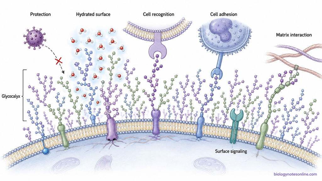
- Protection of cell surface- The cell coat forms a protective covering over the plasma membrane. It protects the surface against mechanical and chemical damage and acts as a physical barrier between the membrane and external environment.
- Prevention of unwanted surface interactions- The carbohydrate coat keeps some foreign particles and other cells away from direct contact with the membrane. Thus, undesirable protein-protein interactions at cell surface can be reduced.
- Cell recognition- One of the important function of glycocalyx is recognition of cells. The oligosaccharides exposed on cell surface are highly variable and act as surface markers. By these carbohydrate patterns, different types of cells can be recognized from one another.
- Cell-cell adhesion- Carbohydrates of the cell coat participate in adhesion between cells. In some cases, membrane proteins called lectins recognize particular oligosaccharides present on another cell. The interaction of selectins with carbohydrate ligands during adhesion of leukocytes to vascular endothelial cells is an example.
- Cell-matrix interaction- The glycans present on cell surface can interact with molecules of the extracellular matrix. Such interactions help in association of cells with their surrounding extracellular material. Cell-surface glycans and glycan-binding proteins are involved in these specific contacts.
- Binding and specific biological interactions- The sugar chains of cell coat provide binding sites for different glycan-binding proteins. These interactions are not all of one type. They are involved in processes such as sperm-egg interaction, lymphocyte movement and inflammatory responses, while particular carbohydrate structures also participate during blood cell adhesion.
Examples of the Cell Coat in Different Cell Types
The cell coat is present over many different types of animal cells, but its thickness and carbohydrate components are not same in every cell. Some of the important examples are-
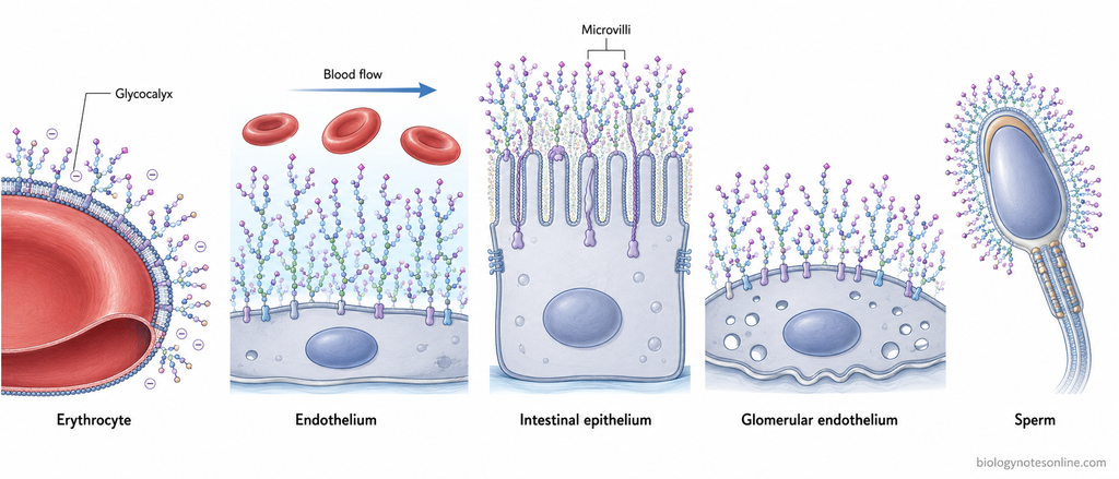
- Red blood cells (Erythrocytes)- The surface of erythrocytes contains a carbohydrate-rich coat with large number of terminal sialic acid residues. These negatively charged sugars are mainly carried by membrane glycoproteins and produce much of the negative charge of red blood cell surface.
- Vascular endothelial cells- A well-developed glycocalyx is present on the luminal surface of endothelial cells, directly facing the circulating blood. It is a meshwork of membrane-bound proteoglycans, glycoproteins and associated soluble molecules. This layer may be quite extensive. It participates in vascular permeability, interaction with blood cells and sensing the shear force produced by blood flow.
- Intestinal epithelial cells- The apical surface of intestinal epithelial cells also contains a distinct glycocalyx. Here, glycosylated transmembrane mucins form an important component of the coat over the epithelial surface and microvillar region. It provides the interface between intestinal cells and the material present in intestinal lumen, including microorganisms.
- Glomerular endothelial cells- In the capillaries of kidney glomerulus, endothelial cells have a carbohydrate-rich surface layer. The glycocalyx covers their luminal surface and forms a part of the specialized glomerular filtration barrier. Damage or removal of this coat increases permeability of the glomerular barrier to macromolecules such as albumin.
- Sperm cells- Mammalian spermatozoa possess a thick and highly modified glycocalyx over their plasma membrane. The coat is not fixed throughout the life of sperm. Glycans are formed during sperm development, further changed during passage through the epididymis and additional glycoconjugates can be received from surrounding reproductive tract secretions. During capacitation, considerable modification of this surface coat again takes place. The sperm glycocalyx is involved in survival of sperm in female reproductive tract and different interactions required before fertilization.
How Is the Cell Coat Observed and Studied?
The cell coat or glycocalyx is a very delicate and highly hydrated structure. Ordinary light microscopy does not show its fine structure properly. Therefore, special stains, carbohydrate-binding probes and different microscopic techniques are used for its study. The following are some of the important methods-
- Transmission Electron Microscopy (TEM)- Electron microscopy is one of the important methods used to observe the cell coat. The glycocalyx can be made electron dense by using cationic stains such as ruthenium red. Alcian blue and some other electron-dense probes have also been used. Under TEM, the stained material is seen extending outside the plasma membrane.
- Ruthenium red staining- Ruthenium red binds with acidic carbohydrate material of the surface coat and was used in early electron microscopic observation of glycocalyx. It makes this otherwise poorly visible layer electron dense. The method, however, requires fixation and processing of the specimen.
- Lectin labeling- Lectins are proteins which bind with particular carbohydrate groups. Fluorescently labeled lectins can therefore be used for locating sugars present over the cell surface. Wheat germ agglutinin (WGA) is one commonly used lectin for glycocalyx staining. Different lectins can give different binding pattern depending on the carbohydrates present in the coat.
- Fluorescence and confocal microscopy- After binding of fluorescent lectins or other fluorescent probes, the glycocalyx can be studied by fluorescence microscope. Confocal laser scanning microscopy (CLSM) is also used. It gives optical sections through the labeled surface and several sections can be combined for studying the three-dimensional distribution of the coat.
- Antibody labeling- Specific components of cell coat can be detected with antibodies. For example, antibodies or binding proteins against heparan sulfate, hyaluronan and particular proteoglycans are used when an individual component has to be studied rather than staining the complete carbohydrate layer. These labels are commonly observed by fluorescence microscopy.
- Intravital microscopy- This is used particularly for study of the glycocalyx on vascular endothelial cells in a living animal. The layer can be studied by fluorescent labeling, or indirectly from the region where circulating cells and some tracer molecules are excluded near the endothelial surface. Thus, changes in endothelial glycocalyx can be followed under more physiological conditions.
- Super-resolution and two-photon microscopy- These advanced optical methods are also used for cell coat study. Super-resolution microscopy can show the organization of selected surface glycans at a much smaller scale, while two-photon microscopy has been applied for imaging labeled glycocalyx in intact tissues and vessels.
- Enzymatic treatment- Specific carbohydrate components can be removed with enzymes and the treated surface is then compared with the normal surface. Hyaluronidase, neuraminidase or other glycan-degrading enzymes have been used depending upon the component being examined. Reduction of the labeled or visible coat after treatment helps in studying the contribution of that carbohydrate component.
The glycocalyx is easily altered during fixation, dehydration and other preparation steps. This creates an important problem particularly during electron microscopy, because the coat may collapse and appear much thinner than it was in living condition. Hence preservation of cell coat is an important part during its microscopic study.
Cell Coat vs Cell Wall vs Bacterial Glycocalyx
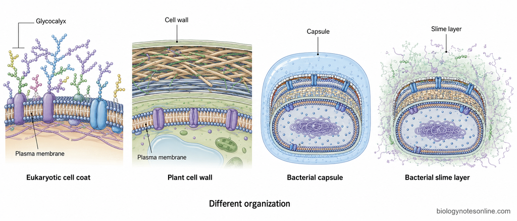
The terms cell coat, cell wall and bacterial glycocalyx describe structures present outside the plasma membrane, but they are not the same structure. The word glycocalyx is also used for bacterial capsule and slime material. This sometimes creates confusion. Their composition and structural organization are different.
Cell Coat vs Cell Wall
| Features | Cell Coat (Glycocalyx) | Cell Wall |
|---|---|---|
| Basic nature | It is a carbohydrate-rich zone present over the outer surface of plasma membrane, especially described in animal cells. It is not a rigid wall. | Cell wall is a strong extracellular structure present in plants, fungi and many other organisms. It gives mechanical support and is much more rigid than the cell coat. |
| Relation with plasma membrane | Much of the coat is formed by carbohydrates attached with intrinsic membrane glycoproteins, glycolipids and membrane proteoglycans. Some secreted molecules are also adsorbed on the surface. | It lies outside the plasma membrane but forms a separate extracellular structural framework. The wall is not simply the carbohydrate portions of membrane proteins and lipids. |
| Major composition | Oligosaccharides of glycoproteins and glycolipids, polysaccharide chains of proteoglycans and some adsorbed glycoproteins or proteoglycans. | Plant cell wall contains mainly cellulose microfibrils, cross-linking glycans and pectins. Secondary walls may also contain lignin. Fungal walls are different and contain chitin, glucans and other glycan polymers with glycoproteins. |
| Rigidity | It forms a surface covering. No rigid load-bearing shell is formed by the glycocalyx itself. | Rigidity is an important property. Plant walls withstand internal turgor pressure, while fungal walls also provide mechanical stability and protection against osmotic lysis. |
| Major role | Protection of membrane surface and cell-cell recognition are some of its important functions. The exposed carbohydrate chains also participate in cell interaction. | It mainly gives shape, strength and mechanical support. Protection against osmotic stress is another major function. |
| Are they equivalent? | No. The cell coat should not be considered as a thin form of cell wall. | No. A plant or fungal cell wall is a different extracellular structure with its own polymer framework. |
Eukaryotic Cell Coat vs Bacterial Capsule and Slime Layer
The term glycocalyx can also be used in bacteriology for extracellular material present around bacterial cells. However, bacterial capsule or slime layer should not be considered structurally identical with the eukaryotic cell coat.
| Features | Eukaryotic Cell Coat | Bacterial Capsule | Bacterial Slime Layer |
|---|---|---|---|
| Organization | It is mainly made from carbohydrate chains of molecules associated with the plasma membrane. Secreted and re-adsorbed macromolecules may also become its part. | Capsule is a more organized and distinct external layer. It remains closely associated with bacterial surface. | It is less organized. The material is diffuse and loosely attached, and therefore can be removed more easily from the cell surface. |
| Composition | Mainly oligosaccharides of glycoproteins and glycolipids, together with proteoglycan polysaccharide chains. | Most bacterial capsules are polysaccharide in nature, although some capsules can contain protein or polypeptide material. | Slime layers may contain polysaccharides and other extracellular glycoconjugate material. Their exact composition varies among bacteria. |
| Position | Present immediately at the extracellular face of plasma membrane and closely related with membrane glycoconjugates. | In bacteria having a cell wall, capsule occurs external to the wall or outer cell-envelope structures. It may form the outermost layer. | Also present outside the bacterial surface, but it spreads more loosely around the cells instead of forming a clearly bounded capsule. |
| Attachment | Many of its carbohydrates are covalently attached to membrane proteins and lipids. | More firmly associated with the bacterial cell. | Loosely attached. This is one of the major points separating a slime layer from a capsule. |
| Functions | It is involved in surface protection, recognition and different cell-cell interactions. | Capsule can help in adhesion and protection. In pathogenic bacteria, some capsules also protect the organism from phagocytosis. | Slime material helps bacterial cells to attach with surfaces and participate in biofilm formation. It can also retain water and decrease drying of cells. |
| Meaning of the term glycocalyx | Here, glycocalyx refers to the carbohydrate-rich cell coat of a eukaryotic cell. | Capsule is one form of bacterial glycocalyx. | Slime layer is another form. Thus, use of the same term does not indicate that their molecular arrangement is identical with the eukaryotic cell coat. |
References
- Alberts, B., Johnson, A., Lewis, J., Raff, M., Roberts, K., & Walter, P. (2002). Molecular biology of the cell (4th ed.). Garland Science. https://www.ncbi.nlm.nih.gov/books/NBK21054/
- Ambrogi, M., & Vezina, C. M. (2024). Roles of airway and intestinal epithelia in responding to pathogens and maintaining tissue homeostasis. Frontiers in Cellular and Infection Microbiology, 14, 1346087. https://doi.org/10.3389/fcimb.2024.1346087
- Chevalier, L., Selim, J., Genty, D., Baste, J. M., Piton, N., Boukhalfa, I., Hamzaoui, M., Pareige, P., & Richard, V. (2017). Electron microscopy approach for the visualization of the epithelial and endothelial glycocalyx. Morphologie, 101(333), 55–63. https://doi.org/10.1016/j.morpho.2017.04.001
- Colley, K. J., Varki, A., & Kinoshita, T. (2017). Cellular organization of glycosylation. In A. Varki, R. D. Cummings, J. D. Esko, P. Stanley, G. W. Hart, M. Aebi, A. G. Darvill, T. Kinoshita, N. H. Packer, J. H. Prestegard, R. L. Schnaar, & P. H. Seeberger (Eds.), Essentials of glycobiology (3rd ed., Chapter 4). Cold Spring Harbor Laboratory Press. https://doi.org/10.1101/glycobiology.3e.004
- Cook, G. M. W. (2016). Glycobiology of the cell surface: Its debt to cell electrophoresis 1940–65. Electrophoresis, 37(11), 1399–1406. https://doi.org/10.1002/elps.201500476
- Cooper, G. M. (2000). The cell: A molecular approach (2nd ed.). Sinauer Associates. https://www.ncbi.nlm.nih.gov/books/NBK9839/
- Dane, M. J. C., van den Berg, B. M., Lee, D. H., Boels, M. G. S., Tiemeier, G. L., Avramut, M. C., van Zonneveld, A. J., van der Vlag, J., Vink, H., & Rabelink, T. J. (2015). A microscopic view on the renal endothelial glycocalyx. American Journal of Physiology-Renal Physiology, 308(9), F956–F966. https://doi.org/10.1152/ajprenal.00532.2014
- Freeze, H. H., Kinoshita, T., & Schnaar, R. L. (2017). Genetic disorders of glycan degradation. In A. Varki, R. D. Cummings, J. D. Esko, P. Stanley, G. W. Hart, M. Aebi, A. G. Darvill, T. Kinoshita, N. H. Packer, J. H. Prestegard, R. L. Schnaar, & P. H. Seeberger (Eds.), Essentials of glycobiology (3rd ed., Chapter 44). Cold Spring Harbor Laboratory Press. https://doi.org/10.1101/glycobiology.3e.044
- Fu, B. M., & Tarbell, J. M. (2013). Mechano-sensing and transduction by endothelial surface glycocalyx: Composition, structure, and function. Wiley Interdisciplinary Reviews: Systems Biology and Medicine, 5(3), 381–390. https://doi.org/10.1002/wsbm.1211
- Gagneux, P., Hennet, T., & Varki, A. (2022). Biological functions of glycans. In A. Varki, R. D. Cummings, J. D. Esko, P. Stanley, G. W. Hart, M. Aebi, D. Mohnen, T. Kinoshita, N. H. Packer, J. H. Prestegard, R. L. Schnaar, & P. H. Seeberger (Eds.), Essentials of glycobiology (4th ed., Chapter 7). Cold Spring Harbor Laboratory Press. https://doi.org/10.1101/glycobiology.4e.7
- Lewis, A. L., Chen, X., Schnaar, R. L., & Varki, A. (2022). Sialic acids and other nonulosonic acids. In A. Varki, R. D. Cummings, J. D. Esko, P. Stanley, G. W. Hart, M. Aebi, D. Mohnen, T. Kinoshita, N. H. Packer, J. H. Prestegard, R. L. Schnaar, & P. H. Seeberger (Eds.), Essentials of glycobiology (4th ed., Chapter 15). Cold Spring Harbor Laboratory Press. https://www.ncbi.nlm.nih.gov/books/NBK579976/
- Möckl, L., Pedram, K., Roy, A. R., Krishnan, V., Gustavsson, A.-K., Dorigo, O., Bertozzi, C. R., & Moerner, W. E. (2019). Quantitative super-resolution microscopy of the mammalian glycocalyx. Developmental Cell, 50(1), 57–72.e6. https://doi.org/10.1016/j.devcel.2019.04.035
- Parker, N., Schneegurt, M., Tu, A.-H. T., Lister, P., & Forster, B. M. (2016). Microbiology. OpenStax, Rice University. https://openstax.org/details/books/microbiology
- Reitsma, S., Slaaf, D. W., Vink, H., van Zandvoort, M. A. M. J., & oude Egbrink, M. G. A. (2007). The endothelial glycocalyx: Composition, functions, and visualization. Pflügers Archiv – European Journal of Physiology, 454(3), 345–359. https://doi.org/10.1007/s00424-007-0212-8
- Salton, M. R. J., & Kim, K.-S. (1996). Structure. In S. Baron (Ed.), Medical microbiology (4th ed., Chapter 2). University of Texas Medical Branch at Galveston. https://www.ncbi.nlm.nih.gov/books/NBK8477/
- Tecle, E., & Gagneux, P. (2015). Sugar-coated sperm: Unraveling the functions of the mammalian sperm glycocalyx. Molecular Reproduction and Development, 82(9), 635–650. https://doi.org/10.1002/mrd.22500
- Varki, A., Cummings, R. D., Esko, J. D., Stanley, P., Hart, G. W., Aebi, M., Mohnen, D., Kinoshita, T., Packer, N. H., Prestegard, J. H., Schnaar, R. L., & Seeberger, P. H. (Eds.). (2022). Essentials of glycobiology (4th ed.). Cold Spring Harbor Laboratory Press. https://www.ncbi.nlm.nih.gov/books/NBK579918/
- Walter, F. R., Santa-Maria, A. R., Mészáros, M., Veszelka, S., Dér, A., & Deli, M. A. (2021). Surface charge, glycocalyx, and blood-brain barrier function. Tissue Barriers, 9(3), 1904773. https://doi.org/10.1080/21688370.2021.1904773
Very useful information