

Blood cells are the cellular and cell-derived formed components present in circulating blood. They are also referred to as blood corpuscles. A broader and more suitable term is formed elements, which includes erythrocytes, leukocytes and platelets together.
These elements remain suspended in plasma, the fluid extracellular matrix of blood. Thus, whole blood consists of plasma along with its formed elements, and the term does not indicate that every formed element is a true complete cell.
The three major formed elements are erythrocytes (red blood cells or RBCs), leukocytes (white blood cells or WBCs) and platelets. Erythrocytes are specialized biconcave cells which lose their nucleus during maturation. They contain hemoglobin and are mainly used for transport of oxygen. Leukocytes are nucleated complete cells, with their morphology varying among the different types, and they mainly take part in defense and immune functions.
Platelets are different. They are small, anucleate cytoplasmic fragments derived from megakaryocytes, rather than true complete cells. In resting condition they are generally discoid and after blood vessel injury they become activated. Their major role is hemostasis, where platelets help to limit blood loss and take part in formation of the platelet plug.
Types of Blood Cells
The formed elements of blood are mainly of three types, red blood cells (RBCs or erythrocytes), white blood cells (WBCs or leukocytes) and platelets (thrombocytes). RBCs mainly carry respiratory gases, whereas WBCs take part in defense of the body. Platelets are different. They are small fragments derived from megakaryocytes and mainly involved in hemostasis.

1. Red Blood Cells (erythrocytes)
Red blood cells (RBCs), also called erythrocytes, are specialized blood cells used mainly for transport of oxygen and carbon dioxide. In mature form, they are biconcave cells without nucleus and remain in the blood circulation for about 120 days.
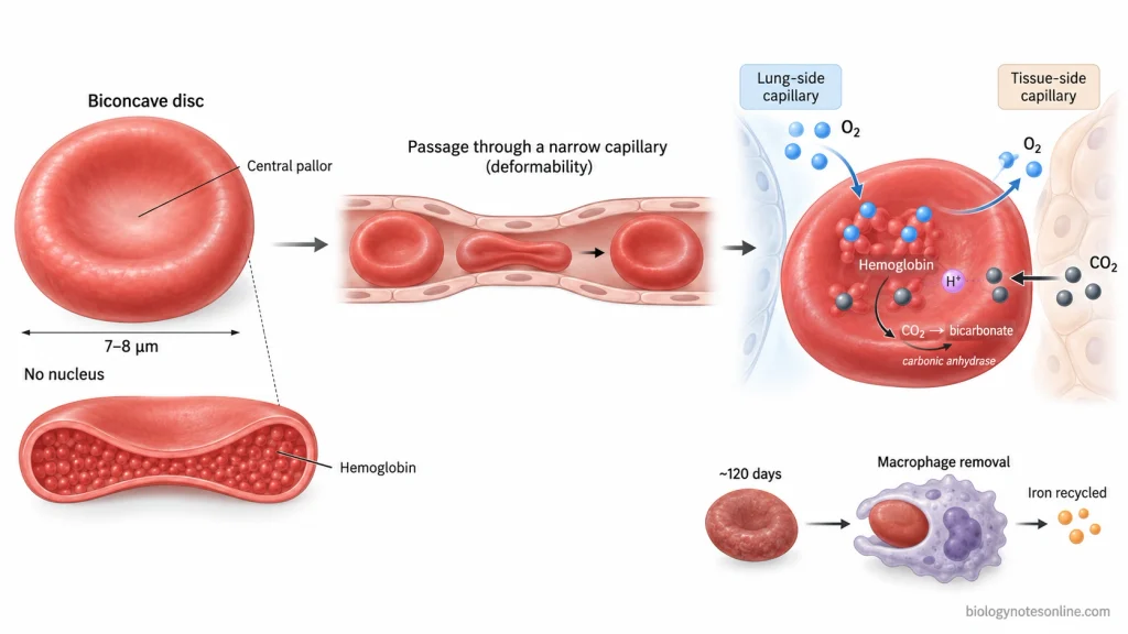
- Mature human erythrocytes are biconcave disc-shaped cells of about 7–8 µm diameter. Nucleus is absent, and most of the cell organelles are also lost during maturation. A major part of the erythrocyte is occupied by hemoglobin.
- Under the light microscope, normal RBCs in a stained peripheral blood smear appear round and pink to reddish in colour. The middle portion appears lighter because the biconcave cell is thinner at its center. This is referred to as central pallor (approximately one-third of the cell diameter).
- Due to its biconcave form, erythrocytes have a high surface area in relation to cell volume and are highly deformable. During movement through narrow capillaries, the RBCs can squeeze through the small spaces and again return back to their normal shape.
- The average life span of a mature human erythrocyte is about 120 days. During this period, repeated mechanical stress and gradual changes occur in its membrane and other cellular components. Replacement of damaged proteins is very limited because mature RBCs have no nucleus and protein-synthesizing organelles.
- After becoming old or damaged, erythrocytes are removed by macrophages, mainly in the spleen. Liver and bone marrow also take part in this process. During removal, hemoglobin is broken down, iron is recovered and reused for the formation of new erythrocytes, while the heme portion undergoes further degradation which contributes to bilirubin formation.
- The major function of red blood cells is transport of oxygen. In the lungs, oxygen binds with hemoglobin and is carried through blood circulation. It is then released to different tissues of the body.
- RBCs also take part in carbon dioxide transport from the tissues towards the lungs. Some carbon dioxide is carried by binding with hemoglobin. A large fraction entering into erythrocytes is converted into bicarbonate with the help of carbonic anhydrase (CA).
- Hemoglobin also binds hydrogen ions formed during carbon dioxide transport. In this way, erythrocytes take part in buffering of blood and maintenance of the acid-base condition.
2. White Blood Cells
White blood cells (WBCs), also called leukocytes, are nucleated cells of blood involved mainly in defense of the body. They are of different types and take part in innate as well as adaptive immune responses.

- Unlike mature red blood cells, leukocytes contain a nucleus and the normal cell organelles. Their size and shape are not same in all types. Shape of the nucleus and cytoplasmic granules also varies, which are used for identifying different WBCs in a stained blood smear.
- White blood cells are present in the blood but are not restricted only to blood circulation. During infection or injury, these cells can pass through the blood vessel wall and enter into the tissues. This movement of leukocytes from blood into tissues is referred to as diapedesis (extravasation).
- The major function of WBCs is protection against microorganisms, foreign materials and damaged cells. Some leukocytes engulf microorganisms by phagocytosis. Others take part in antibody-mediated and cell-mediated immune responses, while different WBCs are also involved during inflammation and allergic reactions.
- Based on the appearance of specific granules in their cytoplasm, leukocytes are divided into two major groups, granulocytes and agranulocytes. Granulocytes contain conspicuous cytoplasmic granules. Their nucleus is generally lobed. The following are the three granulocytes, neutrophils, eosinophils and basophils.
- Neutrophils are phagocytic cells and provide an important defense against microorganisms, particularly bacteria. Eosinophils are mainly involved in defense against parasites and also take part in allergic responses. Basophils, on the other hand, participate in inflammatory and allergic reactions by releasing different mediators from their granules.
- Agranulocytes do not show the prominent specific granules found in granulocytes. Lymphocytes and monocytes come under this group, and their nuclei do not have the typical multiple lobes of granulocytes.
- There are different populations of lymphocytes involved in immune responses. B lymphocytes mainly take part in antibody-mediated immunity, whereas T lymphocytes are involved in cell-mediated immunity. Natural killer cells (NK cells) are another lymphocyte population. These cells can kill certain infected or abnormal cells.
- Monocytes are large circulating leukocytes with an indented or kidney-shaped nucleus. After moving into tissues, many of them differentiate into cells of the mononuclear phagocyte system (including macrophages). These cells take up microorganisms, dead cells and cellular debris and also take part in antigen presentation.
3. Platelets
Platelets, also called thrombocytes, are small anucleate formed elements of blood which take part mainly in hemostasis. They are not complete cells. These are cytoplasmic fragments produced from large megakaryocytes present in bone marrow.

- Platelets are very small, about 2–4 µm in diameter. In resting condition, they are discoid in shape and nucleus is absent. Different granules and other cellular components are present in their cytoplasm, which become important when platelet is activated.
- The formation of platelets takes place from megakaryocytes of bone marrow. These are large cells. During this process, extensions of megakaryocyte cytoplasm (proplatelets) are formed towards the bone marrow sinusoids and finally platelets are released into blood circulation. Thus, platelets are fragments derived from megakaryocyte cytoplasm and not true complete cells.
- The major function of platelets is hemostasis. After injury of a blood vessel, platelets attach to the damaged region. They become activated and other platelets now start to collect at the same area. Aggregation takes place and a platelet plug is formed.
- During activation, substances stored inside platelet granules are released. These help to bring in and activate more platelets. The platelet membrane also provides surface for different reactions of blood coagulation.
- Platelets remain in the blood circulation for only a short period, generally about 7–10 days. New platelets are therefore continuously released from megakaryocytes.
- Old platelets are removed from the circulation mainly through the liver and spleen. Activated or damaged platelets can also be cleared. Macrophages and hepatic clearance mechanisms take part in their removal, while changes occurring on platelet surface during aging can promote this process.
Formation of Blood Cells
The formation of blood cells is referred to as hematopoiesis (hemopoiesis). During this process, erythrocytes, leukocytes and platelets are formed from hematopoietic stem cells (HSCs). In adults, it mainly takes place in the red bone marrow.

The process takes place in following steps-
Step 1- Hematopoietic stem cell
The process starts from a multipotent hematopoietic stem cell (HSC) present in bone marrow. These cells have the ability of self-renewal. Some cells are therefore maintained as stem cells, whereas others proceed towards blood cell formation.
Step 2- Formation of progenitor cells
HSCs now form more restricted progenitor cells. During this process, their self-renewing ability gradually decreases and cells become committed towards particular blood cell lineages.
Several intermediate precursor cells are formed.
Step 3- Formation of myeloid and lymphoid lineages
The progenitor cells mainly proceed through two pathways, myeloid lineage and lymphoid lineage.
Myeloid lineage gives rise to erythrocytes, megakaryocytes, neutrophils, eosinophils, basophils and monocytes. The lymphoid lineage forms B lymphocytes, T lymphocytes and natural killer (NK) cells.
Step 4- Formation of red blood cells
The formation of erythrocytes is called erythropoiesis. In this process, erythroid cells pass through different erythroblast stages. The nucleus becomes condensed and finally removed.
An immature reticulocyte is then formed. It enters into blood and develops into mature erythrocyte. Erythropoietin (EPO), produced mainly by kidneys, is the major hormone involved in this process.
Step 5- Formation of white blood cells
White blood cells are formed through different developmental pathways. Myeloid progenitors form neutrophils, eosinophils, basophils and monocytes.
Lymphocytes develop from lymphoid progenitors. B lymphocytes continue their development mainly in bone marrow, while immature T lymphocytes move to the thymus for further maturation. Different colony-stimulating factors (CSFs) and interleukins take part in regulation of these processes.
Step 6- Formation of platelets
Platelet formation is referred to as thrombopoiesis. Cells of myeloid lineage develop into megakaryocyte precursors and finally large megakaryocytes are formed in bone marrow.
During this process, megakaryocyte cytoplasm forms long extensions called proplatelets. Small platelet fragments are released from these extensions into blood. The major regulator involved is thrombopoietin (TPO).
Platelets formed here are cytoplasmic fragments, not complete cells.
Step 7- Maturation and release into blood
After sufficient development, formed blood cells enter the blood circulation through bone marrow sinusoids. Megakaryocytes remain mainly inside the marrow, but their platelet fragments are released into blood.
Blood cell formation continues throughout life because circulating formed elements have limited life spans and are continuously replaced.
Regulation of Blood-Cell Production
Blood-cell production is controlled by different hormones, cytokines and signals present inside the bone marrow microenvironment. These factors act on hematopoietic stem cells and progenitor cells and regulate their survival, multiplication and development into different blood cells.
- Erythropoietin (EPO)- It is the major hormone involved in formation of red blood cells. In adults, the major source of EPO is kidney. When oxygen level becomes low, its production is increased and erythroid progenitor cells in bone marrow are stimulated. Thus, erythropoiesis increases.
- Thrombopoietin (TPO)- The major regulator involved in formation of megakaryocytes and platelets. It acts through MPL (c-Mpl) receptor present on cells of megakaryocyte lineage. During this process, proliferation and maturation of these cells are supported.
- Colony-stimulating factors (CSFs)- These are glycoprotein factors which act on different hematopoietic progenitor cells. G-CSF mainly acts on neutrophil lineage. M-CSF is associated with monocyte-macrophage lineage, whereas GM-CSF acts on granulocyte and monocyte progenitors. Their effects can overlap with other cytokines.
- Interleukins- Different interleukins also take part in blood-cell formation. IL-3 acts on relatively early hematopoietic progenitor cells and can support formation of several blood-cell lineages. Some other interleukins have more restricted action.
- Bone marrow niche- Hematopoietic stem cells remain in specialized regions of bone marrow. Stromal cells, endothelial cells and other surrounding cells provide different signals. These regulate HSC survival, quiescence, self-renewal and differentiation. Cell to cell interaction is also involved.
- Feedback regulation- Blood-cell production changes according to requirement of the body. Low oxygen level increases EPO production and brings up erythrocyte formation. Platelet number and availability of TPO similarly affect formation of megakaryocytes and platelets.
Functions of Blood Cells
Blood cells perform different functions depending upon their type. Erythrocytes mainly carry respiratory gases, leukocytes are involved in defense of the body, while platelets take part in hemostasis and blood clotting.
- Oxygen transport- The major function of red blood cells is transport of oxygen. In the lungs, oxygen binds with hemoglobin present inside RBCs. It is carried through blood and released to the body tissues.
- Carbon dioxide transport- Erythrocytes also carry carbon dioxide from tissues towards the lungs. Some CO₂ is transported by binding with hemoglobin, while much of the carbon dioxide entering blood is converted into bicarbonate.
- Blood buffering- Hemoglobin present in RBCs can take up or release hydrogen ions. In this way, erythrocytes also take part in maintaining the pH of blood.
- Body defense- White blood cells protect the body against microorganisms and other foreign materials. Different WBCs perform different roles. They are also involved in removal of damaged cells and cellular debris.
- Phagocytosis- Some leukocytes engulf and destroy microorganisms. Neutrophils are important phagocytic cells during bacterial infection, whereas monocytes can develop into macrophages after entering tissues.
- Immune response- Lymphocytes are mainly involved in specific immune responses. Some produce antibodies, while other lymphocytes take part in cellular defense and can destroy infected or abnormal cells.
- Inflammation and allergy- Different leukocytes also take part during inflammatory and allergic reactions. Eosinophils and basophils are particularly involved in these responses.
- Hemostasis- Platelets have a major function in preventing excessive blood loss after injury. At the damaged blood vessel, platelets adhere and become activated. More platelets collect at the region and a platelet plug is formed.
- Blood clotting and repair- During platelet activation, substances are released from their granules and the process of clot formation is supported. Platelets also release factors involved in repair of injured tissue.
References
- Adewoyin, A. S., & Nwogoh, B. (2014). Peripheral blood film: A review. Annals of Ibadan Postgraduate Medicine, 12(2), 71–79. https://pmc.ncbi.nlm.nih.gov/articles/PMC4415389/
- Alberts, B., Johnson, A., Lewis, J., Raff, M., Roberts, K., & Walter, P. (2002). Molecular biology of the cell (4th ed.). Garland Science. https://www.ncbi.nlm.nih.gov/books/NBK26919/
- An, O., & Deppermann, C. (2024). Platelet lifespan and mechanisms for clearance. Current Opinion in Hematology, 31(1), 6–15. https://doi.org/10.1097/MOH.0000000000000792
- Betts, J. G., Young, K. A., Wise, J. A., Johnson, E., Poe, B., Kruse, D. H., Korol, O., Johnson, J. E., Womble, M., & DeSaix, P. (2022). Anatomy and physiology 2e. OpenStax. https://openstax.org/books/anatomy-and-physiology-2e/pages/1-introduction
- Blumenreich, M. S. (1990). The white blood cell and differential count. In H. K. Walker, W. D. Hall, & J. W. Hurst (Eds.), Clinical methods: The history, physical, and laboratory examinations (3rd ed., Chapter 153). Butterworths. https://www.ncbi.nlm.nih.gov/books/NBK261/
- Carminita, E., Becker, I. C., & Italiano, J. E. (2024). What it takes to be a platelet: Evolving concepts in platelet production. Circulation Research, 135(4), 540–549. https://doi.org/10.1161/CIRCRESAHA.124.323579
- Clark, M. A., Douglas, M., & Choi, J. (2018). Biology 2e. OpenStax. https://openstax.org/books/biology-2e/pages/1-introduction
- Dean, L. (2005). Blood and the cells it contains. In Blood groups and red cell antigens. National Center for Biotechnology Information. https://www.ncbi.nlm.nih.gov/books/NBK2263/
- Ernstmeyer, K., & Christman, E. (Eds.). (2024). Medical terminology (2nd ed.). Chippewa Valley Technical College. https://www.ncbi.nlm.nih.gov/books/NBK607436/
- Janeway, C. A., Jr., Travers, P., Walport, M., & Shlomchik, M. J. (2001). Immunobiology: The immune system in health and disease (5th ed.). Garland Science. https://www.ncbi.nlm.nih.gov/books/NBK10779/
- Kurzrock, R. (2003). Hematopoietic growth factors. In D. W. Kufe, R. E. Pollock, R. R. Weichselbaum, R. C. Bast, Jr., T. S. Gansler, J. F. Holland, & E. Frei III (Eds.), Holland-Frei cancer medicine (6th ed., Chapter 58). BC Decker. https://www.ncbi.nlm.nih.gov/books/NBK13175/
- Passegué, E., Jamieson, C. H. M., Ailles, L. E., & Weissman, I. L. (2003). Normal and leukemic hematopoiesis: Are leukemias a stem cell disorder or a reacquisition of stem cell characteristics? Proceedings of the National Academy of Sciences of the United States of America, 100(Suppl. 1), 11842–11849. https://doi.org/10.1073/pnas.2034201100
- Rumbaut, R. E., & Thiagarajan, P. (2010). General characteristics of platelets. In Platelet-vessel wall interactions in hemostasis and thrombosis. Morgan & Claypool Life Sciences. https://www.ncbi.nlm.nih.gov/books/NBK53455/
- Schoener, B., & Borger, J. (2024). Erythropoietin stimulating agents. In StatPearls. StatPearls Publishing. https://www.ncbi.nlm.nih.gov/books/NBK536997/
- Seshadri, M., & Qu, C.-K. (2016). Microenvironmental regulation of hematopoietic stem cells and its implications in leukemogenesis. Current Opinion in Hematology, 23(4), 339–345. https://doi.org/10.1097/MOH.0000000000000251
- Smith, B. R. (1990). Regulation of hematopoiesis. Yale Journal of Biology and Medicine, 63(5), 371–380. https://pmc.ncbi.nlm.nih.gov/articles/PMC2589354/
- Thiagarajan, P., Parker, C. J., & Prchal, J. T. (2021). How do red blood cells die? Frontiers in Physiology, 12, 655393. https://doi.org/10.3389/fphys.2021.655393
- Tigner, A., Ibrahim, S. A., & Murray, I. V. (2026). Histology, white blood cell. In StatPearls. StatPearls Publishing. https://www.ncbi.nlm.nih.gov/books/NBK563148/
- Vives-Corrons, J.-L., Berga Casafont, L., & Feliu Frasnedo, E. (2021). Concise review: How do red blood cells born, live, and die? Annals of Hematology, 100(10), 2425–2433. https://doi.org/10.1007/s00277-021-04575-z
- Yu, M., & Cantor, A. B. (2012). Megakaryopoiesis and thrombopoiesis: An update on cytokines and lineage surface markers. In J. M. Gibbins & M. P. Mahaut-Smith (Eds.), Platelets and megakaryocytes: Volume 3, additional protocols and perspectives (pp. 291–303). Humana Press. https://doi.org/10.1007/978-1-61779-307-3_20