

Mitochondria are double membrane-bound cell organelles present in most eukaryotic cells. They are mainly involved in aerobic cellular respiration and energy production. Hence, mitochondria are commonly referred to as the powerhouse of the cell.
The number of mitochondria is not same in all cells. It depends on the type of cell and its energy requirement. Their shape also varies.
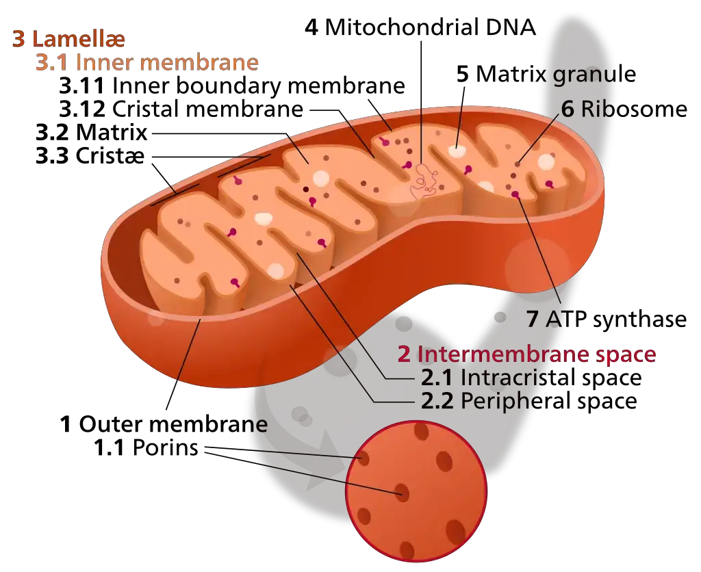
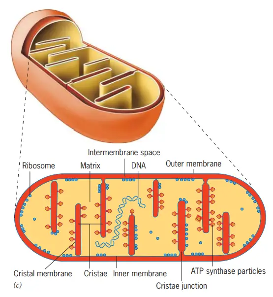
A mitochondrion is made up of two membranes, an outer mitochondrial membrane and an inner mitochondrial membrane. The outer membrane is smooth. Inner membrane is folded into many structures called cristae. Between these two membranes is a narrow intermembrane space. The space inside the inner membrane is known as the mitochondrial matrix.
The matrix contains different metabolic enzymes, mitochondrial ribosomes and mitochondrial DNA (mtDNA). Many enzymes required for the Tricarboxylic Acid (TCA) cycle are also present here. On the other hand, the Electron Transport Chain (ETC) and ATP synthase are located in the inner mitochondrial membrane.
During cellular respiration, electrons from NADH and FADH₂ pass through the electron transport chain. During this process, protons are transported from the matrix to the intermembrane space and a proton gradient is formed. This gradient is then used by ATP synthase. ATP is produced. Oxygen acts as the final electron acceptor and water is formed at the end. This process is referred to as oxidative phosphorylation.
Mitochondria also have their own DNA and ribosomes. But they are not completely independent. Most of the proteins required by mitochondria are encoded by nuclear genes and then transported into the organelle.
The major function of mitochondria is energy production. It also takes part in calcium regulation, different metabolic reactions, formation of reactive oxygen species (ROS) and regulation of apoptosis. Mitochondria are dynamic organelles. They can divide and fuse, and their shape and position inside the cell also changes according to cellular condition.
Characteristics of Mitochondria
- Double membrane – Mitochondria are surrounded by two membranes. The outer membrane is comparatively smooth, while the inner membrane folds inward forming cristae.
- Own DNA – They possess mitochondrial DNA (mtDNA). In animals, it is generally a circular DNA molecule and contains only a small part of the genetic information required for mitochondrial functions.
- Ribosomes – Mitochondria also contain their own ribosomes. These are used for synthesis of proteins encoded by mtDNA.
- Semi-autonomous – They have their own genetic system, but are not completely independent. Most mitochondrial proteins are encoded by nuclear genes and imported into the mitochondria.
- Variable form – The shape and number of mitochondria are not fixed. They can occur as small spherical or elongated structures and may also form a connected mitochondrial network within the cell.
- Dynamic nature – Mitochondria continuously change their shape. Fission divides mitochondria, whereas fusion joins mitochondrial membranes and their contents.
- Cristae – The inner membrane forms cristae which increase its membrane surface and contain important components required for oxidative phosphorylation. Their shape and organization can also change according to mitochondrial condition.
- Energy production – Mitochondria are the major site of aerobic ATP formation in most eukaryotic cells. The respiratory chain and ATP synthase are present in the inner mitochondrial membrane.
Where Mitochondria Are Found and Why Their Number Varies
Mitochondria are present in most eukaryotic cells, but their number is not same in every cell. It can range from few mitochondria to a large mitochondrial population, and in many cells they also remain connected as a network. Their amount and arrangement change with cell type and physiological condition.
Mitochondrial Abundance in Different Cell Types
- Cell requirement – Mitochondrial abundance varies according to the function and metabolic activity of cells. Cells depending strongly on aerobic ATP production generally have greater mitochondrial content.
- Cardiac muscle – Cardiomyocytes contain a large number of mitochondria. These cells contract continuously and need a constant supply of ATP.
- Skeletal muscle – Mitochondrial content is also high in many skeletal muscle fibres, but it is not equal in all muscle types. It can change with muscle activity, fibre type and metabolic demand.
- Neurons – Neurons have many mitochondria distributed through the cell body, axons and dendrites. Some are transported for long distances. At active synaptic regions they provide ATP and also help in handling Ca²⁺.
- Low abundance – Cells having less dependence on mitochondrial respiration may contain fewer mitochondria. Mature mammalian red blood cells are an extreme case, they lose mitochondria during maturation.
The number is therefore not permanently fixed. Mitochondrial biogenesis, division and removal can change the mitochondrial population when cellular conditions are changed.
Shape and Distribution Inside Cells
- Variable shape – Mitochondria are not always small oval structures as commonly shown in diagrams. They may be spherical, short rods, elongated tubules or highly branched structures. Sometimes an extensive mitochondrial network is formed.
- Fission and fusion – Their shape changes continuously by mitochondrial fission and fusion. The balance between these processes determines whether mitochondria remain fragmented or form long interconnected networks.
- Cell position – Mitochondria are also not distributed randomly in all cells. They can be moved and retained at particular regions where ATP production or Ca²⁺ regulation is required.
- Neuronal distribution – In neurons, mitochondria move along cytoskeletal tracks from the cell body into axons and other distant regions. Many become positioned near active synapses having immediate energy and calcium-handling requirements.
- Muscle arrangement – Muscle mitochondria have a more organized distribution. In skeletal muscle they can form interconnected mitochondrial structures around the contractile system, helping to support energy use throughout the fibre.
Their shape, number and position can therefore change even within the same type of cell under different physiological conditions.
Structure of a Mitochondrion
- Membranes – Mitochondria have a double membrane system. It consists of an outer mitochondrial membrane and an inner mitochondrial membrane.
- Outer membrane – It forms the outer covering of mitochondrion. The membrane is comparatively smooth and contains different proteins including Voltage-Dependent Anion Channels (VDACs), which allow passage of many small ions and metabolites.
- Intermembrane space – It is the narrow space present between outer and inner mitochondrial membranes.
- Inner membrane – The inner membrane is folded towards the inner side. It is less permeable to ions and contains different transport proteins, respiratory chain complexes and ATP synthase.
- Cristae – The folds of inner mitochondrial membrane are called cristae. They extend into the mitochondrial matrix. Their number and arrangement vary in different cells, and cristae are connected with the inner boundary membrane through narrow regions called crista junctions.
- Matrix – The inner membrane encloses a central region called the mitochondrial matrix. It contains different soluble enzymes and metabolites. Most of the enzymes of the Tricarboxylic Acid (TCA) cycle are present here.
- mtDNA – The matrix contains mitochondrial DNA (mtDNA). It occurs with associated proteins forming structures called mitochondrial nucleoids.
- Ribosomes – Mitochondrial ribosomes are also present in the matrix. They are involved in synthesis of proteins encoded by mitochondrial DNA.
Mitochondrial DNA
Mitochondrial DNA (mtDNA) is the genetic material present inside mitochondria. It is located mainly in the mitochondrial matrix. Unlike nuclear DNA, several copies of mtDNA can occur in a single cell.
- In humans, mitochondrial DNA is a small circular, double-stranded DNA molecule. It contains 16,569 base pairs.
- There are 37 genes in human mtDNA, among which 13 genes encode proteins required for oxidative phosphorylation, 22 genes encode transfer RNAs and 2 genes encode ribosomal RNAs.
- Mitochondrial DNA is not present freely throughout the matrix. It occurs together with different proteins forming DNA-protein structures called mitochondrial nucleoids.
- Only a small number of mitochondrial proteins are encoded by mtDNA. Most of the proteins required for mitochondrial structure, replication and other functions are encoded by nuclear genes and are transported into mitochondria after their synthesis.
- A cell contains many mtDNA molecules. Their number is not same in every type of cell and can also change according to mitochondrial content and metabolic condition.
- In humans, mitochondrial DNA is inherited almost completely from the mother. The mtDNA present in the egg is passed to the developing offspring.
- All copies of mitochondrial DNA are not always identical. When only one mtDNA type is present, this condition is called homoplasmy. Presence of more than one mtDNA variant within the same cell or individual is referred to as heteroplasmy.
- mtDNA can undergo its own replication. Its replication is not strictly linked with nuclear DNA replication, although proteins encoded by nuclear genes are required for this process.
- Mitochondria also have their own transcription and translation system. Mitochondrial genes are transcribed inside the organelle and the encoded proteins are synthesized by mitochondrial ribosomes.
- Mutations can occur in mtDNA. Some mutations have little effect when present in low proportion, but when mutant mtDNA becomes sufficiently high, mitochondrial functions can be affected and this is especially important in tissues having greater energy requirement.
How Mitochondria Make ATP
The major amount of ATP in mitochondria is produced by oxidative phosphorylation. It takes place in the inner mitochondrial membrane. Electrons from NADH and FAD-linked reactions are passed through the electron transport chain, and the released energy is first stored in the form of a proton gradient. The following are the steps-
Step 1
NADH is oxidized at Complex I. The electrons are transferred through different electron carriers and finally reach Coenzyme Q (CoQ). During this process, H⁺ ions are moved from the matrix to the intermembrane space.
NAD⁺ is formed.
Step 2
Electrons also enter through Complex II, which is the enzyme succinate dehydrogenase of the TCA cycle. These electrons are transferred to CoQ. Complex II does not pump H⁺ across the membrane.
Step 3
CoQ receives electrons and becomes reduced to CoQH₂ (ubiquinol). It then carries these electrons to Complex III.
In this step, electrons are passed further to cytochrome c and H⁺ ions are transferred towards the intermembrane space.
Step 4
Cytochrome c carries the electrons to Complex IV. Oxygen acts as the final electron acceptor. It receives electrons and together with protons forms water.
Complex IV also contributes to proton movement across the inner membrane.
Step 5
As electron transfer continues, Complexes I, III and IV move H⁺ from the mitochondrial matrix to the intermembrane space. Complex II does not.
A high amount of H⁺ is therefore accumulated on the outer side of inner mitochondrial membrane, while the matrix has a lower H⁺ concentration. An electrical difference is also present. This forms the proton-motive force.
Step 6
The accumulated H⁺ ions now flow back into the matrix through ATP synthase (Complex V).
This proton movement causes rotation of the membrane portion of ATP synthase and changes the catalytic portion of the enzyme. The stored energy of proton gradient is used here.
Step 7
ADP and inorganic phosphate (Pi) are converted into ATP by ATP synthase.
ADP + Pi → ATP
ATP is formed on the matrix side of the inner mitochondrial membrane.
Step 8
For continuous ATP production, ADP and phosphate must enter the matrix. ATP formed inside is transported out in exchange for ADP, while phosphate enters through its own carrier. Another cycle of ATP formation can then occur.
Evolutionary Origin of Mitochondria
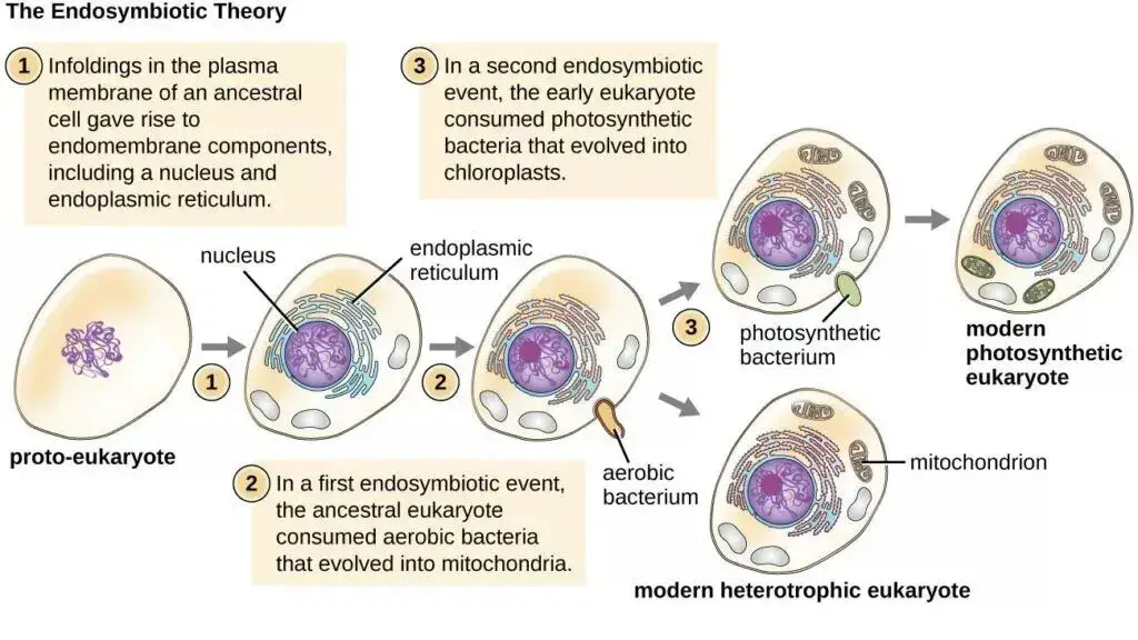
- Endosymbiosis – Mitochondria are considered to have originated from a bacterial endosymbiont. The bacterium entered into a stable association with an ancestral host cell and was gradually converted into the mitochondrial organelle.
- Bacterial ancestor – The mitochondrial ancestor was related to Alphaproteobacteria. Its exact position among the presently known alphaproteobacterial groups is still uncertain, with different phylogenetic studies placing mitochondria either within or close to this bacterial lineage.
- Host cell – The host had an archaeal ancestry. Current studies strongly connect the eukaryotic lineage with Asgard archaea, although the exact nature of the ancestral host and the sequence of early events during eukaryogenesis are still under study.
- Permanent association – The bacterial cell was no longer an independent organism after long evolutionary integration. Many of its original functions were lost or transferred to the host, while respiratory and other metabolic functions became part of the developing mitochondrion.
- Gene transfer – A large number of genes from the mitochondrial ancestor were transferred to the nuclear genome. The mitochondrial genome was therefore greatly reduced. Most proteins required by present mitochondria are now encoded by nuclear genes and transported back into the organelle.
- Evidence – Mitochondria still contain their own DNA and genetic machinery. Sequence similarities between mitochondrial genes and bacterial genes, together with their bacterial-type ancestry, provide major evidence for an endosymbiotic origin.
- Common origin – Mitochondria are generally considered to have originated from a single ancient endosymbiotic event. By the time of the Last Eukaryotic Common Ancestor (LECA), a mitochondrion or mitochondrion-derived system was already present.
The Endosymbiotic Hypothesis
The endosymbiotic hypothesis explains the origin of mitochondria from an ancestral bacterium that became permanently associated with an early host cell. With time, the bacterium lost its independent existence and developed into the mitochondrion.
- Bacterial ancestor – The ancestor of mitochondria was related to Alphaproteobacteria. Its exact position among known alphaproteobacterial groups is still debated.
- Host association – An ancestral bacterial cell entered into a close and stable association with the host. The early host had an archaeal-related ancestry. The exact cellular events by which this association first developed are not completely known.
- Endosymbiosis – The bacterium remained inside the host rather than continuing as an independent cell. A permanent endosymbiotic relationship was established.
- Energy benefit – The bacterial partner had an efficient system of oxidative phosphorylation. Its respiratory machinery became useful for energy metabolism of the developing eukaryotic cell.
- Gene transfer – During long evolutionary association, many bacterial genes were transferred to the host nuclear genome. Some genes were also lost. Therefore, present mitochondria retain only a small part of the original bacterial genetic system.
- Organelle formation – The endosymbiont gradually became dependent on the host cell. Proteins encoded by transferred nuclear genes were now transported back into mitochondria, producing a highly integrated cellular organelle.
- Common ancestry – Mitochondria of modern eukaryotes are generally considered to descend from this ancient endosymbiotic event. The mitochondrial system was already established in the ancestor from which the major present-day eukaryotic lineages developed.
Evidence for Endosymbiotic Origin
The endosymbiotic origin of mitochondria is supported by several bacterial-like features. The following are some of the important evidences-
- Own DNA – Mitochondria contain their own mitochondrial DNA (mtDNA), separate from the nuclear DNA. In most animals it is circular. The genome is much smaller now.
- Bacterial genes – Mitochondrial genes show their evolutionary relationship with bacteria. Phylogenetic studies associate the mitochondrial ancestor with Alphaproteobacteria, although its exact closest living bacterial group is not certain.
- Ribosomes – They also have their own ribosomes. These mitoribosomes are used for synthesis of proteins encoded by mtDNA, and several parts of mitochondrial translation have a bacterial origin.
- Division – New mitochondria are produced from pre-existing mitochondria by growth and division. They are not formed newly from other cell organelles.
- Double membrane – Mitochondria have an outer and an inner membrane. Such a membrane arrangement is consistent with the engulfment and retention of an ancestral bacterial cell.
- Gene transfer – A large number of genes of the original endosymbiont were transferred to the nuclear genome during evolution. Therefore, most proteins required by present mitochondria are now encoded in the nucleus. After synthesis in cytosol, they are transported back into mitochondria.
- Respiratory system – The inner mitochondrial membrane contains the electron transport chain and ATP synthase. Many components of this respiratory system are evolutionarily related to those present in bacteria.
- Antibiotic effect – Some antibiotics that act on bacterial translation can also inhibit mitochondrial protein synthesis. This similarity occurs because parts of the mitochondrial translation system came from its bacterial ancestor.
- Reduced genome – Present mitochondrial DNA contains only a small fraction of the genes possessed by the original bacterial ancestor. Many genes were lost, while others moved to the nucleus during long endosymbiotic association.
Functions of Mitochondria
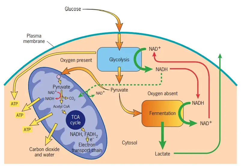
- ATP production – The major function of mitochondria is production of ATP. Most of the ATP during aerobic respiration is formed here by oxidative phosphorylation. The electron transport chain and ATP synthase are involved in this process.
- TCA cycle – The TCA cycle takes place in the mitochondrial matrix. During this cycle, acetyl-CoA is oxidized and NADH, FADH₂ are formed. These are further used in electron transport chain.
- Fat oxidation – Mitochondria are also the major site for β-oxidation of fatty acids. During this process, fatty acids are broken down into acetyl-CoA units along with formation of NADH and FADH₂.
- Calcium regulation – Mitochondria can take up and release Ca²⁺. It is important in maintaining cellular calcium level and also affects different metabolic activities of the cell.
- ROS production – The electron transport chain produces small amount of reactive oxygen species (ROS) during normal respiration. These molecules can act in cellular signalling, while excess formation may damage cellular components.
- Apoptosis – Mitochondria have an important role in apoptosis or programmed cell death. During intrinsic apoptosis, cytochrome c is released from mitochondria into cytoplasm and activates the further reactions of cell death.
- Heme synthesis – Some steps of heme synthesis occur in mitochondria. The first reaction and the final reactions of this pathway take place in mitochondrial compartments.
- Heat production – In brown adipose tissue, mitochondria are also used for heat production. UCP1 allows protons to return across the inner mitochondrial membrane without normal ATP formation. Energy is released as heat during this process.
Mitochondrial Dysfunction and Clinical Significance
Mitochondrial dysfunction refers to the condition in which mitochondria fail to perform their normal functions properly. It mainly affects energy production. Defects in oxidative phosphorylation can decrease the formation of ATP, which is more serious in cells having greater energy requirement.
Some of the important effects and clinical significance are-
- Energy failure – Defective electron transport and oxidative phosphorylation decrease ATP production. Cells may not get sufficient energy for their normal activities.
- Genetic defects – Mitochondrial disorders can result from mutations in mtDNA or nuclear genes associated with mitochondrial functions. A large number of genes can be involved, hence the clinical features are also highly variable.
- High-energy tissues – Brain, skeletal muscles, heart and nerves are commonly affected. These tissues require continuous energy supply and are more sensitive to mitochondrial defects.
- Neurological effects – Seizures, neuropathy, ataxia and encephalopathy can occur in mitochondrial diseases. Some disorders also produce stroke-like episodes.
- Muscle defects – Muscle weakness and exercise intolerance are common. In some patients, myopathy becomes one of the major clinical features.
- Heart involvement – Mitochondrial dysfunction can also affect cardiac muscles. Cardiomyopathy is present in several mitochondrial disorders.
- ROS formation – Abnormal electron transport may increase the formation of reactive oxygen species (ROS). Excess ROS can damage proteins, membrane lipids and DNA, including mitochondrial DNA.
- Calcium balance – Mitochondria are involved in cellular calcium regulation. Defective mitochondrial function can disturb this balance and affects different cellular processes.
- Cell death – Mitochondria also take part in the intrinsic pathway of apoptosis. Abnormal regulation of this process may result in excessive cell death or failure to remove damaged cells.
- Other symptoms – Hearing loss, visual abnormalities, ptosis, external ophthalmoplegia and diabetes can also occur. The symptoms are not same in every patient.
- Secondary dysfunction – Mitochondrial abnormalities are also observed in several diseases that are not primary mitochondrial disorders. Neurodegenerative diseases are some important examples, where mitochondrial dysfunction can form one part of the disease process.
- Clinical variation – A mitochondrial disease may affect only one tissue or several organ systems together. Age of onset and severity also vary considerably, which can make their diagnosis difficult.
References
- Ahmad, M., Wolberg, A., & Kahwaji, C. I. (2023). Biochemistry, electron transport chain. In StatPearls. StatPearls Publishing. https://www.ncbi.nlm.nih.gov/books/NBK526105/
- Alberts, B., Johnson, A., Lewis, J., Raff, M., Roberts, K., & Walter, P. (2002). The genetic systems of mitochondria and plastids. In Molecular biology of the cell (4th ed.). Garland Science. https://www.ncbi.nlm.nih.gov/books/NBK26924/
- Barbagallo, F., La Vignera, S., Cannarella, R., Aversa, A., Calogero, A. E., & Condorelli, R. A. (2020). Evaluation of sperm mitochondrial function: A key organelle for sperm motility. Journal of Clinical Medicine, 9(2), 363. https://doi.org/10.3390/jcm9020363
- Boore, J. L. (1999). Animal mitochondrial genomes. Nucleic Acids Research, 27(8), 1767–1780. https://doi.org/10.1093/nar/27.8.1767
- Deshpande, O. A., & Mohiuddin, S. S. (2023). Biochemistry, oxidative phosphorylation. In StatPearls. StatPearls Publishing. https://www.ncbi.nlm.nih.gov/books/NBK553192/
- Harrington, J. S., Ryter, S. W., Plataki, M., Price, D. R., & Choi, A. M. K. (2023). Mitochondria in health, disease, and aging. Physiological Reviews, 103(4), 2349–2422. https://doi.org/10.1152/physrev.00058.2021
- Hessenberger, M., Zerbes, R. M., Rampelt, H., Kunz, S., Xavier, A. H., Purfürst, B., Lilie, H., Pfanner, N., van der Laan, M., & Daumke, O. (2017). Regulated membrane remodeling by Mic60 controls formation of mitochondrial crista junctions. Nature Communications, 8, 15258. https://doi.org/10.1038/ncomms15258
- Ogun, A. S., Joy, N. V., & Valentine, M. (2023). Biochemistry, heme synthesis. In StatPearls. StatPearls Publishing. https://www.ncbi.nlm.nih.gov/books/NBK537329/
- Osellame, L. D., Blacker, T. S., & Duchen, M. R. (2012). Cellular and molecular mechanisms of mitochondrial function. Best Practice & Research Clinical Endocrinology & Metabolism, 26(6), 711–723. https://doi.org/10.1016/j.beem.2012.05.003
- Pfeiffer, K., Gohil, V., Stuart, R. A., Hunte, C., Brandt, U., Greenberg, M. L., & Schägger, H. (2003). Cardiolipin stabilizes respiratory chain supercomplexes. Journal of Biological Chemistry, 278(52), 52873–52880. https://doi.org/10.1074/jbc.M308366200
- Pinke, G., Zhou, L., & Sazanov, L. A. (2020). Cryo-EM structure of the entire mammalian F-type ATP synthase. Nature Structural & Molecular Biology, 27, 1077–1085. https://doi.org/10.1038/s41594-020-0503-8
- Rossmann, M. P., Dubois, S. M., Agarwal, S., & Zon, L. I. (2021). Mitochondrial function in development and disease. Disease Models & Mechanisms, 14(6), dmm048912. https://doi.org/10.1242/dmm.048912
- Spinelli, J. B., & Haigis, M. C. (2018). The multifaceted contributions of mitochondria to cellular metabolism. Nature Cell Biology, 20, 745–754. https://doi.org/10.1038/s41556-018-0124-1
- Stewart, J. B., & Chinnery, P. F. (2015). The dynamics of mitochondrial DNA heteroplasmy: Implications for human health and disease. Nature Reviews Genetics, 16, 530–542. https://doi.org/10.1038/nrg3966
- Tábara, L.-C., Segawa, M., & Prudent, J. (2025). Molecular mechanisms of mitochondrial dynamics. Nature Reviews Molecular Cell Biology, 26, 123–146. https://doi.org/10.1038/s41580-024-00785-1
- Vafai, S. B., & Mootha, V. K. (2012). Mitochondrial disorders as windows into an ancient organelle. Nature, 491, 374–383. https://doi.org/10.1038/nature11707
- Wiedemann, N., & Pfanner, N. (2017). Mitochondrial machineries for protein import and assembly. Annual Review of Biochemistry, 86, 685–714. https://doi.org/10.1146/annurev-biochem-060815-014352
- Zick, M., Rabl, R., & Reichert, A. S. (2009). Cristae formation—Linking ultrastructure and function of mitochondria. Biochimica et Biophysica Acta (BBA) – Molecular Cell Research, 1793(1), 5–19. https://doi.org/10.1016/j.bbamcr.2008.06.013
- Zong, S., Wu, M., Gu, J., Liu, T., Guo, R., & Yang, M. (2018). Structure of the intact 14-subunit human cytochrome c oxidase. Cell Research, 28, 1026–1034. https://doi.org/10.1038/s41422-018-0071-1