

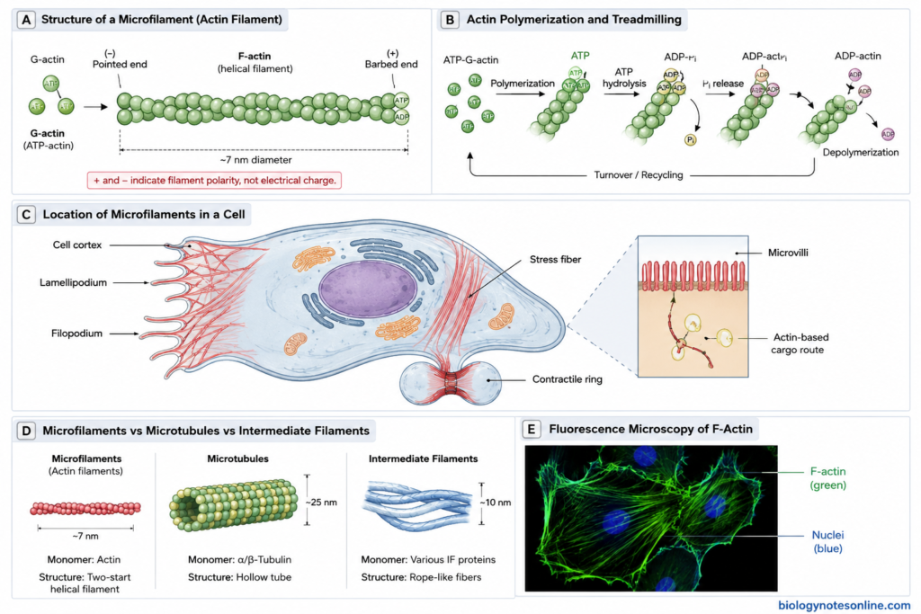
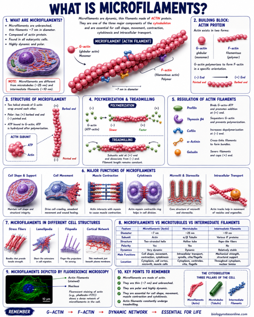
Microfilaments, also known as actin filaments, are thin protein filaments present as a part of the cytoskeleton in eukaryotic cells. They are made up of actin protein. Actin is present as globular actin (G-actin) which joins together to form filamentous actin (F-actin). These actin filaments are about 7 nm in diameter.
The actin filaments are polar structures having two different ends. These filaments are not fixed structures and their formation and breakdown takes place continuously inside the cell. Microfilaments have a role in maintaining the cell shape, movement of cells and division of the cell cytoplasm during cytokinesis. They also interact with myosin and take part in different contractile movements including muscle contraction.
What Are Microfilaments?
Microfilaments or actin filaments are thin filaments of the cytoskeleton present in eukaryotic cells. They are about 7 nm in diameter. These filaments are mainly made up of actin protein. Actin occurs as globular G-actin, and these actin molecules join to form filamentous F-actin.
Microfilaments are polar filaments with two different ends. They are also highly dynamic because actin molecules are continuously added and removed from the filament. These filaments have a major role in maintaining cell shape and movement of the cell. They are also involved in cytokinesis, formation of different cell surface structures and contraction with myosin.
| Quick facts | Microfilaments |
|---|---|
| Other name | Actin filaments |
| Diameter | About 7 nm |
| Main protein | Actin |
| Forms of actin | G-actin and F-actin |
| Nature | Polar and dynamic |
| Present in | Eukaryotic cells |
| Major functions | Cell shape, cell movement, cytokinesis and contraction |
Structure of Microfilaments
The following are the main structural features of microfilaments–
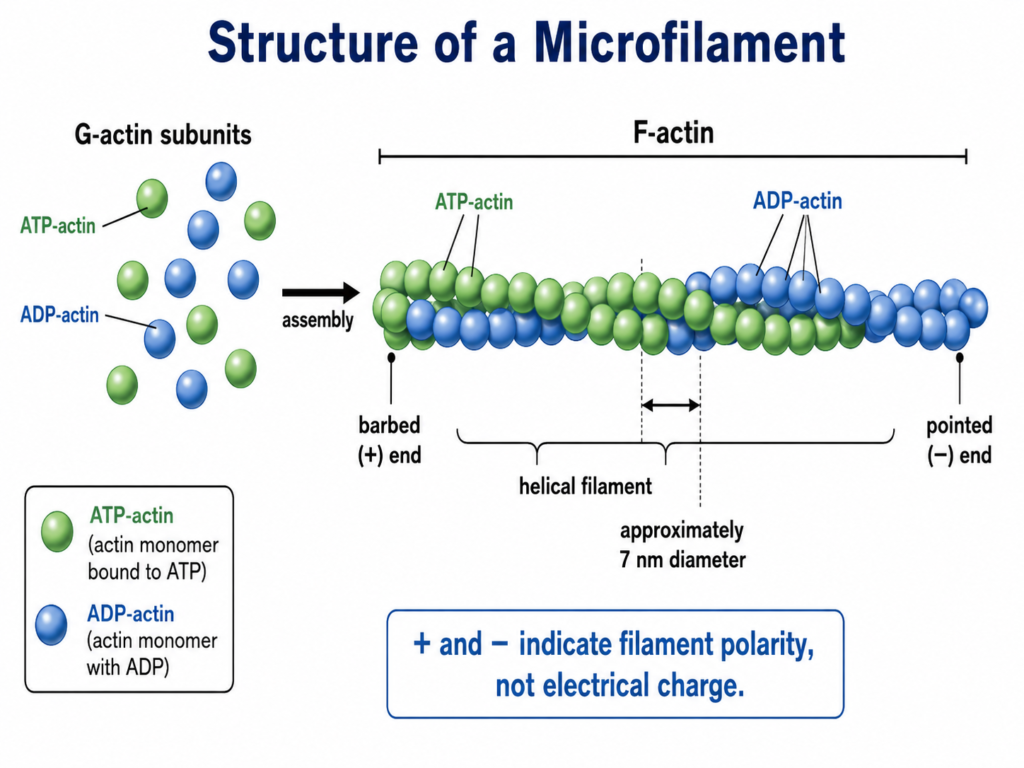
- Microfilaments are very thin fibers of the cytoskeleton, about 7 nm in diameter. They are mainly formed of actin protein.
- Actin occurs in two forms, globular actin (G-actin) and filamentous actin (F-actin). G-actin is the individual globular unit. Many of these units polymerize together to produce F-actin.
- The F-actin is made up of two chains of actin molecules which are twisted around one another forming a helical filament, giving the characteristic thin and thread-like appearance of microfilaments.
- All the actin units are arranged in the same direction. Because of this arrangement, two different ends are formed in an actin filament.
- One end is called barbed or (+) end, while another is called pointed or (−) end. Addition of actin units takes place more rapidly at the (+) end. The filament is therefore polar in nature.
- Each G-actin molecule has a site for binding ATP. During formation of F-actin, ATP-actin is incorporated into the growing filament and ATP is hydrolyzed after its addition. Addition and loss of actin units makes these filaments highly dynamic.
- Inside the cells, microfilaments are not always present as single fibers. They may form parallel bundles, contractile bundles or a network of filaments with the help of different actin-binding proteins.
- A dense network of actin filaments is present just below the plasma membrane in many cells. This region forms the cell cortex, which provides mechanical support to the cell surface and is associated with changes in cell shape.

How Microfilaments Form and Remodel
Formation of microfilaments takes place by the polymerization of actin molecules. It is a highly dynamic process. The different steps involved in their formation and remodeling are as follows-
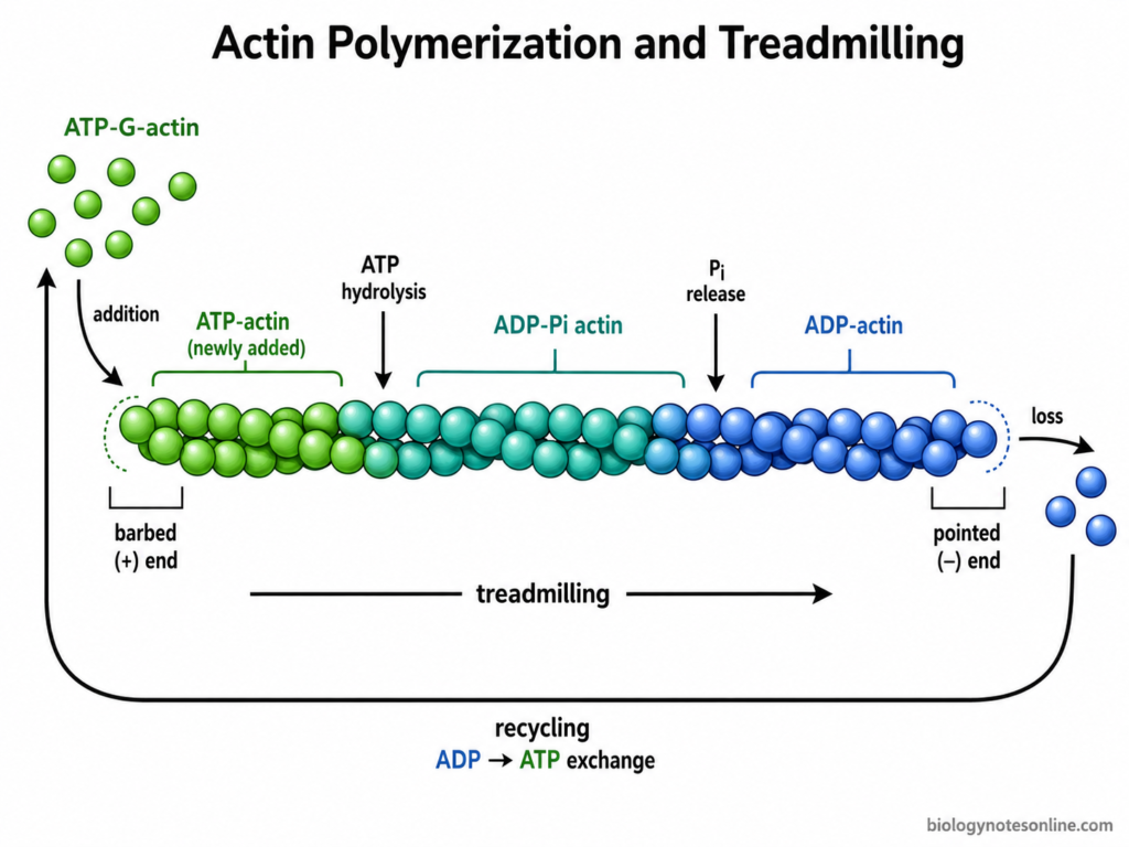
- At first, globular actin or G-actin molecules bind ATP and a few of them come together. A small stable group of actin subunits is formed. This initial process is called nucleation.
- After the formation of nucleus, more G-actin molecules are added and the filament begins to increase in length. This is called elongation. Addition of actin takes place more rapidly at the barbed (+) end, while it is slower at the pointed (−) end.
- ATP present in the newly added actin is hydrolyzed after polymerization. Thus, the older portion of an actin filament generally contains more ADP-actin. These regions are less stable and can be removed more easily during filament turnover.
- Sometimes actin molecules are added at one end and removed from another end of the same filament. This process is referred to as treadmilling. It allows continuous replacement of actin subunits without complete disappearance of the filament.
- The formation of actin filaments inside a cell is controlled by several proteins. Arp2/3 complex helps in nucleation and formation of branched actin networks. Formins are mainly associated with the formation of long unbranched actin filaments.
- Remodeling also involves cutting and breakdown of already formed filaments. Cofilin binds mainly to older ADP-rich regions of F-actin, increasing filament severing and disassembly. In this way old actin can be rapidly removed and reused.
- Some actin-binding proteins cap the ends and prevent further addition or loss of actin. Other proteins cross-link the filaments and arrange them into bundles or networks. The arrangement is therefore not permanent.
- During cell movement, change in cell shape or cytokinesis, actin filaments may be rapidly formed at one region and removed from another. This continuous formation, breakdown and rearrangement of actin filaments is known as actin remodeling.
How Cells Control Microfilament Behavior
The behavior of microfilaments inside the cell is controlled by different actin-binding proteins and signaling molecules. These control where actin filaments are formed, their growth and breakdown. In this way, the arrangement of actin can be changed rapidly according to the activity of the cell.
Actin polymerization is also controlled by the availability of G-actin. Profilin binds with actin monomers and helps their addition into growing filaments, especially during formin-dependent growth. Thymosin-β4, on the other hand, binds free actin and maintains a reserve pool of actin monomers inside the cell.
Different proteins control the type of actin structure formed. Formins are mainly involved in formation of long unbranched filaments. The Arp2/3 complex helps in formation of branched actin networks from pre-existing filaments. Thus, actin may be arranged differently in different parts of the same cell.
The length of microfilaments is not permanent. Capping proteins can bind to filament ends and stop further growth. Cofilin acts mainly on older ADP-actin regions, where it promotes cutting and disassembly of the filaments. Actin units released from these filaments can again be used for formation of new microfilaments.
Cell signals also regulate these changes. Rho family GTPases are important regulators of actin organization. RhoA is associated with formation of stress fibers and actomyosin contraction, Rac1 mainly promotes lamellipodia, while Cdc42 is involved in formation of filopodia. Their activity allows the cell to change its actin cytoskeleton during movement, adhesion, cytokinesis and changes in cell shape.
Actin-Binding Proteins and Regulation
Actin-binding proteins (ABPs) control the formation, breakdown and arrangement of microfilaments inside the cell. Some proteins help in filament formation, while others cut, cap or organize the already formed actin filaments. The major actin-binding proteins include-
| Actin-binding protein | Role in microfilaments |
|---|---|
| Arp2/3 complex | It is a seven-subunit protein complex involved in the formation of branched actin filaments. Arp2/3 binds to the side of an already existing actin filament and nucleates a new daughter filament. This produces the characteristic branched actin network. High-resolution cryo-EM studies have shown its arrangement at the actin branch junction. |
| Formins | Formins help in nucleation and growth of mainly unbranched actin filaments. After nucleation, the formin remains associated with the growing barbed end. Its FH1 region also interacts with profilin-actin and helps rapid addition of new actin units. |
| Profilin | Profilin is an actin monomer-binding protein. It binds G-actin and maintains actin in a form that can be supplied to growing filaments. Profilin-actin is particularly important during formin-mediated filament elongation. |
| Cofilin | Cofilin is mainly involved in turnover of old actin filaments. It binds preferentially to ADP-rich regions of F-actin and promotes filament severing and disassembly. More filament ends are produced by this process, increasing the rate of actin turnover. |
| Capping proteins | These proteins bind to the ends of actin filaments. The common capping protein binds the barbed (+) end and prevents further addition or loss of actin subunits from that end. In this way, the length and growth of the filament can be controlled. |
| Fascin and α-actinin | These proteins are involved in organization of F-actin into bundles. Fascin forms tightly packed actin bundles, such as those present in filopodia. α-Actinin produces more widely spaced cross-linked actin bundles and is commonly associated with contractile actin structures. |
| Myosin | Myosin is an actin-associated molecular motor. It uses energy from ATP and moves along actin filaments, producing mechanical force. Actin-myosin interaction is involved in muscle contraction, cytokinesis and several forms of cell movement. |
Where Are Microfilaments Found?
Microfilaments are present throughout the cytoplasm of eukaryotic cells. Their distribution is not uniform. In many cells, a large amount of actin is found near the plasma membrane, while other actin filaments form bundles or special structures in different regions of the cell.
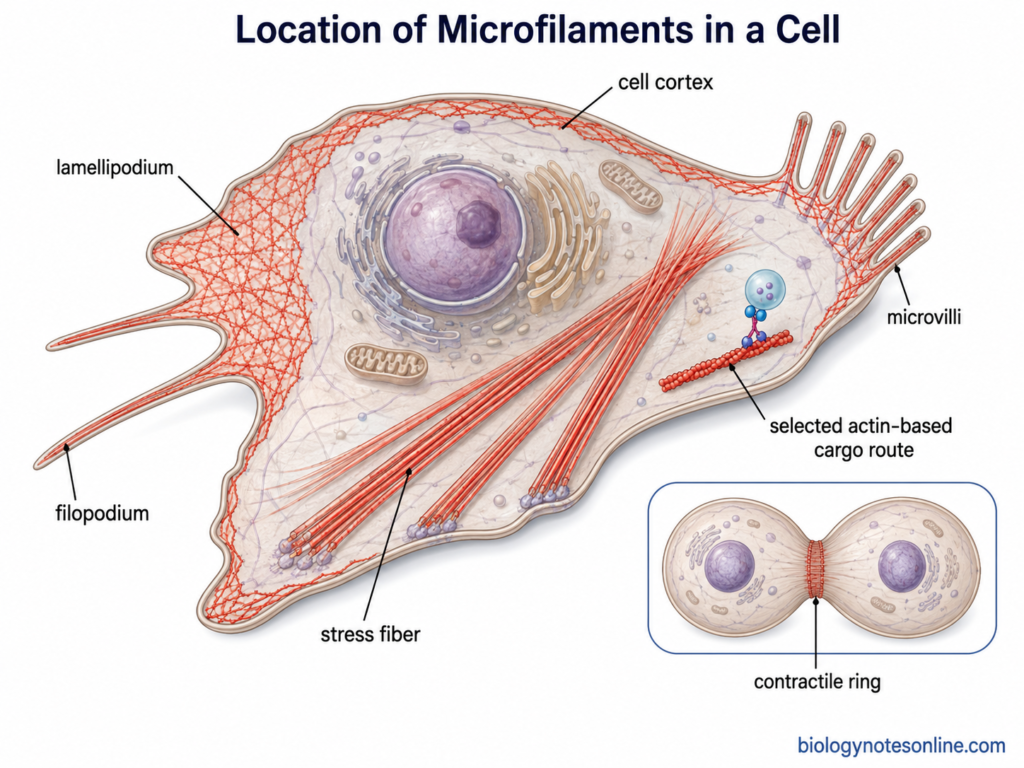
The important locations of microfilaments include-
- Cell cortex – A dense network of actin filaments is present just below the plasma membrane. This region is called the cell cortex. It provides support to the cell surface and is involved in change of cell shape.
- Lamellipodia – These are broad sheet-like extensions, mainly formed at the leading region of migrating cells. They contain a dense branched network of actin filaments.
- Filopodia – Actin is also present in the thin finger-like projections called filopodia. Here the filaments are arranged mainly as long parallel bundles.
- Microvilli – Microvilli have bundles of actin filaments in their central region. These filaments provide structural support to the projections. They are especially prominent in cells having an absorptive surface, such as intestinal epithelial cells.
- Stress fibers – In many adherent non-muscle cells, actin and myosin form contractile bundles called stress fibers. These bundles extend through the cytoplasm and are commonly connected with sites of cell attachment.
- Contractile ring – During cytokinesis of animal cells, actin filaments together with myosin II accumulate around the middle of the cell. They form a contractile ring. Contraction of this ring helps in separation of the two daughter cells.
- Muscle contractile structures – Actin forms the thin filaments of muscle cells. In striated muscle, these filaments are arranged in sarcomeres and interact with myosin during muscle contraction.
- Plant cells – Actin filaments are also widely present in plant cells. They occur as cortical arrays, individual filaments and bundles in the cytoplasm. These actin arrays are highly dynamic and take part in different cellular activities. In Arabidopsis thaliana, highly dynamic actin filaments have been observed in the cortical cytoplasm of epidermal cells.
Functions of Microfilaments
The major functions of microfilaments are as follows-
- Maintenance of cell shape – Microfilaments form a network below the plasma membrane known as the cell cortex. It provides mechanical support to the cell surface. Changes in this actin network can also change the shape of the cell.
- Cell movement – Actin filaments have an important role in movement of cells. Polymerization of actin pushes the plasma membrane forward and forms structures such as lamellipodia and filopodia. These structures are commonly present at the leading region of migrating cells.
- Muscle contraction – In muscle cells, actin forms the thin filaments of the contractile apparatus. These filaments interact with myosin and produce contraction. ATP energy is used by myosin during this process.
- Cytokinesis – Microfilaments take part in separation of animal cells after nuclear division. Actin filaments together with myosin II form a contractile ring around the middle of the cell. Contraction of this ring produces the cleavage furrow and finally separates the two daughter cells.
- Cell adhesion – Actin filaments are connected with different sites of cell attachment. They produce tension and help the cell to remain attached with other cells or with the extracellular matrix. Actin is also present in adhesion belts of epithelial cells.
- Support of cell surface projections – Microfilaments form the internal supporting framework of structures such as microvilli. In intestinal epithelial cells, bundles of actin extend through the core of each microvillus and help in maintaining its shape.
- Intracellular transport – Actin filaments can act as tracks for movement of vesicles and organelles with the help of myosin motors. This type of transport is especially important in many plant cells, where actin-myosin interaction also produces rapid movement of organelles and cytoplasmic streaming.
- Phagocytosis and membrane movement – Rearrangement of actin takes place during extension of pseudopodia and engulfment of particles. The plasma membrane is pushed around the particle and helps in its internalization by the cell.
- Cell growth and development in plants – Actin filaments are involved in cell growth, cytokinesis and intracellular trafficking in plant cells. Their arrangement continuously changes according to the growth and activity of the cell.
Microfilaments vs Microtubules vs Intermediate Filaments
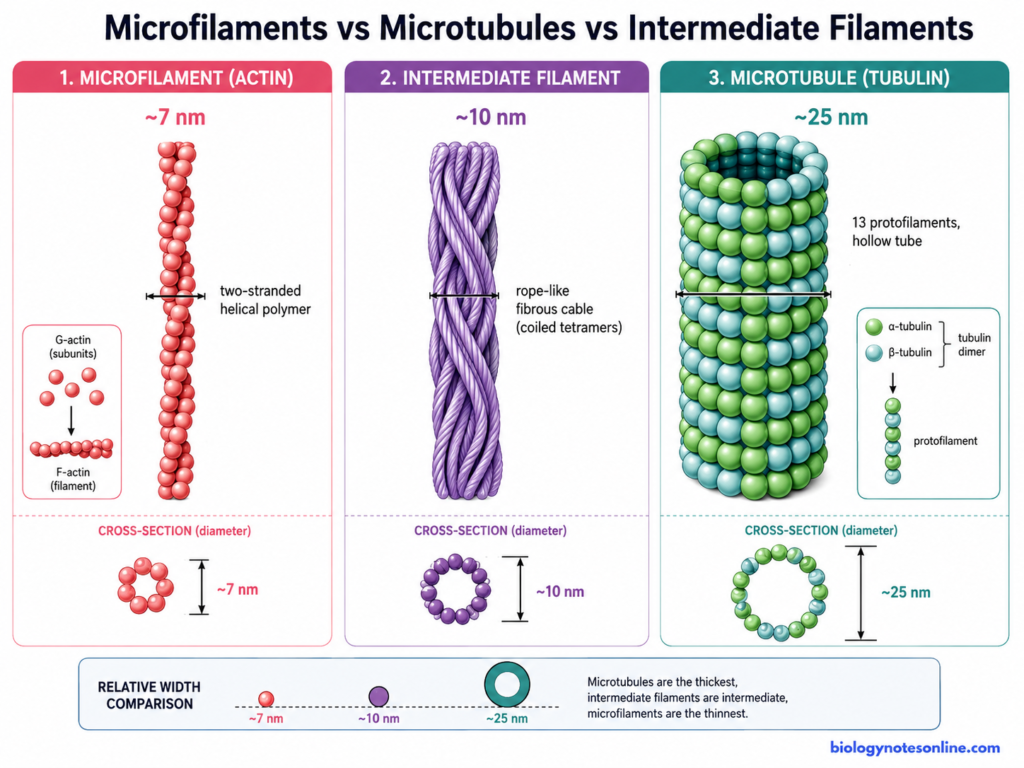
Microfilaments, microtubules and intermediate filaments are the three major filament systems of the cytoskeleton. They differ in their size, protein composition and functions. The major differences are as follows-
| Features | Microfilaments | Microtubules | Intermediate filaments |
|---|---|---|---|
| Diameter | They are the thinnest, about 7 nm in diameter. | These are the largest cytoskeletal filaments, about 25 nm in diameter. | They are about 10 nm in diameter, intermediate in size between actin filaments and microtubules. |
| Main protein | Mainly made up of actin. G-actin units polymerize to form F-actin. | Made up of α- and β-tubulin heterodimers. | Made up of different proteins depending on the cell type, such as keratins, vimentin, desmin, neurofilament proteins and lamins. |
| Basic structure | Thin solid filaments. Actin subunits are arranged into two intertwined strands. | They form hollow tubular structures. | Rope-like fibrous structures. They do not have the hollow structure of microtubules. |
| Polarity | Microfilaments are polar, having a (+) and (−) end. | Microtubules are also polar with (+) and (−) ends. | Intermediate filaments are generally non-polar. |
| Nature | Highly dynamic. Addition and removal of actin units can take place rapidly. | Highly dynamic and may rapidly grow or shrink. | Comparatively more stable and mechanically strong than the other two filament systems. |
| Major functions | Mainly involved in cell shape, cell movement, muscle contraction and cytokinesis. | They have roles in intracellular transport, organization of cell contents and chromosome movement during cell division. Microtubules also form the structural framework of cilia and flagella. | Their major function is mechanical support. They help cells and tissues to withstand stress and also provide structural support to the nucleus. |
| Associated motors | Myosin proteins move along actin filaments. | Kinesin and dynein use microtubules as tracks for movement. | They generally do not serve as tracks for cytoskeletal motor proteins. |
References
- Carlsson, A. E. (2010). Actin dynamics: From nanoscale to microscale. Annual Review of Biophysics, 39, 91–110. https://doi.org/10.1146/annurev.biophys.093008.131207
- Cooper, G. M. (2000). The cell: A molecular approach (2nd ed.). Sinauer Associates. https://www.ncbi.nlm.nih.gov/books/NBK9839/
- Ding, B., Narvaez-Ortiz, H. Y., Singh, Y., Hocky, G. M., Chowdhury, S., & Nolen, B. J. (2022). Structure of Arp2/3 complex at a branched actin filament junction resolved by single-particle cryo-electron microscopy. Proceedings of the National Academy of Sciences of the United States of America, 119(22), e2202723119. https://doi.org/10.1073/pnas.2202723119
- Edwards, M., Zwolak, A., Schafer, D. A., Sept, D., Dominguez, R., & Cooper, J. A. (2014). Capping protein regulators fine-tune actin assembly dynamics. Nature Reviews Molecular Cell Biology, 15(10), 677–689. https://doi.org/10.1038/nrm3869
- Funk, J., Merino, F., Venkova, L., Heydenreich, L., Kierfeld, J., Vargas, P., Raunser, S., Piel, M., & Bieling, P. (2019). Profilin and formin constitute a pacemaker system for robust actin filament growth. eLife, 8, e50963. https://doi.org/10.7554/eLife.50963
- Gautreau, A. M., Fregoso, F. E., Simanov, G., & Dominguez, R. (2022). Nucleation, stabilization, and disassembly of branched actin networks. Trends in Cell Biology, 32(5), 421–432. https://doi.org/10.1016/j.tcb.2021.10.006
- Geitmann, A., & Nebenführ, A. (2015). Navigating the plant cell: Intracellular transport logistics in the green kingdom. Molecular Biology of the Cell, 26(19), 3373–3378. https://doi.org/10.1091/mbc.E14-10-1482
- Goley, E. D., & Welch, M. D. (2006). The ARP2/3 complex: An actin nucleator comes of age. Nature Reviews Molecular Cell Biology, 7(10), 713–726. https://doi.org/10.1038/nrm2026
- Guo, M., Ehrlicher, A. J., Mahammad, S., Fabich, H., Jensen, M. H., Moore, J. R., Fredberg, J. J., Goldman, R. D., & Weitz, D. A. (2013). The role of vimentin intermediate filaments in cortical and cytoplasmic mechanics. Biophysical Journal, 105(7), 1562–1568. https://doi.org/10.1016/j.bpj.2013.08.037
- Narita, A., Oda, T., & Maéda, Y. (2011). Structural basis for the slow dynamics of the actin filament pointed end. The EMBO Journal, 30(7), 1230–1237. https://doi.org/10.1038/emboj.2011.48
- Oosterheert, W., Klink, B. U., Belyy, A., Pospich, S., & Raunser, S. (2022). Structural basis of actin filament assembly and aging. Nature, 611(7935), 374–379. https://doi.org/10.1038/s41586-022-05241-8
- Paul, A. S., & Pollard, T. D. (2009). Review of the mechanism of processive actin filament elongation by formins. Cell Motility and the Cytoskeleton, 66(8), 606–617. https://doi.org/10.1002/cm.20379
- Staiger, C. J., Sheahan, M. B., Khurana, P., Wang, X., McCurdy, D. W., & Blanchoin, L. (2009). Actin filament dynamics are dominated by rapid growth and severing activity in the Arabidopsis cortical array. The Journal of Cell Biology, 184(2), 269–280. https://doi.org/10.1083/jcb.200806185
- Suarez, C., Carroll, R. T., Burke, T. A., Christensen, J. R., Bestul, A. J., Sees, J. A., James, M. L., Sirotkin, V., & Kovar, D. R. (2015). Profilin regulates F-actin network homeostasis by favoring formin over Arp2/3 complex. Developmental Cell, 32(1), 43–53. https://doi.org/10.1016/j.devcel.2014.10.027
- Svitkina, T. (2018). The actin cytoskeleton and actin-based motility. Cold Spring Harbor Perspectives in Biology, 10(1), a018267. https://doi.org/10.1101/cshperspect.a018267
- von der Ecken, J., Müller, M., Lehman, W., Manstein, D. J., Penczek, P. A., & Raunser, S. (2015). Structure of the F-actin–tropomyosin complex. Nature, 519(7541), 114–117. https://doi.org/10.1038/nature14033
- Winkelman, J. D., Suarez, C., Hocky, G. M., Harker, A. J., Morganthaler, A. N., Christensen, J. R., Voth, G. A., Bartles, J. R., & Kovar, D. R. (2016). Fascin- and α-actinin-bundled networks contain intrinsic structural features that drive protein sorting. Current Biology, 26(20), 2697–2706. https://doi.org/10.1016/j.cub.2016.07.080
- Zsolnay, V., Katkar, H. H., Chou, S. Z., Pollard, T. D., & Voth, G. A. (2020). Structural basis for polarized elongation of actin filaments. Proceedings of the National Academy of Sciences of the United States of America, 117(48), 30458–30464. https://doi.org/10.1073/pnas.2011128117