

Microtubules are hollow tubular structures of the cytoskeleton found in the cytoplasm of eukaryotic cells. They are formed by α-tubulin and β-tubulin proteins which combine together as tubulin heterodimers. These dimers arrange into protofilaments and generally 13 protofilaments form the wall of a microtubule. The diameter of a microtubule is about 25 nm.
Microtubules are dynamic structures, and their formation and breakdown continuously occurs inside the cell. This property is known as dynamic instability. They have an important role in maintaining organization of the cell and movement of different materials within the cytoplasm.
They are also involved in chromosome movement during cell division by formation of the mitotic spindle. Microtubules form important structural parts of cilia and flagella and help in their movement. Thus, they are one of the major components of the cytoskeleton having both structural and movement related functions in the cell.
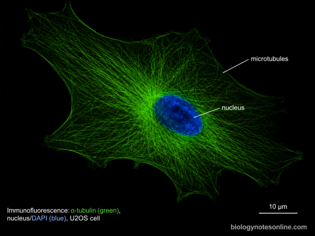
What Are Microtubules?
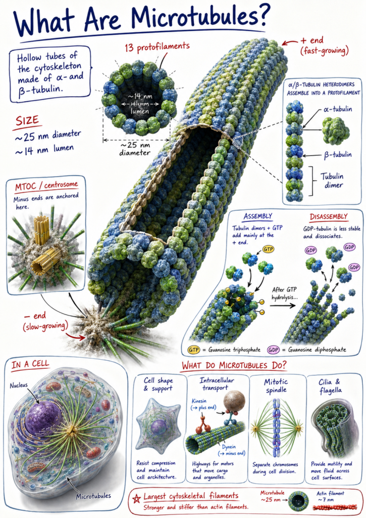
Microtubules are long, hollow tubes present as a major part of the cytoskeleton of eukaryotic cells. They are made up of tubulin proteins, mainly α-tubulin and β-tubulin.
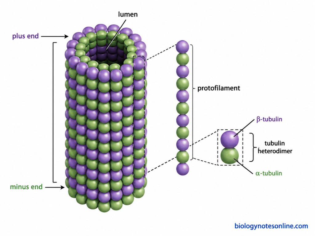
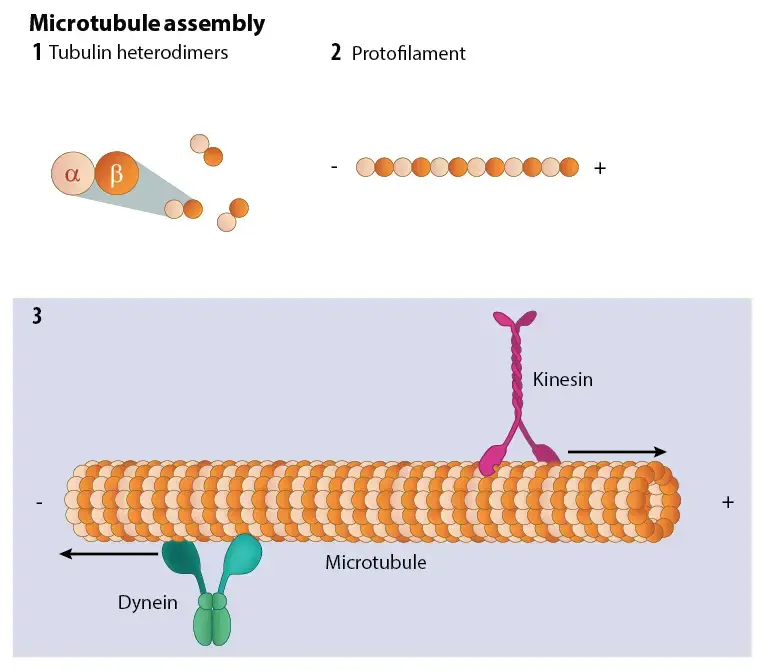
These two tubulins remain joined together forming α/β-tubulin heterodimers. The dimers are arranged one after another into long rows, called protofilaments. Usually 13 protofilaments join side by side and form the cylindrical wall of a microtubule, having a diameter of about 25 nm. Microtubules are therefore the thickest of the three major cytoskeletal filaments.
They are not fixed structures within the cell. Growth and shortening of microtubules can occur rapidly by addition and loss of tubulin units.
Microtubules have two different ends, the plus (+) end and minus (−) end. This structural polarity is important in their assembly and also in movement of cellular materials. They form tracks for intracellular transport, participate in chromosome movement during cell division and form important parts of cilia and flagella.
Structure and Composition of Microtubules
The major structural features and composition of microtubules are-
- Tubulin proteins – Microtubules are mainly made up of α-tubulin and β-tubulin. These are globular proteins. One α-tubulin and one β-tubulin remain associated together, forming an α/β-tubulin heterodimer. It is the basic building unit of the microtubule.
- Protofilaments – The tubulin heterodimers are joined in a head-to-tail manner. This forms a long linear chain called protofilament. Several such protofilaments are present in one microtubule.
- Hollow cylinder – Usually, 13 protofilaments are arranged side by side and form a hollow cylindrical wall. The diameter is about 24–25 nm. The hollow space present at the centre is called the lumen. The number of protofilaments can vary in some organisms and specialized cells.
- Polarity – Due to the head-to-tail arrangement of tubulin dimers, microtubules have two different ends. One is the plus (+) end and another is the minus (−) end. The plus end generally shows faster addition and loss of tubulin than the minus end.
- GTP – Both α-tubulin and β-tubulin bind guanosine triphosphate (GTP). GTP present on β-tubulin can be hydrolyzed after its addition into the microtubule. This has an important role in stability and dynamic behaviour of microtubules.
- Microtubule lattice – Protofilaments are not present separately. They are laterally associated with neighbouring protofilaments, forming the microtubule wall. In the common 13-protofilament microtubule, most lateral contacts form a B-type lattice with a single discontinuity called the seam.
- Tubulin isoforms – Different forms of α- and β-tubulin are present in cells. Tubulin molecules may also undergo modifications such as acetylation, detyrosination, polyglutamylation and glycylation. These modifications are not same in all microtubules.
- Associated proteins – Several proteins can remain attached with microtubules. These are commonly called microtubule-associated proteins (MAPs). They are not the main material forming the hollow wall but can regulate microtubule organization, stability and other activities.
Microtubule Polarity: Plus and Minus Ends
Microtubules are polar structures. This polarity is produced because the α/β-tubulin heterodimers are joined in the same head-to-tail direction throughout the protofilament. As a result, the two ends of a microtubule are different. These are referred to as plus (+) end and minus (−) end.
The major differences between these two ends are as follows-
- Plus (+) end – It is the end where β-tubulin is exposed. Addition of tubulin generally occurs faster at this end, therefore it is also called the faster-growing end. Growth and shortening are frequently seen here. The plus end is highly dynamic.
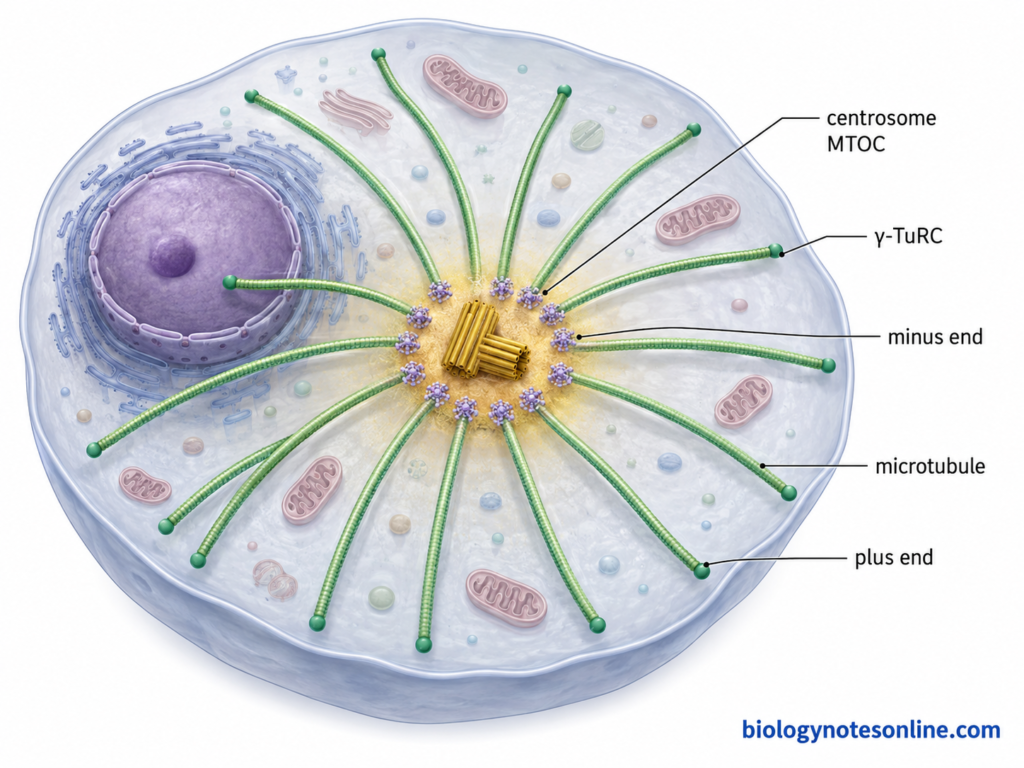
- Minus (−) end – α-tubulin is exposed at the minus end. Its rate of growth is usually slower than the plus end. In many animal cells, this end remains associated with the microtubule-organizing centre (MTOC), where microtubules are nucleated and their minus ends can be retained or stabilized.
- Difference in stability – Both ends do not show the same dynamic behaviour. The plus end commonly undergoes rapid polymerization and depolymerization. Minus ends are often more stable, although free minus ends can also grow and shorten.
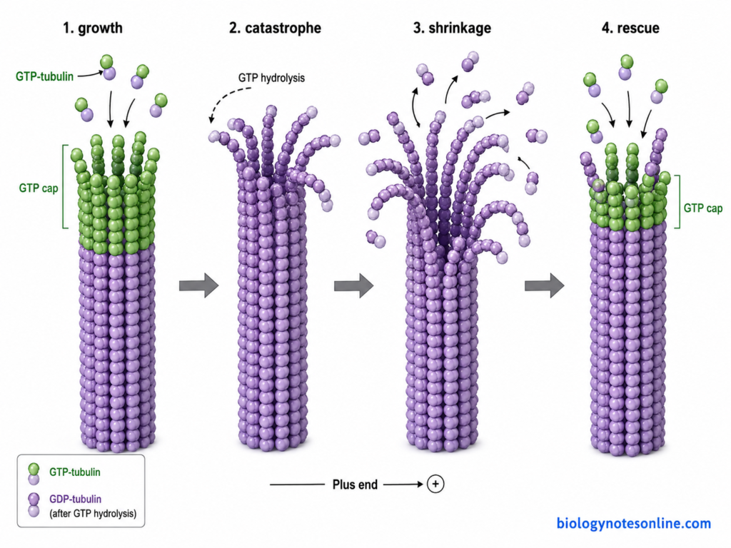
- GTP cap – During growth, GTP-bound tubulin is added to microtubule ends. The larger and more prominent GTP-rich cap is generally present at the growing plus end, helping in its stability. Loss of this stabilizing cap can lead to rapid shortening, called catastrophe. Growing minus ends can also contain a smaller GTP-rich cap.
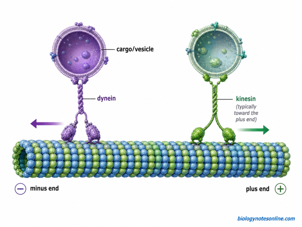
- Direction for motor proteins – Microtubule polarity also gives a fixed direction for intracellular movement. Most kinesins move towards the plus end. Cytoplasmic dynein, on the other hand, normally moves towards the minus end. This arrangement allows cellular materials to move in opposite directions along the microtubules.
- Arrangement inside cells – In many animal cells, microtubule minus ends are located near the centrosome and plus ends extend outward towards the cell periphery. But this type of arrangement is not present in all cells. Acentrosomal microtubule arrays are also common, including in plant cells and several differentiated animal cells.
Microtubule Assembly
Microtubule assembly is the polymerization of α/β-tubulin heterodimers to form protofilaments and then a hollow microtubule. The process is dependent on GTP. In cells, assembly is generally initiated from specific microtubule-nucleating sites.
The process of microtubule assembly is as follows-
- Formation of tubulin dimers – α-tubulin and β-tubulin remain associated as a stable heterodimer. Both tubulins bind GTP. The nucleotide present on β-tubulin is exchangeable and it has an important role during microtubule assembly.
- Nucleation – It is the initial step of microtubule formation. In the cell, nucleation is commonly carried out by γ-tubulin ring complex (γ-TuRC) present at microtubule-organizing sites. It provides a template on which the first tubulin molecules can be arranged. The minus end generally remains associated with the nucleating complex.
- Elongation – After nucleation, more GTP-bound α/β-tubulin dimers are added to the growing microtubule. The dimers first form longitudinal contacts and are also joined laterally with neighbouring protofilaments. Growth can occur at both ends of a free microtubule, but it is usually faster at the plus (+) end. Generally 13 protofilaments form the wall of a typical mammalian microtubule.
- GTP hydrolysis – After the tubulin dimer becomes incorporated into the microtubule lattice, GTP bound with β-tubulin is hydrolyzed to GDP. The GTP of α-tubulin is not exchanged in the same way. Thus, most of the older microtubule lattice contains GDP-tubulin.
- GTP cap formation – During rapid growth, addition of GTP-tubulin can occur faster than its hydrolysis. A region containing GTP or GTP-like tubulin is therefore maintained at the growing end, known as the GTP cap. It helps in keeping the microtubule stable and growing.
- Catastrophe and rescue – The microtubule does not always continue to grow. Loss of the stabilizing cap can cause sudden rapid shortening, called catastrophe. Sometimes a shortening microtubule again changes into growth. This is referred to as rescue. The repeated switching between growth and shortening is known as dynamic instability.
Functions of Microtubules
The following are some of the important functions of microtubules–
- Maintenance of cell shape – Microtubules form an internal supporting framework within the cytoplasm. They help in maintaining the shape and general organization of cell.
- Intracellular transportation – They are used as tracks for movement of vesicles, organelles and other cellular materials. Kinesin and dynein are the major motor proteins associated with this movement. Kinesin generally carries materials towards the plus end, while cytoplasmic dynein moves towards the minus end. This transport is especially important over longer distances within cells.
- Cell division – Microtubules form the mitotic spindle during cell division. Spindle microtubules take part in attachment, positioning and separation of chromosomes, allowing their distribution into daughter cells.
- Formation of cilia and flagella – They form the main internal framework or axoneme of eukaryotic cilia and flagella. In motile forms, interaction of axonemal microtubules with dynein produces bending and movement.
- Cell polarity – The arrangement of microtubules helps to maintain polarity of different cells and directs cellular materials towards particular regions. They are also involved in organization of polarized cells and during some forms of cell movement.
- Positioning of cellular components – Different organelles do not remain randomly distributed inside the cell. Microtubules together with motor proteins help in their movement and positioning at specific regions of the cytoplasm. They also participate in movement of secretory materials.
- Plant cell wall organization – In plant cells, cortical microtubules have an important role in organization of the cell wall. They guide the movement of cellulose synthase complexes at the plasma membrane, affecting the direction in which cellulose is deposited. Thus microtubule arrangement is also associated with the direction of plant cell expansion.
Microtubules vs Microfilaments vs Intermediate Filaments
The major differences between microtubules, microfilaments and intermediate filaments are as follows-
| Features | Microtubules | Microfilaments (Actin filaments) | Intermediate filaments |
|---|---|---|---|
| Structure | Hollow cylindrical tubes. | Thin, solid filaments formed by two twisted actin chains. | Rope-like fibrous structures. |
| Diameter | About 25 nm. | About 7 nm. | About 10 nm. |
| Basic protein | Made up of α-tubulin and β-tubulin. | Mainly formed from actin protein. | Made from different fibrous proteins depending on the cell type. |
| Basic subunit | α/β-tubulin heterodimer. | Globular G-actin, which polymerizes into F-actin. | Intermediate filament proteins form dimers, tetramers and then filament. |
| Polarity | Polar. It has plus (+) and minus (−) ends. | Also polar with plus and minus ends. | They do not have structural polarity. |
| Nucleotide used | GTP is associated with tubulin during assembly. | ATP is bound with actin during polymerization. | No ATP or GTP is directly required for polymerization. |
| Dynamic nature | Highly dynamic. Growth and shortening can occur rapidly. | Dynamic filaments and undergo continuous assembly and disassembly. | Generally more stable than microtubules and microfilaments, although their organization can also change. |
| Motor proteins | Kinesin and dynein move along microtubules. | Myosin moves along actin filaments. | Usually do not act as tracks for conventional motor proteins. |
| Major location | Extend throughout cytoplasm and also form spindle, cilia and flagella. | Commonly concentrated beneath the plasma membrane and in structures involved in cell movement. | Distributed through cytoplasm and also present in the nuclear lamina. |
| Cell shape | Help in maintaining cell organization and resist compression. | Help in cell shape and changes of cell surface. | Provide mechanical strength and resist stretching forces. |
| Cell division | Form the mitotic spindle and help in chromosome movement. | Form the contractile ring during cytokinesis in animal cells. | Nuclear lamins disassemble and reassemble during mitosis. |
| Cell movement | Important in movement of cilia and flagella. | Important in cell crawling, muscle contraction and formation of cellular projections. | They mainly provide mechanical support rather than producing cell movement. |
| Intracellular transport | Major tracks for transport of vesicles and organelles over long distances. | Involved in short-range transport with myosin and movement near cell cortex. | Not a major pathway for vesicle transport. |
| Examples | Tubulin microtubules of mitotic spindle and axoneme. | Actin filaments of microvilli, contractile ring and muscle cells. | Keratins, vimentin, desmin, neurofilaments and nuclear lamins. |
References
- Ashaduzzaman, M., Tang, Y., Okada, K., Fried, S. D., McKenney, R. J., & Al-Bassam, J. (2026). Structural basis of kinesin-1 autoinhibition and its control of microtubule-based motility. Science Advances, 12(29), eaeg1267. https://doi.org/10.1126/sciadv.aeg1267
- Benoit, B., & Poüs, C. (2019). Microtubule reorientation in the blue spotlight: Cutting and CLASPing at dynamic hot spots. Journal of Cell Biology, 218(1), 8–9. https://doi.org/10.1083/jcb.201812063
- Desai, A., & Mitchison, T. J. (1997). Microtubule polymerization dynamics. Annual Review of Cell and Developmental Biology, 13, 83–117. https://doi.org/10.1146/annurev.cellbio.13.1.83
- Ehrhardt, D. W., & Shaw, S. L. (2006). Microtubule dynamics and organization in the plant cortical array. Annual Review of Plant Biology, 57, 859–875. https://doi.org/10.1146/annurev.arplant.57.032905.105329
- Ganguly, A., Zhu, C., Chen, W., & Dixit, R. (2020). FRA1 kinesin modulates the lateral stability of cortical microtubules through cellulose synthase-microtubule uncoupling proteins. The Plant Cell, 32(8), 2508–2524. https://doi.org/10.1105/tpc.19.00700
- Goodson, H. V., & Jonasson, E. M. (2018). Microtubules and microtubule-associated proteins. Cold Spring Harbor Perspectives in Biology, 10(6), a022608. https://doi.org/10.1101/cshperspect.a022608
- Hurd, D. D. (2018). Tubulins in C. elegans. In The C. elegans Research Community (Ed.), WormBook: The online review of C. elegans biology. WormBook. https://doi.org/10.1895/wormbook.1.182.1
- Iyer, S. S., & Akhmanova, A. (2026). Slow but steady: Molecular mechanisms controlling microtubule growth in centrioles and cilia. Journal of Cell Biology, 225(6), e202601174. https://doi.org/10.1083/jcb.202601174
- Lawrence, E. J., & Zanic, M. (2019). Rescuing microtubules from the brink of catastrophe: CLASPs lead the way. Current Opinion in Cell Biology, 56, 94–101. https://doi.org/10.1016/j.ceb.2018.10.011
- Liu, J. S., Schubert, C. R., & Walsh, C. A. (2012). Rare genetic causes of lissencephaly may implicate microtubule-based transport in the pathogenesis of cortical dysplasias. In J. L. Noebels, M. Avoli, M. A. Rogawski, R. W. Olsen, & A. V. Delgado-Escueta (Eds.), Jasper’s basic mechanisms of the epilepsies (4th ed.). National Center for Biotechnology Information (US). https://www.ncbi.nlm.nih.gov/books/NBK98194/
- Markus, S. M. (2025). Microtubule motors of opposite polarity cooperate rather than compete in cargo transport. Nature Structural & Molecular Biology, 32(4), 595–597. https://doi.org/10.1038/s41594-025-01524-6
- Matković, J., Ghosh, S., Ćosić, M., Eibes, S., Barišić, M., Pavin, N., & Tolić, I. M. (2022). Kinetochore- and chromosome-driven transition of microtubules into bundles promotes spindle assembly. Nature Communications, 13, 7307. https://doi.org/10.1038/s41467-022-34957-4
- McIntosh, J. R. (2025). Understanding microtubule dynamics: The synergy of technology, theory, and experiment. Journal of Cell Biology, 224(11), e202505046. https://doi.org/10.1083/jcb.202505046
- McKenna, E. D., Sarbanes, S. L., Cummings, S. W., & Roll-Mecak, A. (2023). The tubulin code, from molecules to health and disease. Annual Review of Cell and Developmental Biology, 39, 331–361. https://doi.org/10.1146/annurev-cellbio-030123-032748
- Park, J. H., Woo, Y. M., Ko, J. Y., & Kim, D. Y. (2015). Autosomal dominant polycystic kidney disease induced by ciliary defects. In X. Li (Ed.), Polycystic kidney disease. Codon Publications. https://doi.org/10.15586/codon.pkd.2015.ch15
- Paumier, C., Sauvanet, C., & Gigant, B. (2025). Microtubule nucleation: How the NEDD1:MZT1 trio captures the γ-TuRC. Journal of Cell Biology, 224(8), e202506019. https://doi.org/10.1083/jcb.202506019
- Wordeman, L. (2019). GTP-tubulin loves microtubule plus ends but marries the minus ends. Journal of Cell Biology, 218(9), 2822–2823. https://doi.org/10.1083/jcb.201908039