What is Plasmodesmata?
- Plasmodesmata (singular: plasmodesma) are specialized microscopic channels that facilitate communication and transport between plant cells and certain algal cells. These structures play a critical role in maintaining cellular connectivity, allowing for the exchange of various substances such as water, proteins, and small RNA molecules. Plasmodesmata are crucial for cellular communication and metabolic coordination in plant tissues.
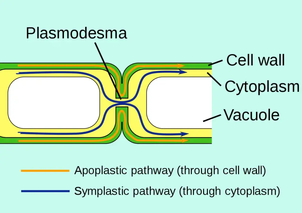
- Originating in multiple evolutionary lineages, plasmodesmata are found in a range of organisms, including members of the Charophyceae, Charales, Coleochaetales, and Phaeophyceae, as well as all embryophytes, commonly known as land plants. Unlike animal cells, which lack rigid cell walls, plant cells are encased in a polysaccharide cell wall that separates them from adjacent cells. This configuration leads to the formation of an extracellular space known as the apoplast, which is delineated by cell walls and the intervening middle lamella. While the cell walls permit the diffusion of small solutes, plasmodesmata enable a more regulated and efficient symplastic transport mechanism, allowing substances to traverse directly between adjacent cells.
- There are two primary forms of plasmodesmata: primary plasmodesmata, which emerge during the process of cell division, and secondary plasmodesmata, which can develop later between mature cells. Each plant cell is interconnected to its neighbors through these fine cytoplasmic channels, effectively creating a continuous cytoplasmic network throughout plant tissues. In contrast, animal cells utilize analogous structures known as gap junctions to facilitate intercellular communication.
- First described by botanist Eduard Strasburger in 1901, the term “plasmodesma” derives from the Latin word plasmo, meaning fluid, and the Greek word desma, meaning bond. This nomenclature reflects the fundamental nature of these structures in linking the cytoplasm of adjacent cells. Notably, plasmodesmata are integral to plant survival, enabling the movement of essential biomolecules and ions, thereby influencing processes such as growth, development, and defense signaling.
- The functionality of plasmodesmata is closely linked to their structural composition. Transport through these channels is highly regulated and can involve signaling molecules such as calcium ions (Ca²⁺) and protein phosphorylation. Generally, plasmodesmata allow the passage of molecules with a molecular weight of less than 800 daltons. The density of plasmodesmata can vary significantly between different cell types; for instance, some cells may contain upwards of 15 plasmodesmata per square micrometer of cell wall surface, while others may have fewer than one.
- In a typical plant cell, the number of plasmodesmata can range from 10³ to 10⁵, translating to approximately 1-10 plasmodesmata per square micrometer. They are particularly abundant in specific tissues, such as the endosperm of seeds from genera like Phoenix, Diospyros, and Aesculus, where they can be readily observed.
- Callose, a β-1,3-glucan polymer, plays a dual role as both a structural component and a regulatory factor within plasmodesmata. Its presence can influence the opening and closing of these channels, thus affecting the movement of substances between cells. In conclusion, plasmodesmata are essential features of plant and algal cells, facilitating intercellular communication and playing a vital role in the overall functioning of plant physiology.
Definition of Plasmodesmata
Plasmodesmata are microscopic channels that traverse the cell walls of plant and algal cells, allowing for direct transport and communication between adjacent cells. These structures enable the movement of water, proteins, small RNAs, and other metabolites, playing a crucial role in cellular connectivity and plant physiology.
Structure of Plasmodesmata
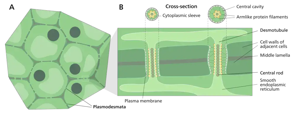
The structure of plasmodesmata is intricate and essential for facilitating intercellular communication in plants and algae. These microscopic channels vary in complexity, enabling direct transport between adjacent cells. The following points detail the structural components and characteristics of plasmodesmata:

- Variety in Structure: Plasmodesmata exhibit a range of structures, from simple forms characterized by a single sheath to more complex arrangements such as branched, H-shaped, and twinned structures. Typically, young plant tissues contain simpler plasmodesmata, while more complex configurations develop later as the cells expand.
- Dimensions: The diameter of a plasmodesma generally measures between 20 to 40 nanometers, forming a cylindrical, membrane-lined channel that connects plant cells.
- Main Compartments: Plasmodesmata consist of three primary components:
- Plasma Membrane (Plasmalemma): This membrane extends from the cell membrane and shares a structural similarity to the phospholipid bilayer found in cellular membranes.
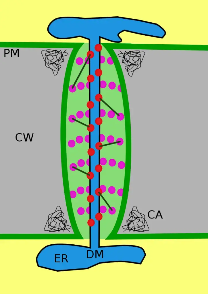
- Cytoplasmic Sleeve: This fluid-filled space, surrounded by the plasmalemma, serves as a continuous extension of the cytosol. Within the cytoplasmic sleeve, myosin-like proteins and actin filaments are localized, contributing to the movement of materials. Additionally, proteinaceous spike-like projections are regularly spaced, forming nanochannels of varying sizes that aid in transport.
- Desmotubule: This dense, cylindrical structure runs through the center of plasmodesmata, connecting adjacent cells. Derived from the smooth endoplasmic reticulum of the connected cells, the desmotubule serves as a central rod facilitating intercellular transport. Initially referred to as the axial component, the term “desmotubule” is now widely accepted.
- Cytosolic Annulus: Surrounding the desmotubule is the annulus, which constitutes the space between the desmotubule and the cylindrical plasma membrane. This annulus contains 8-10 microchannels that allow for the movement of ions and smaller molecules.
- Electron Microscopy Observations: Electron microscopy provides detailed insights into the structure of plasmodesmata. The plasma membrane appears as a tripartite structure with a width of approximately 7.2 nanometers. The dense central rod of the desmotubule measures around 1.4 nanometers in radius, while the pale ring surrounding it has a width of 2.2 nanometers.
- Transport Mechanism: The cytoplasmic sleeve plays a crucial role in the trafficking of ions and molecules through plasmodesmata. Smaller molecules, including amino acids, can pass through the cytoplasmic sleeve via diffusion, facilitating intercellular communication.
Types of Plasmodesmata
The types of plasmodesmata are categorized based on their origin and developmental processes. Understanding these types is essential for grasping how plant cells communicate and exchange materials. The two primary types of plasmodesmata are as follows:
- Primary Plasmodesmata:
- These structures are formed during the process of cell division.
- They emerge as part of the cellular development in certain plant species, such as Chara zelanica, which produces both primary and secondary plasmodesmata.
- The initial structure of primary plasmodesmata is relatively simple, providing a direct link between newly divided cells.
- Secondary Plasmodesmata:
- In contrast to primary plasmodesmata, secondary plasmodesmata are developed de novo within the existing cell wall, meaning they are created anew rather than arising from cell division.
- The development of secondary plasmodesmata can be influenced by external factors, such as the hormone cytokinin, which has been shown to stimulate their formation.
- An example of a species that produces only secondary plasmodesmata is Chara corallina.
- Initially, secondary plasmodesmata, like primary ones, are also simple in structure but can evolve into more complex forms.
Formation of Plasmodesmata
The formation of plasmodesmata (PD) is a complex and essential process in plant biology, serving as vital channels for intercellular communication and transport. Understanding how these structures originate sheds light on their functions and roles within plant tissues.
- Primary Formation During Mitosis:
- Plasmodesmata are generally formed during mitotic cell division. During this process, the protoplasm of newly formed daughter cells is divided by the cell plate. However, the endoplasmic reticulum (ER) remains connected across the newly formed cell plate.
- This connection is crucial as it prevents the deposition of wall-forming substances at the junction, thereby maintaining a continuous channel between the two separated cells.
- Under the pressure exerted by the cell plate or membrane, the ER transforms into plasmodesmata, establishing a pathway for transport between the daughter cells.
- Formation Between Non-Sister Cells:
- Interestingly, plasmodesmata can also form between non-sister cells, indicating that their formation is not limited to mitotic processes. The mechanisms and molecular origins of this secondary formation, however, remain less well understood and are an area of ongoing research.
- Structural Characteristics of Plasmodesmata:
- Research by Ding et al. employed advanced techniques such as rapid freezing and high-resolution electron microscopy to reveal new structural details about plasmodesmata. They proposed a structural model wherein each PD consists of two enlarged openings at either end, connected by a cylindrical body formed by appressed ER, termed the desmotubule.
- The desmotubule exhibits a tightly constricted structure, with minimal space, suggesting that it plays a crucial role in the selective transport of molecules between cells.
- Cytoskeletal Components:
- Cytoskeletal proteins, including actin and myosin, are commonly found within plasmodesmata. These proteins contribute to the structural integrity and functionality of the PD, and their arrangement indicates their importance in the transport processes.
- Protein particles approximately 3 nm in size are embedded between the appressed ER and the plasma membrane. Electron-dense radial fibrils connect these particles, forming a network that may facilitate transport through the PD. Notably, in cross-sectional views, 7 to 9 particles can be observed, further emphasizing the complexity of the PD structure.
- Control of Permeability:
- The permeability of plasmodesmata is regulated through two primary mechanisms: actin filaments and callosum sphincters. Actin filaments are situated near and inside the PD channel, and their polymerization state can significantly influence the permeability of plasmodesmata.
- Additionally, callose aggregates can form at the neck region of the plasmodesmata, effectively narrowing the space between the cell wall and the desmotubule. This regulation mechanism enables the plant to control the flow of substances, maintaining homeostasis and responding to environmental changes.

Transport in Plasmodesmata
Transport in plasmodesmata is a critical process that enables the movement of various molecules between plant cells, facilitating communication and resource sharing essential for plant growth and development. This intricate system allows for the exchange of proteins, nucleic acids, and other metabolites, thereby playing a vital role in physiological processes such as flowering and viral spread.

- Types of Molecules Transported:
- Plasmodesmata are responsible for transporting a wide array of biomolecules, including proteins (such as transcription factors), short interfering RNA, messenger RNA, viroids, and viral genomes.
- A notable example of a viral movement protein is the tobacco mosaic virus MP-30, which binds to the virus’s genome and facilitates its movement from infected to uninfected cells through the plasmodesmata.
- Another significant transport role is played by the Flowering Locus T protein, which moves from leaves to the shoot apical meristem via plasmodesmata to trigger flowering in plants.
- Symplastic Transport in Phloem:
- In addition to transporting various biomolecules, plasmodesmata are utilized by phloem cells for symplastic transport. This process is essential for regulating sieve-tube cells by companion cells, ensuring efficient nutrient distribution within the plant.
- Size Exclusion Limit (SEL):
- The capacity for molecules to pass through plasmodesmata is regulated by the size exclusion limit (SEL), which varies and can be actively modified.
- For instance, the movement protein MP-30 can increase the SEL from 700 daltons to 9,400 daltons, facilitating the transport of larger molecules through the plasmodesmata.
- Furthermore, increasing calcium concentrations in the cytoplasm, whether by direct injection or through cold-induction, has been shown to constrict the openings of adjacent plasmodesmata, thereby limiting transport.
- Active Transport Mechanisms:
- Several models have been proposed to explain potential active transport mechanisms through plasmodesmata. These models suggest that transport could be mediated by protein interactions localized on the desmotubule or by chaperones that partially unfold proteins, allowing them to traverse the narrow channels.
- Similar mechanisms may be implicated in the transport of viral nucleic acids, enhancing the understanding of how viruses exploit plasmodesmata for propagation.
- Mathematical Models of Transport:
- To quantify and predict transport across plasmodesmata, several mathematical models have been developed. These models typically treat transport as a diffusion problem, incorporating variables that account for hindrances due to the complex structure of the plasmodesmata.
The cytoskeletal components of plasmodesmata
The cytoskeletal components of plasmodesmata play a crucial role in facilitating intercellular transport and communication within plant tissues. By examining the interplay between these components—specifically actin microfilaments, myosin proteins, and microtubules—it becomes evident how they contribute to both normal cellular functions and the spread of viral infections.
- Actin Microfilaments:
- Actin microfilaments are integral to the transport of viral movement proteins to plasmodesmata, facilitating cell-to-cell communication.
- Research involving fluorescent tagging in tobacco leaves has demonstrated that these actin filaments are responsible for transporting viral proteins to the plasmodesmata.
- When the polymerization of actin was inhibited, there was a noticeable decrease in the targeting of movement proteins to the plasmodesmata. Consequently, this alteration allowed for smaller components, specifically 10-kDa molecules, to pass between tobacco mesophyll cells rather than the larger 126-kDa proteins. Such changes significantly impacted the overall cell-to-cell movement of molecules within the plant.
- Viruses and Actin Interaction:
- Certain viruses, like the cucumber mosaic virus (CMV), exploit the plant’s cytoskeletal system by breaking down actin filaments within the plasmodesmata channels, enabling their movement throughout the plant.
- The viral movement proteins facilitate the transport of the virus from cell to cell. When tobacco leaves were treated with phalloidin, a drug that stabilizes actin filaments, the CMV movement proteins could not expand the plasmodesmata’s size exclusion limit (SEL), thereby hindering viral spread.
- Myosin Proteins:
- High concentrations of myosin proteins are found at the sites of plasmodesmata, where they play a significant role in directing viral cargoes toward these channels.
- Studies involving mutant forms of myosin in tobacco plants indicated that such mutations adversely affected the targeting of viral proteins to plasmodesmata.
- Additionally, permanent binding of myosin to actin—induced by certain drugs—resulted in decreased cell-to-cell movement of materials. This suggests that myosin proteins may also selectively bind to viruses, further influencing viral transport mechanisms.
- Microtubules:
- Microtubules are crucial for the transport of viral RNA between cells. Viruses have evolved various strategies to associate with microtubules for their movement.
- For instance, in tobacco plants infected with tobacco mosaic virus (TMV) under elevated temperatures, a significant correlation was observed between GFP-labeled TMV movement proteins and microtubules. This association led to an increased spread of viral RNA through the plant tissues.
Associated Proteins of Plasmodesmata
Plasmodesmata (PD) are essential structures that facilitate intercellular communication in plants. They are composed of channels that traverse the cell walls, allowing the passage of various molecules between adjacent cells. A critical aspect of PD functionality lies in the associated proteins that contribute to their structural integrity and regulatory mechanisms. These proteins are integral for the maintenance and modulation of PD permeability, impacting processes such as nutrient transport and defense responses.
- PD-Associated Structural Proteins:
- Structural proteins like actin, myosin, tubulin, and calreticulin are integral components of PD.
- Actin and myosin form part of the cell’s cytoskeletal network, playing significant roles in intracellular transport.
- Tubulin, composed of heterodimers, is crucial for cell division and the organization of intracellular structures.
- Although these proteins are not classified as cell-wall proteins, they are relevant to the architecture of PD, as shown in the summarized table of associated proteins.
- Actin:
- Actin is distributed throughout PD, confirmed through studies using fluorescence probes and advanced microscopy techniques.
- The organization of actin filaments within PD is not entirely understood; they may reside in the lumen, connecting the cytoskeletal elements between adjacent cells.
- Actin filaments may also facilitate vesicular trafficking along PD. Disruption of actin with cytochalasin D leads to reduced transport efficiency of larger molecules, suggesting that actin influences PD permeability.
- Myosin:
- Myosin has been identified as a component of PD, with localization studies using immunochemistry revealing its presence.
- It is classified into 15 families, with only some members identified in plants; the specific myosin associated with PD belongs to the eighth family.
- However, the role of myosin in regulating PD function remains an area of ongoing research.
- Tubulin:
- Tubulin’s presence in PD was supported by studies showing its isolation from PD-containing cell walls, indicating a potential role in structural support.
- Its specific function in PD has not been characterized extensively, but it may contribute indirectly to long-distance transport within the plant.
- PD-Associated Regulatory Proteins:
- Callose is a crucial component deposited at the neck of PD, regulating the size exclusion limit (SEL) and thereby controlling intercellular communication.
- Two primary enzymes, callose synthase (GSL) and β-1,3-glucanase (BG), manage the levels of callose within PD, affecting their permeability.
- Callose Synthases:
- Callose synthases, such as GSL8 (CALS10) and GSL12 (CALS3), play pivotal roles in depositing callose and influencing plant development and responses to auxin.
- A high level of callose at the PD neck restricts permeability, whereas lower levels enhance it, thereby facilitating the transport of macromolecules.
- Callose Hydrolases:
- Class I β-1,3-glucanase (GLU I) is critical for degrading callose, thereby promoting communication between cells and enhancing PD permeability.
- Studies demonstrated that GLU I deficiency in tobacco mutants reduced viral spread, implicating this enzyme in both callose metabolism and pathogen defense.
- PD-Associated Callose Binding Proteins (PDCBs):
- PDCBs such as PDCB1, PDCB2, and PDCB3 are involved in stabilizing callose at PD, influencing the diffusion of signaling molecules and maintaining cellular communication.
- PDCB1, in particular, was shown to associate closely with callose and modulate PD permeability in response to varying callose levels.
- PD-Located Receptor-Like Proteins (PDLPs):
- PDLPs are transmembrane proteins localized at PD that play roles in signaling and defense responses.
- Studies have indicated that PDLPs can regulate intercellular transport, with specific members influencing responses to pathogens.
- Other PD-Related Proteins:
- Various proteins and lipids, including the sterol carrier protein GHSCP2D and expansins such as NbEXPA1, have been identified as being associated with PD.
- These proteins have been shown to influence PD permeability and are implicated in responses to viral infections, highlighting the multifaceted nature of PD function in plant biology.
Functions of Plasmodesmata
Plasmodesmata serve several crucial functions within plant cells, acting as essential conduits for communication and transport. Their role is particularly significant given the rigid nature of plant cell walls, which can impede the movement of larger molecules. The following points elucidate the key functions of plasmodesmata:
- Facilitating Molecular Entry: Plasmodesmata enable larger molecules and various entities to traverse the cell wall, which would otherwise restrict their passage due to its tough, rigid structure. This capability ensures that essential nutrients and signaling molecules can enter the cell efficiently.
- Cellular Communication: Communication between plant cells is vital for growth, development, and survival. Plasmodesmata are instrumental in mediating this communication, allowing for the exchange of information and molecules between adjacent cells.
- Molecular Translocation: Plasmodesmata play a critical role in the translocation of various molecules, including nutrients and water. They contain both passive and active pores that facilitate the movement of these substances. Passive pores allow for the simple diffusion of nutrients and water, whereas active mechanisms can regulate the transport of larger or more complex molecules.
- Transport of Genetic Material: The presence of actin structures within plasmodesmata aids in the movement of transcription factors, such as messenger RNA, viroids, short interfering RNA, and even plant viruses. This transport mechanism is essential for regulating gene expression and responding to various environmental stimuli.
- Defense Mechanisms: Research has identified plasmodesmata-located protein 5 (PDLP5), which has the ability to produce salicylic acid. This compound enhances plant defenses against pathogenic attacks, providing a protective mechanism against harmful bacteria and other threats.
- Role in Phloem Functionality: In addition to their roles in other plant tissues, plasmodesmata are also present in phloem cells. They are crucial for the movement of nutrients and signaling molecules throughout the plant.
- Viral Movement: Plasmodesmata are involved in the short-distance movement of viruses within plant tissues. This function highlights their dual role in both facilitating plant health and being exploited by pathogens.
FAQ
What are plasmodesmata?
Plasmodesmata are microscopic channels that connect adjacent plant cells and allow for the transport of molecules and communication between them.
How are plasmodesmata formed?
Plasmodesmata are formed when specific regions of the cell wall are removed, creating a channel between adjacent cells.
What is the function of plasmodesmata?
The primary function of plasmodesmata is to facilitate the movement of water, nutrients, signaling molecules, and other substances between adjacent plant cells.
What types of molecules can pass through plasmodesmata?
Small molecules such as ions, sugars, and amino acids can pass through plasmodesmata, as well as larger molecules such as proteins and RNA molecules.
How are plasmodesmata regulated?
The opening and closing of plasmodesmata are regulated by various factors, including developmental signals, environmental cues, and plant hormones.
Can viruses and pathogens move through plasmodesmata?
Yes, some viruses and pathogens can move through plasmodesmata to infect neighboring cells, which can contribute to the spread of diseases within the plant.
How do plasmodesmata contribute to plant growth and development?
Plasmodesmata play an important role in plant growth and development by enabling cell-to-cell communication, transport of nutrients and signaling molecules, and regulation of cell differentiation and fate.
How are plasmodesmata related to plant stress responses?
Plasmodesmata are involved in plant stress responses, as they allow for the movement of stress-related signaling molecules between cells, enabling the plant to respond to environmental challenges.
How can plasmodesmata be visualized and studied?
Plasmodesmata can be visualized and studied using various techniques, such as electron microscopy, fluorescent microscopy, and live cell imaging.
Are plasmodesmata unique to plants?
Yes, plasmodesmata are unique to plants and are not found in other organisms. However, similar structures called gap junctions are found in animals and allow for cell-to-cell communication.
References
- Han X, Huang LJ, Feng D, Jiang W, Miu W, Li N. Plasmodesmata-Related Structural and Functional Proteins: The Long Sought-After Secrets of a Cytoplasmic Channel in Plant Cell Walls. Int J Mol Sci. 2019 Jun 17;20(12):2946. doi: 10.3390/ijms20122946. PMID: 31212892; PMCID: PMC6627144.
- https://micro.magnet.fsu.edu/cells/plants/plasmodesmata.html
- https://www.news-medical.net/life-sciences/What-are-Plasmodesmata.aspx
- https://www.cell.com/current-biology/pdf/S0960-9822(08)00093-6.pdf
- https://study.com/learn/lesson/what-is-plasmodesmata.html
- https://www.thoughtco.com/plasmodesmata-the-bridge-to-somewhere-419216
- https://www.biologyonline.com/dictionary/plasmodesmata