What is the Endoplasmic Reticulum?
- The endoplasmic reticulum (ER) is a pivotal cell organelle, predominantly found in eukaryotic cells, and is integral to numerous cellular processes. It is the largest membrane-bound organelle, originating from the cell’s nuclear envelope. The ER’s primary roles encompass protein synthesis, modification, lipid synthesis, and the regulation of calcium homeostasis and secretion. This organelle is distinguished by its extensive network of membranous tubules and sheets, which are integral to its function.
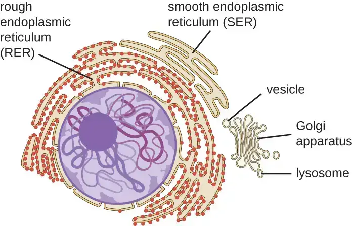
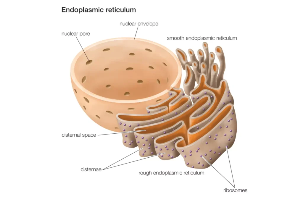
- There are two distinct forms of the endoplasmic reticulum: the rough endoplasmic reticulum (RER) and the smooth endoplasmic reticulum (SER). The RER is characterized by the presence of ribosomes on its surface, which are essential for protein synthesis. This form of ER is particularly prominent in cells such as hepatocytes. Conversely, the SER lacks ribosomes and is primarily involved in lipid synthesis, steroid hormone production, and detoxification processes. The SER is notably abundant in liver and gonad cells.
- The ER’s structure is composed of interconnected membranous tubules, which perform crucial cellular functions like protein synthesis, carbohydrate breakdown, lipid synthesis, and calcium storage. The membranes of the ER form continuous folds, eventually connecting to the outer layer of the nuclear membrane. This interconnectedness is vital for the ER’s role as the cell’s transportation system, facilitating the movement of molecules within the cell.
- Historically, the ER was first observed by light microscopy in 1897 by Garnier, who termed it ergastoplasm. Later, its intricate membranous structure was revealed through electron microscopy by Keith R. Porter, Albert Claude, and Ernest F. Fullam in 1945. The term “endoplasmic reticulum,” which translates to “network within the cytoplasm,” was coined by Porter in 1953 to describe this complex organelle.
- Functionally, the ER is multifaceted. It acts as a secretory, storage, and circulatory system within the cell, and is also involved in the biogenesis of cellular membranes. The ER’s dynamics and distribution are influenced by its interactions with other organelles and the cytoskeleton. It undergoes continuous rearrangements, including tubule branching and membrane fusion, to maintain its network throughout the cell.
- The medical significance of the ER is highlighted by its association with various diseases. Disruptions in ER function can lead to conditions such as Parkinson’s disease and Cystic Fibrosis. Moreover, the ER’s morphology is crucial for many intracellular events, and abnormalities in ER morphogenesis are linked to neurologic disorders like hereditary spastic paraplegias.
- In summary, the endoplasmic reticulum is a complex, dynamic organelle that plays a critical role in various cellular functions. Its unique structure and extensive network facilitate its multifunctional roles, underscoring its importance in cell biology and medicine.
Endoplasmic Reticulum (ER) Definition
The endoplasmic reticulum (ER) is a large, membrane-bound organelle found in eukaryotic cells. It plays a crucial role in the synthesis, folding, modification, and transport of proteins and lipids. The ER is divided into two types: the rough ER, studded with ribosomes and involved in protein synthesis, and the smooth ER, which is involved in lipid synthesis and does not have ribosomes. This organelle is essential for numerous cellular functions, including the production of hormones and enzymes, and the regulation of calcium levels.
Types of Endoplasmic Reticulum (ER)
1. Rough Endoplasmic Reticulum
- Structural Composition The rough endoplasmic reticulum (RER), also known as granular endoplasmic reticulum, is characterized by its rough appearance due to the presence of ribosomes on its cytoplasmic surface. These ribosomes are integral to the RER’s function in protein synthesis. The RER is composed of phospholipid bilayers, similar to the plasma membrane, and contains numerous flattened sacs called cisternae.
- Protein Synthesis and Processing
- Ribosome Attachment: Ribosomes are attached to the RER’s membrane, specifically to transmembrane glycoproteins known as ribophorins I and II. These ribosomes engage in polypeptide synthesis.
- Protein Translocation: Proteins destined for secretion begin their synthesis in the cytosol. A signal peptide directs the ribosome to the RER, where the nascent protein is translocated into the lumen.
- Signal Peptide and Processing: The signal peptide is cleaved by a signal peptidase in the ER lumen, and the protein undergoes further folding and modification.
- Membrane Structure and Dynamics
- Connection to Nuclear Envelope: The RER’s membrane is continuous with the outer layer of the nuclear envelope, forming large double-membrane sheets.
- Vesicular Transport: Proteins synthesized in the RER are packaged into transport vesicles, which are then shuttled to the Golgi apparatus for further processing.
- Functional Diversity
- Lysosomal Enzyme Manufacture: The RER is involved in the manufacture of lysosomal enzymes marked with a mannose-6-phosphate tag.
- Secretory Protein Production: It produces secreted proteins, either constitutively or in a regulated manner.
- Membrane Protein Synthesis: Integral membrane proteins are synthesized and remain embedded in the membrane as vesicles exit the RER.
- Glycosylation: Initial N-linked glycosylation occurs in the RER, where a sugar backbone is added to specific amino acid sequences in the nascent protein.
- Cellular Distribution The RER is found abundantly in cells active in protein synthesis, such as pancreatic cells, plasma cells, goblet cells, and liver cells. Its extensive network allows for efficient synthesis and transport of proteins within the cell.
- Inter-organellar Coordination
- Membrane Contact Sites: The RER coordinates with other organelles through membrane contact sites, facilitating the transfer of lipids and small molecules.
- Integration with Golgi Apparatus: The RER works in concert with the Golgi complex, ensuring that newly synthesized proteins are correctly targeted to their destinations.
2. Smooth Endoplasmic Reticulum
The smooth endoplasmic reticulum (SER), also known as the agranular endoplasmic reticulum, is a critical component of the eukaryotic cell. Unlike the rough endoplasmic reticulum (RER), the SER is characterized by its smooth walls, a result of the absence of ribosomes on its membrane. This organelle is predominantly found in cells involved in lipid and glycogen metabolism, such as adipose cells, liver cells, and certain muscle fibers.
- Structural Characteristics Structurally, the SER shares similarities with the RER, comprising a network of tubules and cisternae. However, its defining feature is the lack of ribosomes, which imparts a smooth appearance. The SER’s structure allows for an increased surface area, facilitating the action and storage of key enzymes and their products.
- Primary Functions The SER plays several vital roles in cellular function:
- Lipid Synthesis: It is responsible for producing phospholipids and steroids, essential for cell membrane formation.
- Detoxification: The SER contains enzymes that detoxify drugs and toxins.
- Calcium Ion Storage: It stores and releases calcium ions, crucial for muscle contraction, cell signaling, and enzyme activation.
- Metabolic Processes: The SER is involved in carbohydrate metabolism and serves as a storage site for molecules like glucose.
- Transitional ER and Its Role In most cells, the SER is limited, with regions where the ER is partly smooth and partly rough, known as the transitional ER. This area is crucial for the formation of transport vesicles that carry lipids and proteins from the ER to the Golgi apparatus.
- Cell Type Specific Functions In specialized cells, the SER has additional functions:
- Synthesis of Specific Products: In cells like those in the testes, ovaries, and sebaceous glands, the SER synthesizes lipids, phospholipids, and steroids.
- Regulation of Calcium in Muscle Cells: In muscle cells, it regulates calcium ion concentration, playing a key role in muscle contraction.
- Sarcoplasmic Reticulum: A Specialized Form of SER The sarcoplasmic reticulum (SR), a form of smooth ER found in muscle cells, differs from the SER mainly in the composition of its proteins. Its primary function is to store calcium ions and release them into the sarcoplasm to facilitate muscle fiber contraction. This process is essential for excitation-contraction coupling in muscle fibers.
Structure of Endoplasmic Reticulum (ER)

- General Structure and Composition The endoplasmic reticulum (ER) is an essential organelle in eukaryotic cells, characterized by its complex membranous structure. The ER membrane, approximately 50 to 60 Å thick, exhibits a fluid-mosaic composition akin to the plasma membrane. This membrane is continuous with the plasma membrane, nuclear membrane, and Golgi apparatus, facilitating inter-organelle communication and transport.
- Enzymatic Composition
- Enzyme Diversity: The ER membrane houses a variety of enzymes crucial for numerous synthetic activities. Key enzymes include stearases, NADH-cytochrome C reductase, NADH diaphorase, glucose-6-phosphatase, and Mg²⁺ activated ATPase.
- Functional Implications: These enzymes play pivotal roles in metabolic processes, including lipid synthesis, detoxification, and energy metabolism.
- Internal Cavity The ER’s internal cavity is well-developed, serving as a conduit for the transport of secretory products. This feature is integral to the ER’s role in protein and lipid trafficking within the cell.
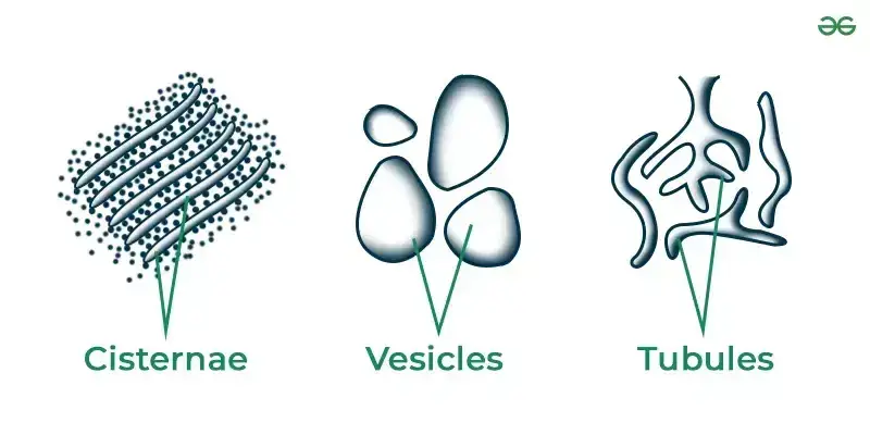
- Morphological Forms of ER The ER manifests in three distinct forms, each with specific structural and functional attributes:
- Cisternae: These are long, flattened, sac-like structures, typically found in cells with high synthetic activity, such as pancreatic and brain cells. Cisternae are arranged in parallel arrays and are about 40 to 50 µm in diameter.
- Vesicles: These are oval, membrane-bound structures, ranging from 25 to 500 µm in diameter. Vesicles are particularly abundant in the smooth endoplasmic reticulum (SER) and play a crucial role in the transport of cellular materials.
- Tubules: Tubules are branched structures that form a reticular system along with cisternae and vesicles. They vary in diameter from 50 to 190 µm and are dynamic, involved in membrane movements and the fission and fusion of the cytocavity network.
- Functional Diversity of ER Components
- Cisternae: In the rough ER (RER), cisternae are the primary sites for protein synthesis, folding, and modification. Their proximity to the nucleus and stacked arrangement facilitate these processes.
- Vesicles: These structures are integral to the transport of proteins and lipids within the cell, shuttling these molecules to various destinations, including the cell surface.
- Tubules: Tubules contribute to the structural flexibility of the ER, extending and connecting the ER throughout the cell. They are instrumental in the internal movement of materials within the ER.

Subdomains of the Endoplasmic Reticulum
| ER Domain | Function | Associated Proteins |
| Rough ER | Protein translocation Protein folding and oligomerization Carbohydrate addition ER degradation | Sec61 complex, TRAP, TRAM, BiP PDI, Calnexin, Calreticulin, BiP Oligosaccharide transferase EDEM, Derlin1 |
| Smooth ER | Detoxification Lipid metabolism Heme metabolism Calcium release | Cytochrome P450 enzymes HMG-CoA reductase Cytochrome b5 IP3 receptors |
| Nuclear envelope | Nuclear pores Chromatin anchoring | POM121, GP210 Lamin B receptor |
| ER export sites | Export of proteins and lipids into secretory pathway | Sar1p, Sec12p, Sec16p |
| ER contact zones | Transport of lipids | LTPs |
Differences Between Smooth Endoplasmic Reticulum (SER) and Rough Endoplasmic Reticulum (RER)
- Appearance
- Rough Endoplasmic Reticulum: Characterized by a rough appearance due to the presence of ribosomes on its surface.
- Smooth Endoplasmic Reticulum: Exhibits a smooth texture as it lacks ribosomes.
- Structural Composition
- RER: Comprises flattened sacs known as cisternae, which are integral to its structure.
- SER: Formed by a network of tubules and vesicles, differing from the cisternal structure of the RER.
- Location within the Cell
- RER: Predominantly situated near the nucleus, although it can extend to other cellular regions.
- SER: Distributed throughout the cell, often found in proximity to the nucleus.
- Primary Functions
- RER: Plays a crucial role in protein synthesis.
- SER: Involved in lipid synthesis, detoxification processes, and calcium storage.
- Cellular Examples
- RER: Commonly found in plasma cells, pancreatic cells, and cells of the digestive system.
- SER: Predominantly present in liver cells and cells in reproductive organs such as the ovaries and testes.
- Additional Functions
- RER: Apart from protein synthesis, it is also involved in membrane synthesis and modification as part of the endomembrane system.
- SER: May have roles in carbohydrate metabolism, adding to its multifunctional nature.
| Feature | Rough Endoplasmic Reticulum (RER) | Smooth Endoplasmic Reticulum (SER) |
|---|---|---|
| Appearance | Rough, due to ribosomes on the surface | Smooth, as it lacks ribosomes |
| Structure | Composed of flattened sacs called cisternae | Consists of a network of tubules and vesicles |
| Location | Primarily near the nucleus, but can extend elsewhere | Found throughout the cell, often adjacent to the nucleus |
| Primary Function | Protein synthesis | Lipid synthesis, detoxification, calcium storage |
| Cellular Examples | Plasma cells, pancreatic cells, digestive system cells | Liver cells, cells in ovaries and testes |
| Additional Functions | Involved in membrane synthesis and modification | May be involved in carbohydrate metabolism |
Endoplasmic Reticulum (ER) Diagram

Endoplasmic Reticulum Shape Generation

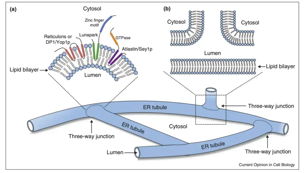
- Role of Specific Proteins in ER Morphology The shape of the endoplasmic reticulum (ER) is intricately determined by various protein classes. These proteins ensure the ER maintains its unique structure, comprising a single polygonal network of tubules and stacked cisternae.
- Formation of ER Tubules
- Key Proteins: Reticulons and DP1/Yop1 proteins are pivotal in mediating ER tubule formation.
- Mechanism: These proteins possess hydrophobic segments predicted to form α-helical hairpins that partially span the lipid bilayer. The insertion of these segments into the cytoplasmic leaflet of the bilayer ER membrane results in the highly curved, tubular ER morphology.
- Stabilization of Curved Membranes Reticulons and DP1/YOP1 proteins also play a role in the formation of nuclear pores by stabilizing curved membranes, further illustrating their multifaceted function in cellular architecture.
- Formation of Three-Way Junctions
- Atlastin Proteins: Dynamin-related GTPases from the Atlastin/RHD3/Sey1p family are crucial for creating three-way junctions, contributing to the polygonal structure of the tubular ER network.
- Structure and Function: Atlastins feature an N-terminal cytoplasmic GTPase domain, a three-helix bundle, two closely spaced transmembrane segments, and a C-terminal amphipathic helix. GTP binding facilitates interactions between atlastin oligomers in adjacent membranes, leading to tethered complex formation.
- Mechanism of ER Tubule Fusion
- GTP Hydrolysis: The fusion of ER tubules is contingent on a conformational change in the cytosolic domain, triggered by GTP hydrolysis.
- GDP Release: Following membrane fusion, GDP is released by atlastin.
- Interactions with Microtubules
- Microtubule-Driven Remodeling: The ER can be remodeled through interactions with microtubules in two ways: being pulled alongside a microtubule by motor proteins, or attaching to +TIP complexes that track the growing ends of microtubules.
- STIM1 and ER Remodeling: STIM1, a transmembrane ER protein, binds directly to the +TIP protein EB1 to facilitate its reach to the plasma membrane and activate Orai.
- Influence of the Actin Cytoskeleton The actin cytoskeleton also drives ER remodeling, highlighting the dynamic interplay between the ER and the cell’s structural framework.
- Clinical Implications of ER Structure Regulation
- Disease Associations: Deficiencies in proteins regulating ER structure often result in diseases. For instance, mutations in atlastin or reticulons are linked to hereditary spastic paraplegias, characterized by the degeneration of axons in corticospinal upper motor neurons.
- Neurological Disorders: These mutations also contribute to the pathogenesis of amyotrophic lateral sclerosis, which involves degeneration of both upper and lower motor neurons.
- Importance in Neurons Given the large size and highly polarized geometry of neurons, the shaping and distribution of the ER network are particularly crucial, underscoring the significance of ER morphology in cellular function and health.
Calcium (Ca2+) Metabolism in the Endoplasmic Reticulum (ER)
- ER as a Major Calcium Store
- The endoplasmic reticulum (ER) is not only pivotal in the synthesis and transport of biomolecules but also serves as a significant reservoir of intracellular Ca2+.
- Concentration Variance: The concentration of Ca2+ in the ER lumen ranges from 100–800 μM, contrasting with the typical cytosolic concentration of ~100 nM and the extracellular concentration of ~2 mM.
- Calcium Channels and Receptors in the ER
- The ER houses several calcium channels, including ryanodine receptors (RyRs) and inositol 1,4,5-trisphosphate (IP3) receptors (IP3R), which are instrumental in releasing Ca2+ from the ER into the cytosol.
- Mechanism of Ca2+ Release
- Activation of phospholipase C (PLC) through G protein-coupled receptor (GPCR) stimulation leads to the cleavage of phosphatidylinositol 4,5 bisphosphate (PIP2) into diacyl-glycerol (DAG) and IP3.
- IP3 subsequently binds to IP3R, triggering Ca2+ release and a transient increase in intracellular Ca2+ levels.
- Role of Ryanodine Receptors
- RyRs facilitate Ca2+-induced Ca2+ release (CICR), responding to elevated cytoplasmic levels of Ca2+.
- Additionally, depolarization of t-tubule membranes can cause conformational changes in voltage-dependent Ca2+ channels, such as dyhydropyridine receptors (DHPRs), which activate RyRs for further Ca2+ release.
- Regulation of Ca2+ in the ER
- Ca2+ can leak from the ER into the cytoplasm and is then pumped back into the ER via sarcoendoplasmic reticular Ca2+ ATPases (SERCAs).
- The cell can also intake Ca2+ from the extracellular medium, adding complexity to the regulation process.
- Store-Operated Ca2+ Entry (SOCE)
- When ER Ca2+ stores are rapidly depleted, particularly through IP3R-mediated release, SOCE is activated.
- STIM1 proteins cluster in ER regions adjacent to the plasma membrane, trapping Orai1 subunits and forming active Ca2+ release-activated channels (CRAC) for extracellular Ca2+ uptake into the ER lumen.
- Unique Aspect of SOCE
- Interestingly, SOCE and CRAC activation do not depend on changes in cytoplasmic Ca2+ levels but respond to alterations in luminal Ca2+ concentration.
- Broad Impact of Ca2+ Signaling
- Calcium is a versatile signaling molecule influencing various processes, including protein localization, function, and interactions.
- Ca2+ release can manifest as a cell-wide wave, a gradient from the release source, or a localized wave from clustered channels, known as a Ca2+ spark.
- Ca2+ in Cellular Functions
- Ca2+ release plays a crucial role in various cellular events, such as muscle contraction, secretion, and neurotransmitter release in neurons.
- Recent evidence suggests that Ca2+ may also influence the reshaping of the ER in response to cellular signals.
Functions of Endoplasmic Reticulum (ER)
- Rough Endoplasmic Reticulum (RER)
- Protein Synthesis: RER facilitates increased protein synthesis by synthesizing proteins.
- Formation of Smooth ER (SER): SER is derived from RER through the loss of ribosomes.
- Nuclear Membrane Reformation: RER plays a role in reforming the nuclear membrane during telophase.
- Vesicle Formation: RER is involved in forming vesicles that transport chemicals from the ER to the Golgi Apparatus.
- Smooth Endoplasmic Reticulum (SER)
- Lipid Synthesis: SER’s primary function is related to lipid synthesis.
- Hormone Synthesis: It synthesizes sex hormones like testosterone and estrogens.
- Glycogenolysis: SER aids in the process of glycogenolysis.
- Muscle Contraction: Sarcoplasmic reticulum, a type of SER in muscle cells, assists in muscle contraction.
- Detoxification: SER helps in detoxifying harmful substances like drugs and carcinogens.
- Common Functions of SER and RER
- Mechanical Support: ER provides structural support to the cell, dividing its fluid content into compartments.
- Transport System: ER functions as a circulatory system, distributing proteins, lipids, enzymes, etc., within the cell.
- Detoxification: ER in liver cells plays a crucial role in detoxifying harmful chemicals.
- Glycogenolysis: ER is involved in converting glycogen to glucose, with key enzymes like glucose-6-phosphatase.
- Additional Functions of SER
- Steroid Hormone Synthesis: SER is involved in synthesizing steroid hormones, carbohydrates, lipids, and cholesterol.
- Plasma Membrane Synthesis: SER contributes to the synthesis of plasma membranes.
- Reproductive Hormone Production: In reproductive cells, SER produces hormones.
- Calcium Storage: In muscle cells, SER stores calcium ions, aiding in muscle contraction.
- Additional Functions of RER
- Efficient Protein Synthesis: The ribosomes on RER are crucial for efficient protein synthesis.
- Secretory Product Production: RER produces secretory products and forms vesicles for their transport.
- Antibody Secretion: In plasma cells, RER is involved in antibody secretion.
- Insulin Secretion: RER in pancreatic cells aids in insulin secretion.
- Glycosylation: The process of glycosylation, linking proteins to sugars to form glycoproteins, occurs in the RER.
FAQ
What is endoplasmic reticulum?
In eukaryotic cells, the endoplasmic reticulum (ER) is a membrane-bound organelle. It is a network of flattened sacs, tubules, and cisternae that stretch throughout the cytoplasm and are attached to the nuclear envelope. Rough endoplasmic reticulum (RER) and smooth endoplasmic reticulum (SER) are the two forms of ER (SER).
Ribosomes cover the surface of RER, giving it a rough appearance. It is indispensable for protein synthesis and processing. Before being delivered to their final destination, newly produced proteins undergo folding, modification, and quality control in the RER lumen.
SER is devoid of ribosomes and has a more uniform look. It participates in a number of metabolic activities, including lipid synthesis, detoxification, and calcium ion storage. It also regulates intracellular calcium levels and is involved in the generation of steroid hormones.
Endoplasmic reticulum is a highly dynamic organelle that is necessary for numerous cellular functions, such as protein synthesis, lipid metabolism, and intracellular signaling.
List the types of endoplasmic reticulum.
There are two types of endoplasmic reticulum:
1. Rough endoplasmic reticulum (RER): It is studded with ribosomes on its surface, giving it a rough appearance. RER plays a crucial role in protein synthesis and processing. Newly synthesized proteins enter the RER lumen, where they undergo folding, modification, and quality control before being transported to their final destination.
2. Smooth endoplasmic reticulum (SER): It lacks ribosomes and has a smoother appearance. SER is involved in various metabolic processes, such as lipid synthesis, detoxification, and calcium ion storage. It also plays a role in the regulation of intracellular calcium levels and the production of steroid hormones.
List the functions of the endoplasmic reticulum.
In eukaryotic cells, the endoplasmic reticulum (ER) is a membrane-bound organelle. It serves multiple crucial tasks within the cell, including:
Protein synthesis: Attached to the membrane of the ER are ribosomes that contribute to the production of proteins. These proteins are then transferred to other cellular compartments or secreted outside the cell.
Lipid synthesis: The ER is also involved in the synthesis of phospholipids and steroids, among other lipids. These lipids have key functions in cell membranes, energy storage, and signaling.
Detoxification: The ER plays an important function in the detoxification of drugs and other foreign substances in the body. In a process known as biotransformation, enzymes in the ER convert these compounds so that they can be removed from the body.
Calcium storage: The endoplasmic reticulum (ER) is also involved in the storage and release of calcium ions, which are essential for numerous cellular functions, including muscle contraction and signal transduction.
Cell signaling: The endoplasmic reticulum (ER) is engaged in multiple signaling pathways, such as the unfolded protein response (UPR) and the ER stress response, which help the cell adapt to stress and maintain homeostasis.
Overall, the ER is critical to the correct functioning of the cell, as it is involved in numerous cellular processes that are essential to its survival.
What is the Endoplasmic Reticulum (ER)?
The Endoplasmic Reticulum (ER) is a cellular organelle that plays a crucial role in the synthesis, modification, and transport of proteins and lipids.
What are the two types of ER?
There are two types of ER: Rough Endoplasmic Reticulum (RER) and Smooth Endoplasmic Reticulum (SER). The RER is covered in ribosomes, while the SER lacks ribosomes.
What is the function of RER?
The RER is responsible for the synthesis and modification of proteins that are destined for secretion or for use in the cell membrane.
What is the function of SER?
The SER is involved in lipid metabolism, detoxification of drugs and toxins, and the storage and release of calcium ions.
How does the ER contribute to protein synthesis?
The ER provides the platform for the synthesis of proteins by ribosomes, which are located on the surface of the RER. Newly synthesized proteins are then transported to the Golgi apparatus for further processing and distribution.
How does the ER play a role in lipid synthesis?
The SER is responsible for the synthesis of lipids, including cholesterol and phospholipids, which are essential components of cell membranes.
How does the ER participate in detoxification?
The SER is involved in the detoxification of drugs and toxins by modifying them to make them more soluble and easier to excrete from the body.
What is the role of ER in calcium storage and release?
The ER plays a critical role in the storage and release of calcium ions, which are important for cell signaling and muscle contraction.
How is ER structure maintained?
The unique morphology of the ER is maintained by a variety of proteins, including reticulons, DP1/Yop1, and atlastins, which ensure that the ER remains a single polygonal network of tubules and stacked cisternae.
What happens when there are defects in ER structure or function?
Defects in ER structure or function can lead to a variety of diseases, including neurodegenerative disorders and metabolic disorders, highlighting the crucial role of the ER in cellular function and health.
References
- Chen, Shuliang; Novick, Peter; Ferro-Novick, Susan (2013). ER structure and function. Current Opinion in Cell Biology, 25(4), 428–433. doi:10.1016/j.ceb.2013.02.006
- Schwarz DS, Blower MD. The endoplasmic reticulum: structure, function and response to cellular signaling. Cell Mol Life Sci. 2016 Jan;73(1):79-94. doi: 10.1007/s00018-015-2052-6. Epub 2015 Oct 3. PMID: 26433683; PMCID: PMC4700099.
- Endoplasmic Reticulum. (2017). Cell Biology, 331–350. doi:10.1016/b978-0-323-34126-4.00020-7
- https://bscb.org/learning-resources/softcell-e-learning/endoplasmic-reticulum-rough-and-smooth/
- https://www.vedantu.com/biology/endoplasmic-reticulum
- http://www.biology4kids.com/files/cell_er.html
- https://biologydictionary.net/rough-endoplasmic-reticulum/
- https://www.geeksforgeeks.org/endoplasmic-reticulum/
- https://www.thoughtco.com/endoplasmic-reticulum-373365
- https://rajusbiology.com/endoplasmic-reticulum-structure-and-functions/