

Membrane proteins are proteins found within the lipid bilayer or attached to the surface of biological membranes. They form an important component of the plasma membrane and the membranes surrounding different cell organelles.
Based on their association with membrane, the proteins are mainly of three types, integral membrane proteins, peripheral membrane proteins, and lipid-anchored proteins. Integral membrane proteins are embedded within the lipid bilayer. Some of them pass completely across the membrane. These are referred to as transmembrane proteins.
On the other hand, peripheral membrane proteins are loosely attached to the inner or outer surface of the membrane and they do not enter into its hydrophobic core. The membrane proteins have different functions in the cell.
They are used in transport of ions and molecules, cell signalling, enzymatic reactions and cell recognition. Some membrane proteins also help in attachment of the membrane with other cellular structures.
How Do Proteins Associate With the Lipid Bilayer?
The lipid bilayer has two different regions. The polar heads of phospholipids are hydrophilic and remain exposed towards water, while the fatty acid chains form a hydrophobic region within the membrane. Due to this arrangement, the hydrophobic region of a protein remains associated with the hydrophobic part of membrane and its hydrophilic region is generally exposed towards the aqueous side. Proteins can therefore be present across the complete membrane, within only one layer or just attached to its surface. The following are the different ways by which proteins associate with the lipid bilayer-
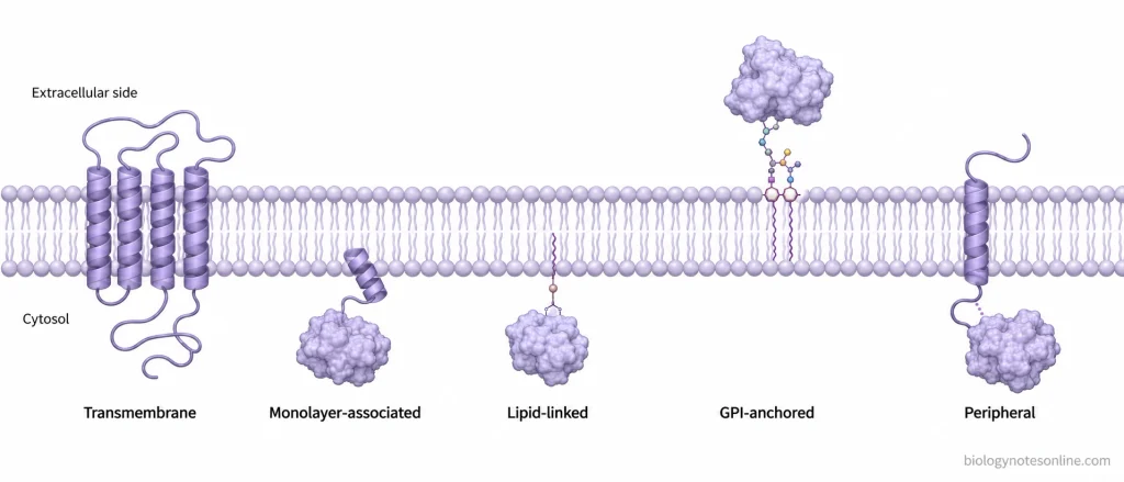
- Transmembrane proteins- These proteins pass through the lipid bilayer and are exposed on both sides of membrane. The membrane spanning portion is mainly composed of nonpolar or hydrophobic amino acids. These amino acids interact with the hydrophobic fatty acid chains present inside the bilayer, whereas the hydrophilic portions of protein remain towards the aqueous environment. Some proteins cross the membrane only once. Others cross several times, forming a number of membrane-spanning regions. Most commonly these regions are present as α-helices, while β-barrel structure is found in some proteins.
- Monolayer-associated proteins- In this type, the complete protein remains on the cytosolic side and does not cross the membrane. An amphipathic α-helix present on the surface of protein is inserted into one monolayer. Its hydrophobic side remains towards the lipid region and the hydrophilic portion faces the cytosol.
- Lipid-linked proteins- Some proteins are attached to the cytosolic surface by a lipid chain. The lipid group is covalently attached with the protein and its hydrophobic part enters into the lipid bilayer. Fatty acid chains and prenyl groups are commonly involved in this type of attachment. Here the protein itself does not need to extend across the membrane.
- GPI-anchored proteins- These proteins are present on the noncytosolic surface of membrane. They are attached with lipid bilayer through glycosylphosphatidylinositol (GPI) anchor, which remains in the outer monolayer of plasma membrane. Thus, the protein is held at the membrane surface without having its polypeptide chain passing through the hydrophobic interior.
- Peripheral membrane proteins- These proteins do not extend into the hydrophobic region of lipid bilayer at all. Instead, they are attached with integral membrane proteins or other membrane components by noncovalent interactions. This association is comparatively weak. Many of these proteins can therefore be separated from membrane by changing ionic strength or pH without breaking the lipid bilayer. This type of proteins are referred to as peripheral membrane proteins.
Types of Membrane Proteins
Membrane proteins are classified mainly based on their association with membrane and how far the protein enters into the lipid bilayer. The classification is not same in all the textbooks. Some descriptions divide them mainly into integral and peripheral membrane proteins, whereas lipid-anchored proteins are included with integral proteins or shown separately depending on the classification. Transmembrane proteins are also not another name for all integral membrane proteins. They are the integral proteins which pass through the complete lipid bilayer.
The following are the major types of membrane proteins-
| Type | Position in membrane | Entry/crossing of hydrophobic core | Major association | Extraction | Examples |
|---|---|---|---|---|---|
| Integral membrane protein | Present within lipid bilayer | Enters the hydrophobic region. It may not cross the complete bilayer. | Mainly hydrophobic interaction with membrane lipids | Difficult. Detergents are generally required. | Cytochrome b5 (monotopic) |
| Transmembrane protein | Extends across the membrane | Crosses the complete hydrophobic core | Hydrophobic regions of protein interact with fatty acid chains | Difficult without disrupting lipid-protein interactions | Glycophorin, band 3 |
| Peripheral membrane protein | Inner or outer membrane surface | Does not enter the hydrophobic core | Noncovalent interaction with lipid head groups or membrane proteins | Comparatively easy in many cases by salt or change in pH | Spectrin |
| Lipid-anchored protein | Protein remains mainly on one membrane surface | Attached lipid enters the bilayer, the polypeptide itself need not cross it | Covalent lipid attachment | High salt usually does not remove it. Anchor cleavage or membrane-disrupting treatment may be required. | Ras proteins, Thy-1 |
| Amphitropic protein | Soluble form or associated with membrane | Variable | Reversible membrane interaction | Association can be reversed | Protein kinase C |
1. Integral Membrane Proteins
Integral membrane proteins are the proteins that are tightly associated with the lipid bilayer. A portion of protein enters into the hydrophobic region of membrane where it interacts with the fatty acid chains of lipids. Due to these hydrophobic interactions, the proteins are not normally removed by high salt concentration or simple change in pH. Detergents are generally used for their extraction from the membrane.
Not all integral proteins cross the whole lipid bilayer. Some proteins remain on one side of the membrane but a hydrophobic part enters into only one monolayer. This is referred to as an integral monotopic protein. Cytochrome b5 is an example of this type of membrane association.
2. Transmembrane Proteins
Transmembrane proteins are integral membrane proteins which pass from one side of the lipid bilayer to the other. The portion lying inside the membrane is mainly hydrophobic, while the regions exposed towards water are hydrophilic. Depending on the number of times the polypeptide crosses the membrane, these proteins are mainly of two forms-
- Single-pass or bitopic proteins- The polypeptide crosses the lipid bilayer only once. Glycophorin of the red blood cell membrane is a single-pass transmembrane protein, having one hydrophobic α-helical region through the membrane.
- Multipass or polytopic proteins- These proteins pass through the lipid bilayer several times. Thus, several membrane-spanning regions are present in the same protein. Band 3 of red blood cells is a multipass membrane protein.
Therefore, transmembrane proteins form a major type of integral membrane proteins, but every integral membrane protein is not necessarily a transmembrane protein.
3. Peripheral Membrane Proteins
Peripheral membrane proteins do not enter into the hydrophobic interior of lipid bilayer. They are present on either surface of the membrane and may be attached with an integral membrane protein or with the polar head groups of membrane lipids. The attachment is mainly noncovalent. Many of these proteins can therefore be separated by high or low ionic strength and extreme pH without breaking the lipid bilayer. Spectrin, which is associated with the cytosolic surface of red blood cell membrane, is an example.
4. Lipid-Anchored
In this type, a lipid group is covalently attached with the protein and the lipid portion gets inserted into the membrane. Myristoyl, palmitoyl and prenyl groups can attach proteins towards the cytosolic surface. Glycosylphosphatidylinositol (GPI) anchor, on the other hand, attaches proteins to the noncytosolic surface of plasma membrane. Ras proteins and Thy-1 are examples of lipid-anchored proteins. The position of these proteins in classification varies. They are included with integral proteins in some descriptions, whereas in others lipid-anchored proteins are written separately.
5. Amphitropic Membrane Proteins
These proteins are not permanently fixed with the membrane. They can remain in soluble form and under suitable conditions become associated with membrane lipids or membrane proteins. The binding is reversible. It can be controlled by Ca2+, ligand binding, phosphorylation, acylation or exposure of membrane-binding regions in different proteins. Protein kinase C is one of the examples of an amphitropic protein.
Structural Features of Membrane Proteins
Membrane proteins have special structural features which allow them to remain stable within the lipid bilayer and at the same time interact with the aqueous environment. Their structure therefore differs in the membrane-spanning region and the portions present outside the membrane. Some of the important structural features are as follows-
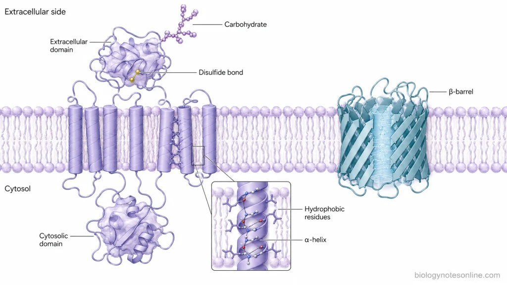
- Amphipathic nature- Most transmembrane proteins are amphipathic, having both hydrophobic and hydrophilic regions. The hydrophobic region lies within the lipid bilayer where it interacts with the fatty acid chains. Hydrophilic portions are exposed towards water on either side of membrane. Thus, different amino acids of the same protein may be present in quite different chemical environments.
- Transmembrane α-helices- The membrane-spanning region of most transmembrane proteins occurs in the form of an α-helix. It is generally made up of about 20-25 amino acid residues, with large number of nonpolar side chains. These hydrophobic residues face the lipid molecules. The α-helical arrangement also allows the polar peptide bonds of the protein backbone to form hydrogen bonds with one another within the water-free membrane interior. Some proteins contain a single α-helix while others have many such helices.
- β-barrel structure- Not every membrane protein crosses the membrane as an α-helix. In some proteins, several β-strands are arranged together forming a closed β-barrel. The nonpolar amino acid side chains generally face towards the surrounding membrane lipids, while polar residues can face the water-filled interior of the barrel. Porins are the common examples. β-barrel proteins are found mainly in the outer membranes of bacteria, mitochondria and chloroplasts.
- Hydrophilic domains and loops- The parts of transmembrane proteins which remain outside the hydrophobic core are generally hydrophilic. These regions may form large domains or short loops on the cytosolic and noncytosolic sides of membrane. In multipass proteins, the membrane-spanning regions are therefore connected with several loops of unequal lengths. Some of these exposed regions form binding sites or interact with other cellular molecules.
- Fixed membrane orientation- A transmembrane protein has a particular orientation in the lipid bilayer. Its cytosolic domain remains towards the cytosol and the noncytosolic domain remains towards the opposite side. Once inserted, the protein does not normally turn over from one side of the bilayer to the other. This arrangement gives the membrane protein an asymmetric structure across the membrane.
- Carbohydrate chains- Many membrane proteins, particularly those of animal plasma membrane, contain carbohydrate chains and are referred to as glycoproteins. The carbohydrate portions are attached during passage through the endoplasmic reticulum and Golgi apparatus. They are found on the noncytosolic surface of intracellular membranes and finally on the extracellular surface of plasma membrane.
- Disulfide bonds- Disulfide bonds can be present in the noncytosolic domains of membrane proteins. These bonds are formed between cysteine residues and help in stabilizing the folded protein or association between different polypeptide chains. On the cytosolic side, such bonds are much less common because the cytosol has a reducing environment.
- Association of protein subunits- Membrane proteins are not always present as a single polypeptide working alone. Many of them associate with identical or different protein chains and form larger protein complexes. The membrane-spanning α-helices themselves can participate in these protein-protein interactions. Glycophorin, for example, can occur as a homodimer where interaction takes place between its transmembrane helices.
How Membrane Protein Structure Determines Function
The function of membrane proteins is based on their structure, position in membrane and arrangement of amino acids. Different folding of protein forms pores, binding regions and sites for interaction with other molecules. The following are some of the important structure-function relationships-
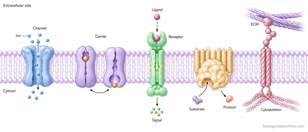
- Membrane-spanning regions- The hydrophobic portion of protein remains within the lipid bilayer, whereas the hydrophilic portion is exposed towards water. It also fixes the protein in a particular position and orientation in the membrane.
- Channel proteins- These proteins form a hydrophilic passage through the membrane. The size, charge and amino acids present in the channel determine the ions or molecules that can pass through it.
- Carrier proteins- Carrier proteins have specific binding sites for the molecule that is to be transported. The molecule first binds with the protein. This changes the shape of protein and helps in its movement across membrane.
- Receptor proteins- It has an outer region for binding of signal molecule and an inner region involved in cellular response. Binding of ligand changes the receptor structure, which then transfers the signal towards inside of the cell.
- Membrane-bound enzymes- Some membrane proteins have catalytic regions and act as enzymes. Their position in membrane allows the protein to remain near the substrates or other proteins involved in the reaction.
- Cell adhesion proteins- These proteins bind extracellular molecules on one side and cytoskeletal proteins on the other side. Thus, they help in cell attachment, maintaining cell shape and organization of tissues.
- Specific binding sites- Folding of protein brings particular amino acids together, forming a specific binding region. Change in this structure can also change ligand binding, transport or enzymatic activity of the protein.
What Are the Major Functions of Membrane Proteins?
Membrane proteins perform most of the specific functions of biological membranes. They are involved in transport, communication, cellular recognition and also provide structural attachment to the cell. Some membrane proteins also act as enzymes. The following are some of the important functions-
- Transport of molecules- Membrane proteins are used to transport ions, sugars, amino acids and other polar molecules across the membrane. Channel proteins form pores while carrier proteins bind and transfer particular molecules.
- Receptors and cell signalling– Some membrane proteins act as receptors for hormones and other signal molecules. The signal binds on the outer surface and produces a response towards inside of the cell.
- Enzymatic activity- A number of membrane proteins function as enzymes. They catalyze reactions taking place on the membrane surface or within membrane-associated pathways.
- Cell recognition- Glycoproteins present on the cell surface can act as identification markers. These are involved in recognition between cells and help cells distinguish different cell surfaces.
- Cell adhesion and joining- Some proteins of adjacent cells attach with one another, forming connections between the cells. It helps in maintaining the arrangement of cells in tissues.
- Attachment with cytoskeleton and extracellular matrix- Membrane proteins can provide a link between cytoskeleton present inside the cell and extracellular matrix outside it. This association helps in maintaining cell shape and structural organization.
- Energy transduction- In mitochondrial and chloroplast membranes, membrane proteins take part in electron transport and ATP production. Hence, these proteins are important components of cellular energy-producing systems.
Examples of Membrane Proteins
The following are some of the important examples of membrane proteins-
- Glycophorin- Glycophorin is a major transmembrane protein of human red blood cell membrane, having a single α-helix that passes through the lipid bilayer. The carbohydrate-rich portion is present towards the outer surface of cell. Its exact function is not clearly known.
- Band 3 protein- Another major transmembrane protein of red blood cells. It is a multipass protein and involved in exchange of bicarbonate (HCO₃⁻) and chloride (Cl⁻) ions across the membrane.
- Na⁺/K⁺-ATPase- It is an integral membrane pump found in animal cells, which uses one ATP for transporting 3 Na⁺ ions outside and 2 K⁺ ions inside the cell.
- Aquaporin 1 (AQP1)- A water channel protein of red blood cell membrane. It allows rapid movement of water across the membrane while ions are not transported through the channel.
- Rhodopsin- Rhodopsin is a seven-pass transmembrane protein found in rod photoreceptor membranes. It contains 11-cis-retinal which is used in the detection of light during vision.
- Integrins- These are transmembrane glycoproteins made up of α and β subunits. They bind extracellular matrix proteins with the cell and also connect with the cytoskeleton. Cell adhesion and signalling are some of the functions.
- Spectrin- It is a peripheral membrane protein present at the cytosolic surface of red blood cell membrane. A flexible protein meshwork is formed by spectrin, helping in maintaining the structural integrity and biconcave shape of RBC.
- Ras proteins- These are membrane-associated signalling proteins. A prenyl group is covalently attached with the C-terminal region of Ras, which helps in association of the protein with membrane.
Membrane Protein Mobility and Organization in the Membrane
The membrane is a fluid structure and many proteins present in it can move within the same plane of lipid bilayer. This movement, however, is not same for all membrane proteins. Some remain freely mobile while movement of others is restricted by proteins, cytoskeleton or particular membrane regions. The following are some of the important features-
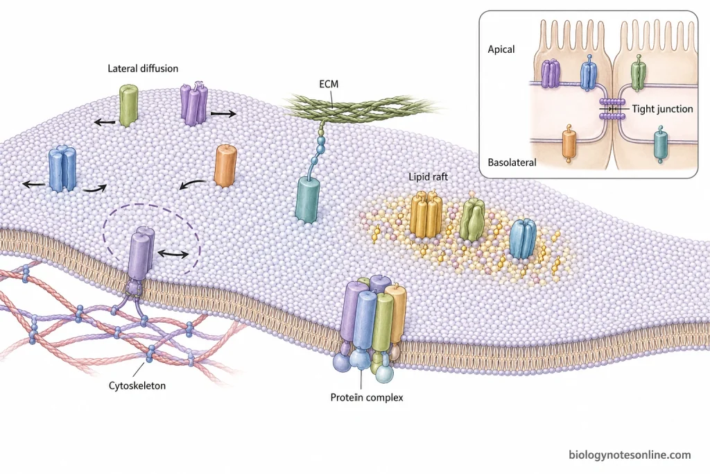
- Lateral movement- Many membrane proteins move sideways within the lipid bilayer. This is referred to as lateral diffusion. Proteins can also rotate within membrane, but movement of a protein from one side of bilayer to the other does not normally occur.
- Restriction by cytoskeleton- Some proteins are attached with the cytoskeleton present below the plasma membrane. Their movement is therefore restricted to a small region or they may remain almost fixed at one position.
- Attachment with extracellular structures- Membrane proteins may also be attached to extracellular matrix or proteins outside the cell. Such attachment reduces their lateral movement in membrane.
- Protein complexes- Some membrane proteins combine with one another forming large protein complexes or aggregates. The proteins present in such groups move slowly as compared to individual proteins. In some cases they remain relatively fixed.
- Membrane domains- Proteins are not always distributed equally throughout the plasma membrane. In epithelial cells, some proteins are present only at the apical region while others remain at basal and lateral regions. Tight junctions act as barriers and prevent mixing of proteins between these membrane regions.
- Lipid rafts- These are small and dynamic membrane regions containing higher amounts of cholesterol and sphingolipids along with certain membrane proteins. Some proteins become concentrated in these regions, which helps in their organization for processes such as cell signalling and membrane transport. The exact structure and organization of lipid rafts is still described with some variation.
- Mobility is different in different membranes- Proteins of some intracellular membranes are highly mobile, whereas many plasma membrane proteins show slower movement because of interaction with cytoskeleton, extracellular matrix and other membrane components.
How Are Membrane Proteins Studied?
Different biochemical, microscopic and structural methods are used for studying membrane proteins. Integral membrane proteins are more difficult to isolate because their hydrophobic regions remain within the lipid bilayer. The following are some of the important methods-
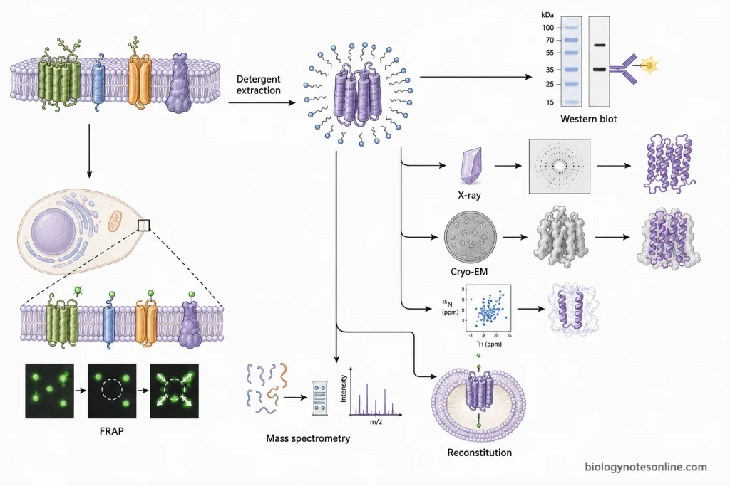
- Detergent extraction and purification- Detergents are used for removing integral proteins from the lipid bilayer. The detergent surrounds the hydrophobic portion of protein and keeps it soluble in water. Mild detergents are generally preferred when protein activity has to be maintained.
- SDS-PAGE and Western blotting- The extracted membrane proteins are separated according to their molecular size by SDS-PAGE. Western blotting is then used for detection of a particular protein with the help of specific antibodies.
- Fluorescence microscopy- Membrane proteins are tagged with fluorescent molecules and their location in the cell can be viewed under fluorescence microscope. FRAP (Fluorescence Recovery After Photobleaching) is used to study lateral movement and diffusion of proteins within membrane.
- X-ray crystallography- It is used to determine three-dimensional structure of membrane proteins. For this method, the purified protein is crystallized after its removal from membrane. Crystallization of membrane proteins is difficult because of their hydrophobic nature.
- Cryo-electron microscopy (Cryo-EM)– This method is used for studying structure of membrane proteins and larger protein complexes at high resolution. It does not require formation of three-dimensional protein crystals.
- NMR spectroscopy- NMR is used to study structure, orientation and molecular movement of membrane proteins. Some proteins can also be examined in lipid bilayer-like environment.
- Mass spectrometry- It is used for identification and analysis of membrane proteins. Protein sequence, post-translational modifications and interaction with other proteins can also be studied by this method.
- Functional reconstitution- Purified membrane proteins are placed again into artificial lipid membranes or liposomes. The activity of protein is then studied, such as transport of ions or molecules through a membrane transporter. This allows the function of a membrane protein to be examined separately from other cellular components.
How Are Membrane Proteins Studied?
Different methods are used for the study of membrane proteins, depending upon whether protein has to be isolated, detected or its structure and activity are to be studied. Integral membrane proteins are comparatively difficult to isolate because of having hydrophobic regions within the lipid bilayer. Some of the important methods include:
- Detergent extraction and purification- Detergents are used for removing integral proteins from membrane. The hydrophobic portion gets surrounded by detergent molecules which allows the protein to remain soluble in aqueous solution. Mild detergents are generally used when protein activity is required to be maintained.
- SDS-PAGE and Western blotting- After extraction, SDS-PAGE separates the membrane proteins according to their molecular size. A particular protein is detected using Western blotting with specific antibodies.
- Fluorescence microscopy- Membrane proteins can be labelled with fluorescent molecules and observed under fluorescence microscope. Their location within the cell can be studied. FRAP (Fluorescence Recovery After Photobleaching) is used for studying the lateral movement and diffusion of protein in membrane.
- X-ray crystallography- A method used for determining three-dimensional structure of purified membrane protein. For this, the protein is removed from membrane and crystallized. Their crystallization is comparatively difficult due to the hydrophobic regions.
- Cryo-electron microscopy (Cryo-EM)- It is used for high-resolution structural study of membrane proteins and large protein complexes without forming the three-dimensional crystals required in X-ray crystallography.
- NMR spectroscopy- NMR is another method for studying membrane protein structure, orientation and molecular movements. Some proteins are also studied in lipid bilayer-like environment.
- Mass spectrometry- It is used in identification and analysis of membrane proteins. Protein sequence and post-translational modifications can be studied by this method, together with interactions of the protein with other proteins.
- Functional reconstitution- Purified membrane protein is again placed into artificial lipid membrane or liposome. Its activity is then studied separately. For example, movement of ions or molecules through a transporter can be examined after reconstitution.
Why Are Membrane Proteins Biologically Important?
Membrane proteins play a vital role in the normal functioning of cells because most of the specific activities of membrane are performed by these proteins. Transport of molecules, reception of signals, cellular attachment and energy production are some of the important functions.
- Transport and cellular environment- Channels and pumps are used in the movement of ions, sugars, amino acids and other molecules across membrane. They also maintain ion gradients. This is necessary for maintaining the internal cellular environment.
- Cell signalling- Receptor proteins present in plasma membrane recognize extracellular signals. The signal is received at the membrane and a response is then produced inside the cell.
- Energy production- In mitochondria and chloroplasts, several membrane protein complexes take part in electron transport and formation of proton gradient. During this process, ATP synthase uses this gradient for synthesis of ATP.
- Electrical activity- Ion channels and transport proteins maintain electrical gradients across membrane, especially in nerve and muscle cells. Production and transmission of electrical signals depend on these proteins.
- Cell adhesion and tissue organization- Some membrane proteins attach neighbouring cells and other proteins bind the cell with extracellular matrix. They also provide connection with cytoskeleton, helping in cell shape and organization of cells in tissues.
- Cell recognition- Surface proteins and glycoproteins act in recognition between different cells. Such recognition is important in cell-cell interactions.
- Membrane-associated reactions- A number of membrane proteins act as enzymes or occur as components of larger enzyme complexes. Electron transfer and ATP synthesis are some reactions carried out by proteins arranged in particular membranes.
References
- Albers, R. W. (1999). Membrane proteins. In G. J. Siegel, B. W. Agranoff, R. W. Albers, S. K. Fisher, & M. D. Uhler (Eds.), Basic neurochemistry: Molecular, cellular and medical aspects (6th ed.). Lippincott-Raven. https://www.ncbi.nlm.nih.gov/books/NBK28193/
- Albers, R. W., & Siegel, G. J. (1999). The ATP-dependent Na+,K+ pump. In G. J. Siegel, B. W. Agranoff, R. W. Albers, S. K. Fisher, & M. D. Uhler (Eds.), Basic neurochemistry: Molecular, cellular and medical aspects (6th ed.). Lippincott-Raven. https://www.ncbi.nlm.nih.gov/books/NBK28174/
- Alberts, B., Johnson, A., Lewis, J., Raff, M., Roberts, K., & Walter, P. (2002). Molecular biology of the cell (4th ed.). Garland Science. https://www.ncbi.nlm.nih.gov/books/NBK26878/
- Anandan, A., & Vrielink, A. (2016). Detergents in membrane protein purification and crystallisation. In I. Moraes (Ed.), The next generation in membrane protein structure determination (Vol. 922, pp. 13–28). Springer. https://doi.org/10.1007/978-3-319-35072-1_2
- Borgnia, M., Nielsen, S., Engel, A., & Agre, P. (1999). Cellular and molecular biology of the aquaporin water channels. Annual Review of Biochemistry, 68, 425–458. https://doi.org/10.1146/annurev.biochem.68.1.425
- Chien, C.-T., Maduke, M., & Chiu, W. (2025). Single-particle cryo-EM structure determination for membrane proteins. Current Opinion in Structural Biology, 92, 103047. https://doi.org/10.1016/j.sbi.2025.103047
- Cooper, G. M. (2000). The cell: A molecular approach (2nd ed.). Sinauer Associates. https://www.ncbi.nlm.nih.gov/books/NBK9898/
- Hong, M., Zhang, Y., & Hu, F. (2012). Membrane protein structure and dynamics from NMR spectroscopy. Annual Review of Physical Chemistry, 63, 1–24. https://doi.org/10.1146/annurev-physchem-032511-143731
- Jacobson, K., Liu, P., & Lagerholm, B. C. (2019). The lateral organization and mobility of plasma membrane components. Cell, 177(4), 806–819. https://doi.org/10.1016/j.cell.2019.04.018
- Janeway, C. A., Jr., Travers, P., Walport, M., & Shlomchik, M. J. (2001). Immunobiology: The immune system in health and disease (5th ed.). Garland Science. https://www.ncbi.nlm.nih.gov/books/NBK27151/
- Johnson, J. E., & Cornell, R. B. (1999). Amphitropic proteins: Regulation by reversible membrane interactions. Molecular Membrane Biology, 16(3), 217–235. https://doi.org/10.1080/096876899294544
- Komath, S. S., Fujita, M., Hart, G. W., Ferguson, M. A. J., & Kinoshita, T. (2022). Glycosylphosphatidylinositol anchors. In A. Varki, R. D. Cummings, J. D. Esko, P. Stanley, G. W. Hart, M. Aebi, D. Mohnen, T. Kinoshita, N. H. Packer, J. H. Prestegard, R. L. Schnaar, & P. H. Seeberger (Eds.), Essentials of glycobiology (4th ed.). Cold Spring Harbor Laboratory Press. https://doi.org/10.1101/glycobiology.4e.12
- Rayan, G., Guet, J.-E., Taulier, N., Pincet, F., & Urbach, W. (2010). Recent applications of fluorescence recovery after photobleaching (FRAP) to membrane bio-macromolecules. Sensors, 10(6), 5927–5948. https://doi.org/10.3390/s100605927
- Rye, C., Wise, R., Jurukovski, V., DeSaix, J., Choi, J., & Avissar, Y. (2016). Biology. OpenStax. https://openstax.org/books/biology/pages/5-1-components-and-structure
- Schey, K. L., Grey, A. C., & Nicklay, J. J. (2013). Mass spectrometry of membrane proteins: A focus on aquaporins. Biochemistry, 52(22), 3807–3817. https://doi.org/10.1021/bi301604j
- Seabra, M. C. (1998). Membrane association and targeting of prenylated Ras-like GTPases. Cellular Signalling, 10(3), 167–172. https://doi.org/10.1016/S0898-6568(97)00120-4
- Shichi, H. (1999). Photoreceptor membranes and visual pigments. In G. J. Siegel, B. W. Agranoff, R. W. Albers, S. K. Fisher, & M. D. Uhler (Eds.), Basic neurochemistry: Molecular, cellular and medical aspects (6th ed.). Lippincott-Raven. https://www.ncbi.nlm.nih.gov/books/NBK27937/
- Takata, K., Matsuzaki, T., & Tajika, Y. (2004). Aquaporins: Water channel proteins of the cell membrane. Progress in Histochemistry and Cytochemistry, 39(1), 1–83. https://doi.org/10.1016/j.proghi.2004.03.001
Hi, some of the pages of write are either Missing or hiding.Also diagrams are not included.Why?
Hi, thank you for informing us. Can you give us links of those pages?, i may be due to we are using some sensor content filter feature, that’s why some words are appearing as **