

A signal transduction pathway is a series of molecular interactions that relays information from an activated receptor to targets inside a cell, producing a specific cellular response. These pathways commonly use signaling proteins, phosphorylation, GTP-binding switches, and second messengers to transmit, amplify, integrate, and regulate incoming signals.
What Is a Signal Transduction Pathway?
A signal transduction pathway is a series of molecular events by which a cell converts a received signal into a specific cellular response. The signal is usually detected by a receptor, followed by activation of intracellular signaling molecules. These molecules carry the information towards particular cellular targets.
In many pathways, the signal passes through relay proteins, protein kinases, G proteins, second messengers, or other signaling molecules. The final effect may be a change in enzyme activity, gene expression, movement, secretion, cell growth or other cellular processes. Actual signaling pathways are not always a single straight chain. They can branch, interact and form signaling networks inside the cell.
A simple pathway can be represented as follows-
Signal (ligand) → Receptor → Intracellular signaling molecules → Target protein → Cellular response
The sentence and paragraph pattern here follows the short definition and direct note-type movement seen throughout the uploaded sample articles, rather than taking biological facts from those samples.
Signal transduction vs. cell signaling
Cell signaling is the broader process by which cells produce, receive and respond to signals. It includes the signaling molecule, its release or presentation, recognition by the target cell, intracellular processing of the signal and the resulting response. Cell signaling is important for communication between cells and also for the response of a cell to its environment.
Signal transduction, on the other hand, mainly refers to the conversion and relay of the received signal into biochemical events within the target cell. For example, binding of an extracellular ligand to a cell-surface receptor can be converted into activation of intracellular proteins or generation of second messengers. This conversion of one form of signal into another is referred to as signal transduction.
Thus, signal transduction is a part of the broader cell signaling process. However, these terms are also sometimes used in a closely related or overlapping manner in biological literature.
Upstream and downstream signaling
The terms upstream and downstream are used to describe the relative position of components within a signaling pathway.
An upstream signaling component acts earlier in the pathway. It usually influences another component that occurs later. A ligand and its activated receptor, for example, are upstream of many intracellular relay proteins.
A downstream signaling component acts after another signaling molecule has been activated. It receives or carries the signal further towards the cellular target. In an RTK-associated pathway, Ras is downstream of the activated receptor, while a MAP kinase acting later in the phosphorylation cascade is further downstream.
The relationship can be shown simply as-
Upstream signal → Receptor → Relay molecules → Downstream effector → Response
These terms are relative. A signaling protein can be downstream of one molecule but upstream of another molecule present later in the same pathway.
Components of a Signal Transduction Pathway
A signal transduction pathway contains different molecules that take part in receiving and transfer of a cellular signal. Some are present outside the cell while others are located on cell membrane or within the cell. Their arrangement depends on the type of signal and receptor involved. The following are the major components of a signal transduction pathway–
- Signals and Ligands. The signal is the molecule which starts the signaling pathway. It binds to a specific receptor of the target cell. These signaling molecules may be hormones, neurotransmitters, growth factors, cytokines and some other small molecules. A signal molecule that binds with receptor is commonly called a ligand. Some signals act outside the cell, while small hydrophobic signals can enter the cell and bind with receptors present inside.
- Receptors. Receptors are specific proteins which receive the signal. They recognize and bind particular signaling molecules. Most receptors are present on the cell surface, whereas some are located in cytoplasm or nucleus. After ligand binding, the receptor becomes activated and further signaling is started. G protein-coupled receptors (GPCRs), enzyme-linked receptors and ion-channel-linked receptors are the major cell-surface receptor groups.
- Intracellular Relay and Signaling Proteins. These are proteins present inside the cell which carry the signal from activated receptor to other signaling components. G proteins, protein kinases, protein phosphatases and small GTP-binding proteins are some of the important proteins involved. Some simply transfer the signal, while some proteins amplify, distribute or regulate it. A number of such proteins can act one after another and form a signaling cascade.
- Second Messengers. These are small molecules or ions formed or released inside the cell after activation of certain receptors. They carry the received signal to different intracellular proteins. Cyclic AMP (cAMP), Ca²⁺, cyclic GMP (cGMP), inositol 1,4,5-trisphosphate (IP₃) and diacylglycerol (DAG) are important second messengers. They are often produced in large amount, therefore one signal can be spread to many molecules inside the cell. All signal transduction pathways do not use second messengers.
- Effector and Target Proteins. At the end of intracellular signaling, the signal acts on particular target proteins. These proteins are responsible for producing the cellular response. The targets may be metabolic enzymes, ion channels, cytoskeletal proteins or proteins controlling gene transcription. Thus, activation of different target proteins can change metabolism, movement, secretion, gene expression or other activity of the cell.
How Does Cell Signaling Progress from Reception to Response?
Cell signaling is generally described in three stages, reception, transduction, and response. In reception, the signal is detected by the target cell. This is followed by transfer of the received signal through different molecules and finally a particular cellular response is produced. The middle stage of this process is more specifically referred to as signal transduction.
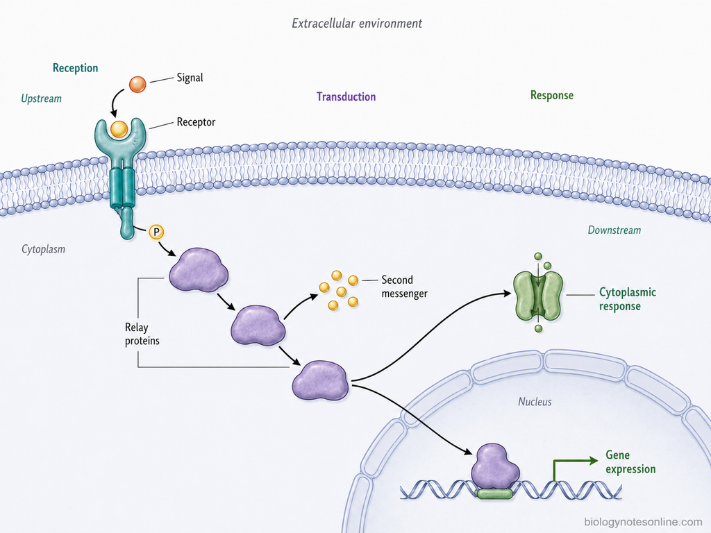
1. Reception — Detection of the Signal
Reception is the first stage of cell signaling where a signaling molecule is recognized by its receptor. Receptors show specificity for particular ligands, therefore a cell responds only when it has the suitable receptor for that signal.
Binding of ligand changes the activity of the receptor. In many receptors this involves change in receptor conformation, association of receptor subunits, or activation of its intracellular region. The activated receptor can now interact with signaling molecules present inside the cell. The exact mechanism is different for different receptor types.
2. Transduction — Relaying the Signal Inside the Cell
After reception, the signal is transferred from the activated receptor to other components present inside the cell. This stage is called signal transduction.
During this process, one signaling molecule activates another molecule and a series of intracellular reactions may be formed. Protein phosphorylation, GTP-binding proteins and production of second messengers are commonly involved in such signal relay. In some pathways, one activated component acts on several molecules and the signal is amplified during the process.
The pathway does not always move as a simple straight line. A signal can be distributed towards more than one cellular target. Different signaling reactions can also be regulated or stopped before the final response is reached.
3. Response — Changing Cell Activity
The last stage is the cellular response. In this step, signaling changes the activity of particular target proteins and this produces an effect in the cell.
Some responses occur rapidly in the cytoplasm. These generally involve modification of proteins already present in the cell, such as enzymes, ion channels or cytoskeletal proteins. The activity of these proteins can therefore be changed without synthesis of new protein.
Other signaling pathways reach the nucleus and regulate transcription factors. This results in increase or decrease in expression of particular genes. Such responses generally develop more slowly because transcription and, in many cases, synthesis of new proteins are involved.
The overall process can be shown as follows-
Molecular Mechanisms Used in Signal Transduction
Signal transduction is carried out by different molecular mechanisms that transfer the information from an activated receptor to intracellular targets. In most pathways, several of these mechanisms work together. They can activate proteins, produce intracellular messengers, change ion concentration or assemble signaling proteins at a particular site of the cell.
The major molecular mechanisms used in signal transduction are as follows-
- Protein conformational change. Binding of a ligand can change the shape or arrangement of its receptor. This change exposes or activates the intracellular region of receptor and allows further signaling proteins to bind. In some receptors, ligand binding also brings two or more receptor molecules together. The activated receptor can now transfer the signal towards intracellular components.
- Protein phosphorylation and dephosphorylation. Phosphorylation is one of the common mechanisms used for transfer and regulation of signals. Protein kinases add phosphate groups to particular amino acid residues of target proteins. This may increase or decrease their activity, or create a site for binding of another signaling protein. Protein phosphatases remove these phosphate groups. Thus, phosphorylation and dephosphorylation act as a reversible molecular switch in many signaling pathways.
- GTP-binding molecular switches. GTP-binding proteins act by changing between GTP-bound and GDP-bound forms. The GTP-bound form is generally active, whereas the GDP-bound form is inactive. Heterotrimeric G proteins and small monomeric GTPases such as Ras use this mechanism. Hydrolysis of bound GTP to GDP helps in switching the protein back to its inactive state.
- Generation of second messengers. Some activated receptors stimulate enzymes that form or release small intracellular signaling molecules. These molecules are called second messengers. cAMP, cGMP, IP₃, DAG and Ca²⁺ are some important examples. They can spread the signal from the membrane towards different intracellular targets and a large number of messenger molecules may be formed from an initial receptor signal.
- Changes in intracellular ion concentration. Movement of ions across cellular membranes is also used for signal transduction. Ca²⁺ is the most important example. Opening of calcium channels or release of Ca²⁺ from intracellular stores increases its concentration in the cytosol. Calcium then binds to different proteins and regulates their activity. Thus, the ion itself acts as an intracellular signaling molecule.
- Protein-protein interaction and signaling complex formation. Many signaling proteins contain regions which allow them to bind with other proteins. After receptor activation, several signaling components can therefore be collected at the receptor or another particular cellular site. Adaptor and scaffold proteins also take part in these interactions. Such arrangement helps different signaling proteins to act in a proper sequence and can connect one activated molecule with its downstream target.
- Enzymatic cascades and signal amplification. An activated signaling enzyme may activate many molecules of the next component. These molecules can further act on other targets and a cascade is formed. Protein kinase cascades are a common example. Because one molecule can affect several molecules at the next stage, the original signal may also become amplified during the process.
These mechanisms finally change the activity of cellular target proteins such as enzymes, ion channels, cytoskeletal proteins or transcription-regulating proteins. A single signaling pathway may use more than one of these mechanisms before a cellular response is produced.
Major Cell-Surface Receptor Signaling Mechanisms
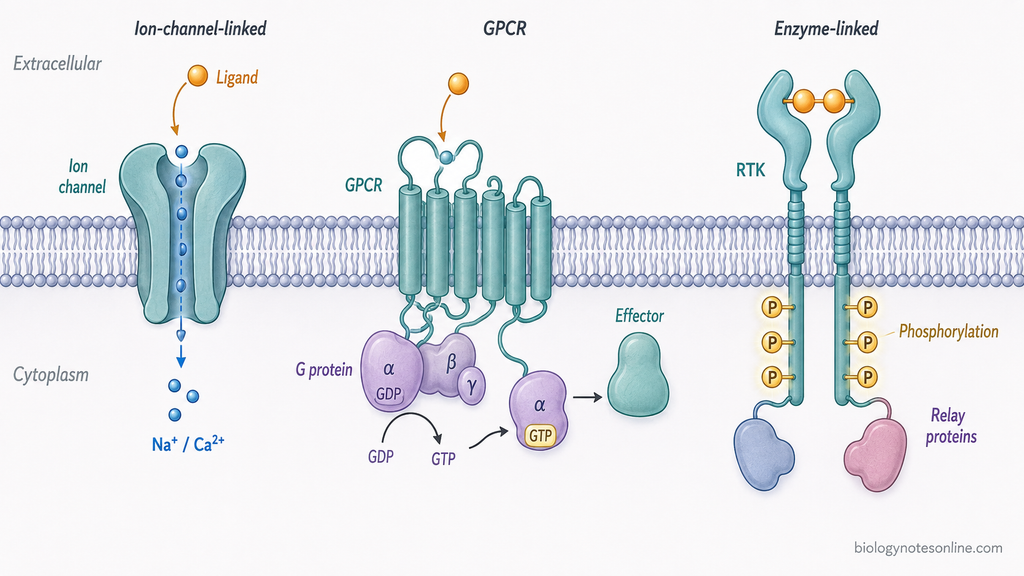
Most cell-surface receptors can be grouped according to the mechanism by which they transfer the extracellular signal across the plasma membrane. The three major classes are ion-channel-linked receptors, G protein-coupled receptors (GPCRs) and enzyme-linked receptors. Their signaling mechanism is different, although several downstream reactions may later interact with one another.
| Receptor type | Activation mechanism | Intracellular mediator | Response speed/type | Representative example |
|---|---|---|---|---|
| Ion-channel-linked receptors | Binding of ligand directly opens or closes an ion channel present in the receptor protein. This changes movement of particular ions across the plasma membrane. | Mainly Na⁺, K⁺, Ca²⁺ or Cl⁻ movement. A separate intracellular relay protein is generally not required for the initial response. | Very rapid, usually in milliseconds. Mainly changes membrane potential or intracellular ion concentration. | Nicotinic acetylcholine receptor (nAChR) |
| G protein-coupled receptors (GPCRs) | Ligand binding changes the conformation of receptor. The activated receptor then activates a heterotrimeric G protein by promoting exchange of GDP for GTP on its α subunit. | Gα and Gβγ can regulate enzymes or ion channels. Depending on the receptor, mediators such as cAMP, IP₃, DAG and Ca²⁺ may then be formed or released. | Usually slower than ligand-gated ion channels. Responses may range from rapid changes in enzymes or ion channels to longer effects on gene expression. | β-adrenergic receptor |
| Enzyme-linked receptors | Ligand binding activates an enzyme associated with the cytoplasmic part of receptor. In receptor tyrosine kinases (RTKs), activation commonly causes receptor rearrangement or dimerization followed by phosphorylation of tyrosine residues. | Phosphorylated receptor sites recruit intracellular signaling proteins. Ras, phospholipase C, PI3K and protein kinase pathways are some common mediators downstream of RTKs. | Commonly minutes to hours. Often involved in growth, survival, differentiation and changes in gene expression, although some responses can occur rapidly. | Epidermal growth factor receptor (EGFR) |
Ion-Channel-Linked Receptor Signaling
Ion-channel-linked receptors produce the most direct type of cell-surface signaling. The receptor itself forms an ion channel. Binding of a neurotransmitter changes its conformation and the channel opens or closes for particular ions. The change in ion flow can rapidly alter membrane potential.
This mechanism is common during rapid synaptic signaling. For example, binding of acetylcholine to the nicotinic acetylcholine receptor opens the channel and allows movement of cations across the membrane. The response develops within milliseconds and does not require formation of a second messenger for the initial ion-channel response.
G Protein-Coupled Receptor Signaling
G protein-coupled receptors (GPCRs) transfer the received signal with the help of heterotrimeric G proteins. These receptors contain seven membrane-spanning regions. After ligand binding, the activated receptor promotes activation of the G protein associated with its cytoplasmic side.
The activated G protein can regulate an enzyme or ion channel. Adenylyl cyclase may be activated to produce cAMP, while another GPCR pathway activates phospholipase C and forms IP₃ and DAG. IP₃ causes release of Ca²⁺ from intracellular stores. Thus, the same receptor class can use different intracellular mediators depending on the G protein and target protein involved.
Enzyme-Linked Receptor Signaling
Enzyme-linked receptors either contain an enzymatic region themselves or are directly associated with an intracellular enzyme. Receptor tyrosine kinases (RTKs) are the major and well-studied group of this receptor class. They are commonly used by growth factors and several other extracellular signals.
In RTKs, ligand binding causes rearrangement of receptor molecules and activation of the kinase domains. Tyrosine residues on the cytoplasmic regions are then phosphorylated. These phosphorylated sites provide binding sites for intracellular signaling proteins. Signals can then pass through pathways involving Ras, protein kinases, phospholipase C or other molecules.
Some cell-surface receptors have no intrinsic kinase activity but remain associated with cytoplasmic kinases. Cytokine receptors using Janus kinases (JAKs) are an important example. Therefore, enzyme-associated signaling includes more than only receptor tyrosine kinases.
Intracellular Receptor Signaling
Some signaling molecules do not bind with the receptors present on cell surface. They enter into the target cell and bind with receptors present in the cytoplasm or nucleus. These receptors are called intracellular receptors. Small hydrophobic signaling molecules generally follow this type of signaling. Therefore, a membrane receptor and long intracellular relay is not always involved in cell signaling.
Lipid-Soluble Signaling Molecules
Lipid-soluble signaling molecules can pass through the plasma membrane because of their hydrophobic nature. Steroid hormones are the common examples. Thyroid hormone, vitamin D and retinoic acid also act through intracellular receptors.
After entering the cell, the signaling molecule combines with its specific intracellular receptor. The receptor now becomes active. In this signaling, the signaling molecule itself reaches inside the cell and hence formation of second messenger at plasma membrane is not necessary.
Cytoplasmic and Nuclear Receptors
Intracellular receptors can be present either in the cytoplasm or inside nucleus. Some steroid hormone receptors are present mainly in cytoplasm in their inactive form. The glucocorticoid receptor is one example. After hormone binding, the receptor-hormone complex enters into the nucleus where it can act on DNA.
On the other hand, some receptors are already present inside nucleus. Thyroid hormone receptors and retinoic acid receptors belong to this group. These receptors can remain associated with DNA even before binding of the ligand. Hormone binding changes their activity and regulation of transcription takes place.
Regulation of Gene Expression
Most of these intracellular receptors act as ligand-regulated transcription factors. They have regions for ligand binding and DNA binding. After activation, receptor binds with specific regulatory DNA sequences of the target genes. Coactivator or corepressor proteins also take part during this process.
The transcription of particular genes is then increased or decreased. This changes the amount of specific proteins produced in the cell and finally changes cellular activity. These responses are generally slower compared to rapid ion-channel or cytoplasmic signaling, and many of their effects remain for longer period.
The process is as follows-
Lipid-soluble signal → Entry into target cell → Intracellular receptor → Binding with DNA/regulatory proteins → Change in gene transcription → Cellular response
Representative Signal Transduction Pathways
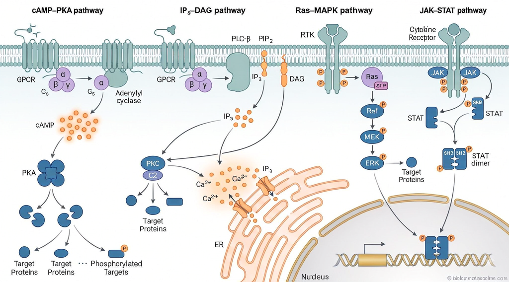
There are different types of signal transduction pathways present in cells. These pathways differ according to receptor, intracellular signaling molecules and type of cellular response. In some pathways, second messengers are formed while in others a series of protein phosphorylation takes place. The following are some important representative signal transduction pathways-
cAMP-PKA Signaling Pathway
The cAMP-PKA signaling pathway is a G protein-coupled receptor pathway in which cyclic AMP acts as a second messenger. Binding of ligand with the receptor activates the heterotrimeric Gs protein. The activated Gαs subunit now stimulates the enzyme adenylyl cyclase.
Adenylyl cyclase converts ATP into cyclic AMP (cAMP). The increased cAMP binds with protein kinase A (PKA) and activates it. PKA then phosphorylates different target proteins present in the cell. Depending upon the target protein, changes in metabolism, ion-channel activity or gene expression can occur.
The pathway is as follows-
Ligand → GPCR → Gs protein → Adenylyl cyclase → cAMP → PKA → Cellular response
IP₃-DAG Signaling Pathway
This pathway is also associated with some G protein-coupled receptors. After binding of ligand, the receptor activates Gq protein, which further activates phospholipase C-β (PLC-β). The enzyme acts on a membrane phospholipid called phosphatidylinositol 4,5-bisphosphate (PIP₂).
PIP₂ is broken down to form inositol 1,4,5-trisphosphate (IP₃) and diacylglycerol (DAG). IP₃ moves into cytoplasm and binds with IP₃ receptors on the endoplasmic reticulum, resulting in release of Ca²⁺. DAG remains in plasma membrane. DAG and Ca²⁺ together participate in activation of protein kinase C (PKC).
The reaction can be summarized as-
Ligand → GPCR → Gq protein → PLC-β → PIP₂ → IP₃ + DAG → Ca²⁺ and PKC activation → Cellular response
Ras-MAPK Signaling Pathway
The Ras-MAPK pathway is commonly activated by receptor tyrosine kinases (RTKs). Growth factors are among the important signals which use this pathway. After ligand binding, receptor activation and phosphorylation takes place and intracellular signaling proteins are recruited to the receptor.
This leads to activation of Ras, a small GTP-binding protein. Ras in its GTP-bound form activates Raf, which is a protein kinase. Raf phosphorylates and activates MEK, and MEK further activates ERK. ERK is a mitogen-activated protein kinase (MAPK) which acts on different cytoplasmic and nuclear proteins.
The pathway is as follows-
Growth factor → RTK → Ras → Raf → MEK → ERK → Target proteins → Cellular response
This pathway is mainly involved in regulation of cell proliferation, differentiation, survival and other cellular activities.
JAK-STAT Signaling Pathway
The JAK-STAT pathway is used by many cytokine receptors. These receptors do not have an intrinsic tyrosine kinase activity. They are associated with cytoplasmic proteins called Janus kinases (JAKs).
After binding of cytokine, receptor-associated JAK proteins become activated. They phosphorylate tyrosine residues of the receptor and form binding sites for STAT proteins. STATs are then phosphorylated by JAKs.
The phosphorylated STAT proteins form dimers and enter into the nucleus. Inside nucleus, they bind with specific DNA regulatory sequences and control expression of target genes. Thus, this pathway provides a relatively direct connection between a cell-surface receptor and regulation of gene transcription.
The pathway can be written as-
Cytokine → Cytokine receptor → JAK → STAT phosphorylation → STAT dimer → Nucleus → Gene expression
How Are Signals Amplified?
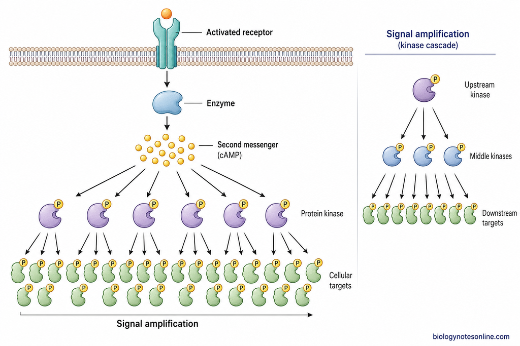
Signal amplification is the process by which a small initial signal produces a much larger intracellular effect. It occurs when one activated signaling component activates or forms many molecules of the next component. Amplification can take place at one or several stages of a pathway. It is not necessary that every step of signal transduction will amplify the signal.
Amplification by Enzyme Activity
Many signaling proteins are enzymes. After activation, a single enzyme molecule can act repeatedly on a large number of substrate molecules. Thus, one activated component can produce many activated molecules in the next step.
For example, activated adenylyl cyclase continuously converts ATP into cAMP while the enzyme remains active. A small number of activated receptors can therefore result in formation of a much larger number of intracellular signaling molecules.
Amplification by Second-Messenger Production
Formation of second messengers is an important mechanism of signal amplification. An activated enzyme can produce large numbers of small messenger molecules within a short period. These molecules then spread inside the cell and act on several target proteins.
cAMP is one common example. One activated adenylyl cyclase can form many cAMP molecules from ATP. Similarly, production of IP₃ and release of Ca²⁺ can increase the number of intracellular molecules carrying the signal. Second-messenger production can therefore provide considerable amplification of the original extracellular signal.
Amplification by Kinase Cascades
Amplification can also occur through a series of protein kinases. In such a cascade, one activated kinase phosphorylates many molecules of the next kinase. Each of these activated molecules can again act on several downstream proteins.
The MAP kinase cascade is one example where protein kinases act one after another. Because several molecules may be activated at each catalytic stage, the number of activated downstream targets can become much greater than the number of molecules that received the original signal.
Signal Specificity, Integration and Crosstalk
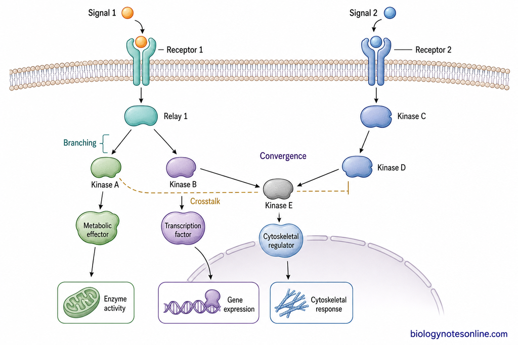
Cell signaling pathways do not always work as separate straight chains. A signal can activate more than one intracellular pathway and different signals can also act on the same signaling component. The final response therefore depends not only on the extracellular signal, but also on receptor and signaling proteins present in that particular cell.
Why the Same Signal Can Produce Different Responses
The same signaling molecule can produce different responses in different types of cells. This occurs because all cells do not contain the same receptors and intracellular signaling proteins.
A cell can respond to a signal only when the suitable receptor is present. Different cells may contain different receptors for the same signaling molecule. For example, acetylcholine produces contraction in skeletal muscle but decreases the rate and force of contraction in heart muscle. Different receptor types are involved in these responses.
Different responses can also occur even when the same type of receptor is present. This is due to differences in intracellular signaling proteins, target proteins and gene regulatory proteins of the cell. Thus, the nature of target cell is also an important factor which determines the final response.
Pathway Branching and Convergence
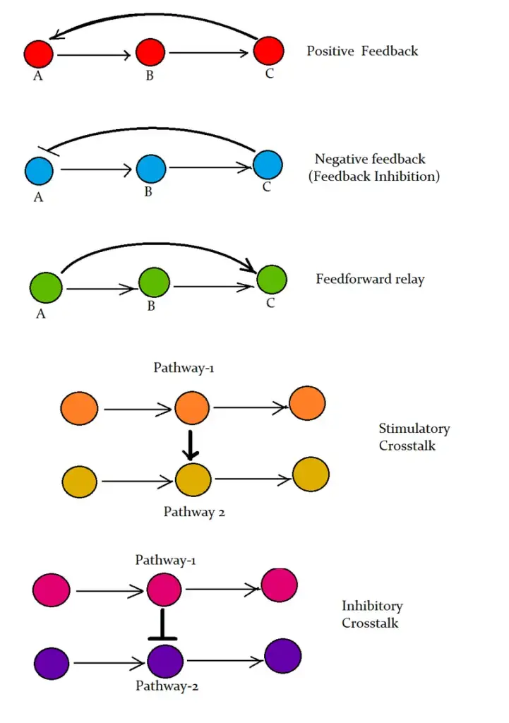
A signaling pathway is not always a single linear sequence. One activated receptor can transfer the signal into two or more downstream pathways. This is called pathway branching. The different branches may control metabolism, movement, gene expression or other cellular activities at the same time.
On the other hand, signals coming from different receptors can act on the same intracellular signaling protein or target. This is referred to as convergence. Such components can receive information from more than one pathway and integrate these signals before further transfer.
Therefore intracellular signaling is better represented as a network of interacting pathways rather than completely separate chains.
Crosstalk Between Signaling Pathways
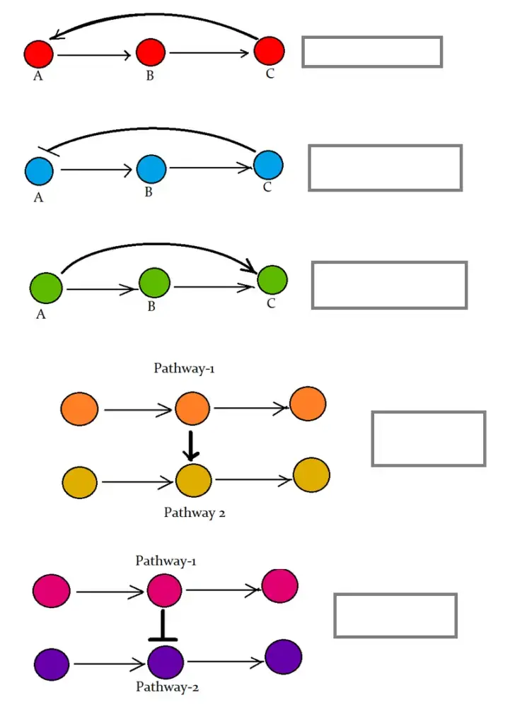
Signaling crosstalk occurs when one signaling pathway affects the activity of another pathway. The interaction may take place at receptors, signaling proteins, protein kinases, second messengers or target proteins.
One pathway may increase the activity of another pathway, while in some cases it can inhibit it. Shared signaling components are also common. Protein kinases activated in one pathway can act on proteins belonging to another signaling pathway.
Scaffold and adaptor proteins also help in controlling these interactions by keeping particular signaling proteins together. This can increase signaling specificity and reduce unwanted interaction between pathways.
At the same time, one pathway can also divide into several branches-
Receptor activation → Signaling component → Pathway 1 / Pathway 2 / Pathway 3 → Different cellular responses
How Are Signal Transduction Pathways Turned Off?
Signal transduction does not continue for unlimited time. After the required response is produced, different components of pathway are inactivated and the signal is stopped. This is important for maintaining proper response of cell and also prevents continuous activation of signaling proteins.
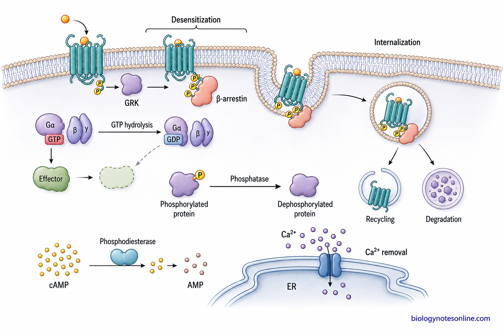
The major mechanisms involved in termination of signal transduction are as follows-
GTP Hydrolysis
GTP-binding proteins remain active when they are bound with GTP. The bound GTP is later converted into GDP by GTPase activity of the protein. After this conversion, the protein becomes inactive and further signal transfer is stopped.
The Gα subunit of heterotrimeric G proteins has its own GTPase activity. Small GTP-binding proteins such as Ras are also switched off by hydrolysis of GTP into GDP. In many cases, regulatory proteins increase this GTP hydrolysis and make the process faster.
Protein Dephosphorylation
Protein phosphorylation is commonly used during signal transduction. Protein kinases add phosphate groups to target proteins and alter their activity. These phosphate groups are removed again when the signaling response has to be decreased.
Protein phosphatases carry out this removal of phosphate groups. The phosphorylated proteins are therefore returned towards their inactive or previous state. In this way, kinase-dependent signaling is controlled by opposite activity of protein phosphatases.
Second-Messenger Degradation or Removal
Second messengers are present only for a limited period during signaling. After their function, they are degraded, removed or returned back to their storage sites.
For example, cAMP is converted into AMP by phosphodiesterase enzymes. This decreases cAMP concentration and activity of cAMP-dependent proteins also decreases. Ca²⁺ present in cytoplasm is pumped back into intracellular stores or transported outside the cell. IP₃ is also rapidly metabolized after its signaling function.
Thus, decrease of second-messenger concentration helps in stopping further intracellular signal transfer.
Receptor Desensitization, Internalization and Degradation
A receptor can also become less responsive when it is continuously exposed to its ligand. This is referred to as receptor desensitization. In this condition, ligand may still be present but receptor produces a reduced response.
In many G protein-coupled receptors (GPCRs), the activated receptor is phosphorylated by G protein-coupled receptor kinases (GRKs). β-arrestin then binds with the phosphorylated receptor and blocks its further interaction with G protein.
The receptor may also be removed from plasma membrane by endocytosis. This process is called receptor internalization. The internalized receptor can be recycled again to cell membrane or transferred to lysosomes for degradation.
When receptors are degraded, the number of receptors present on cell surface decreases. This reduces sensitivity of the cell towards the signal and is called receptor down-regulation.
Cellular Responses Produced by Signal Transduction
Signal transduction causes changes in the activity of target cell. These responses may occur in cytoplasm or nucleus. Some responses are rapid, while changes involving gene expression generally take more time.
The major cellular responses include-
- Metabolic changes. Activity of enzymes can be increased or decreased by signaling. As a result, different metabolic reactions of the cell are changed.
- Ion channel changes. Signaling can cause opening or closing of membrane ion channels. This changes ion movement and membrane potential.
- Secretion. Increase in intracellular Ca²⁺ can stimulate release of substances from secretory vesicles. Hormones, neurotransmitters and other secretory products are released in this way.
- Changes in cell shape and movement. Signaling acts on cytoskeletal proteins and their arrangement can be changed. Cell movement, adhesion and shape are affected by these changes.
- Gene expression. Some intracellular signals reach the nucleus and activate gene regulatory proteins. Transcription of particular genes is then increased or decreased.
- Cell growth and proliferation. Growth factor signaling can stimulate growth and division of cells. Several enzyme-linked receptor pathways are involved in these responses.
- Cell differentiation. Signals can produce changes in gene expression that lead to development of specialized cell characters. It is important during growth and development.
- Cell survival and death. Some signals maintain survival of the cell, whereas other signaling reactions can initiate apoptosis or programmed cell death.
What Happens When Signal Transduction Is Disrupted?
Signal transduction pathways control many normal activities of the cell. Any change in receptor or other signaling components can disturb this process. Sometimes the pathway becomes overactive, while in other condition the signal becomes weak or does not reach the target properly.
The major effects include-
- Cancer. Abnormal activation of growth signaling causes continuous growth and survival of cells. Changes in RAS, PI3K and receptor tyrosine kinase signaling are common in many cancers.
- Insulin resistance. Defective insulin signaling decreases normal action of insulin on target cells. Glucose uptake and metabolism are affected and this is important in type 2 diabetes.
- Immune disorders. Cytokines depend on intracellular signaling for their action. Defects in JAK-STAT signaling are associated with immune deficiency and autoimmune disorders.
- Developmental defects. Signaling is important during formation and differentiation of tissues. Abnormal Notch or Hedgehog signaling can therefore produce developmental abnormalities.
- Abnormal cell survival. Some pathways give survival signal to the cell, whereas some are involved in cell death. Disturbance of these signals can result in survival of unwanted cells or loss of normal cells.
- Changes in cell function. A defective signaling pathway may alter secretion, metabolism, movement or gene expression. The effect depends on the signaling pathway and cell in which the defect occurs.
How to Read a Signal Transduction Pathway Diagram
A signal transduction pathway diagram shows how a signal is received and transferred through different components of the cell. The pathway is generally read from the signaling molecule towards receptor and then to intracellular targets. Some diagrams are simple, while others contain several branches and interacting pathways.
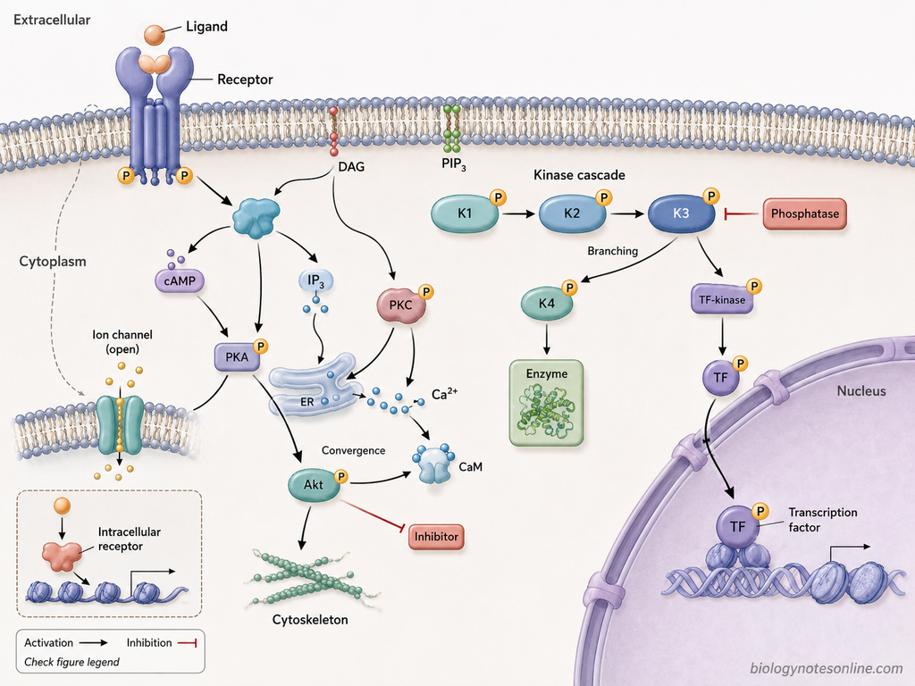
The following points are useful while reading a signaling pathway diagram-
- Start with the signal. First identify the extracellular signaling molecule or ligand. It is generally shown at the beginning of pathway and binds with a specific receptor.
- Identify the receptor. Check whether the receptor is present on plasma membrane or inside the cell. Cell-surface receptors transfer the signal towards intracellular signaling components.
- Follow the arrows. Arrows generally show the direction in which signaling information is transferred. One component may activate the next protein or lead to formation of another signaling molecule.
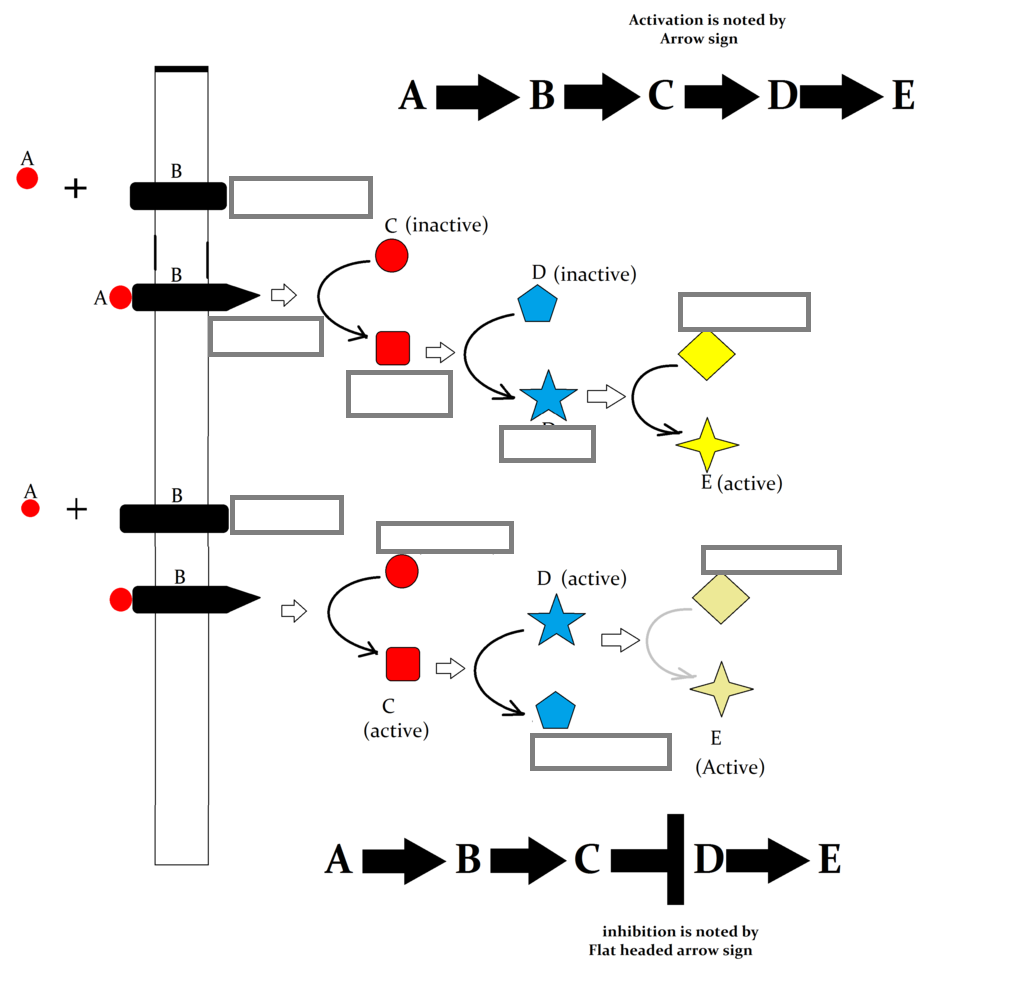
- Look for activation and inhibition. Activation is commonly shown by an arrow. Inhibitory interactions are usually represented by a blunt-ended line or another inhibitory mark. The exact symbol should always be checked from the figure legend because diagram conventions can differ.
- Follow phosphorylation steps. A pathway may show one protein kinase acting on another protein. Labels such as P or phosphorylation arrows indicate addition of phosphate groups. Such reactions are common in kinase cascades.
- Identify second messengers. Small molecules such as cAMP, IP₃, DAG, Ca²⁺ or PIP₃ may appear between receptor activation and downstream targets. These carry or spread the signal inside the cell.
- Check pathway branches. One activated component may act on more than one downstream target. The pathway can therefore divide into separate branches and produce different responses.
- Look for convergence. Two signaling routes may join at the same protein or target. This shows that different signals can use a common intracellular component.
- Find the final target. The last components may be enzymes, ion channels, cytoskeletal proteins or transcription factors. Their change in activity produces the cellular response.
- Check the cellular location. Plasma membrane, cytoplasm and nucleus are often shown separately in the diagram. Movement of a signaling protein into nucleus generally indicates regulation of gene expression.
- Read the figure legend. Symbols are not exactly same in every pathway diagram. Dashed lines, different arrowheads, colours or abbreviations may have a special meaning in that particular figure.
Significance of Signal Transduction Pathway
Signal transduction is important for receiving different signals and producing cellular response. It controls many activities of cell including metabolism, growth, movement and gene expression.
The major significance include-
- Cell communication. It allows cells to receive hormones, neurotransmitters and other signals and respond to them.
- Metabolic control. Activity of different metabolic enzymes is regulated by signaling pathways.
- Gene regulation. Some signals reach the nucleus and alter expression of particular genes.
- Growth and proliferation. Growth factor signaling controls growth and division of cells.
- Cell differentiation. It is involved in development of specialized cells from less specialized cells.
- Cell survival. Different signaling pathways help in survival of normal cells and regulation of apoptosis.
- Movement and adhesion. Signaling can change cytoskeletal arrangement, cell movement and attachment with other cells.
- Immune function. Cytokine signaling is required for activation, differentiation and function of immune cells.
- Response to external conditions. Cells can detect changes in their surroundings and adjust their activity according to the signal.
- Disease development. Abnormal signal transduction is associated with cancer, metabolic diseases, immune disorders and several other conditions.
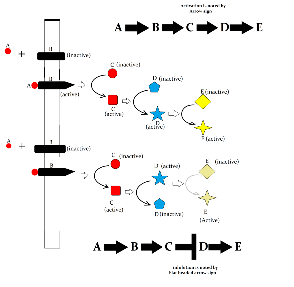
Elements of Signal transduction cascade networking
Signal Transduction Pathway Worksheet
References
- Alberts, B., Johnson, A., Lewis, J., Raff, M., Roberts, K., & Walter, P. (2002). Molecular biology of the cell (4th ed.). Garland Science. https://www.ncbi.nlm.nih.gov/books/NBK21054/
- Boulanger, L. M., & Shatz, C. J. (2004). Immune signalling in neural development, synaptic plasticity and disease. Nature Reviews Neuroscience, 5(7), 521–531. https://doi.org/10.1038/nrn1428
- Bray, S. J., & Bigas, A. (2025). Modes of Notch signalling in development and disease. Nature Reviews Molecular Cell Biology, 26(7), 522–537. https://doi.org/10.1038/s41580-025-00835-2
- Cliff, W. H. (2023). Teaching with core concepts to facilitate the integrated learning of introductory organismal biology. Advances in Physiology Education, 47(3), 562–572. https://doi.org/10.1152/advan.00134.2022
- Cooper, G. M. (2000). The cell: A molecular approach (2nd ed.). Sinauer Associates. https://www.ncbi.nlm.nih.gov/books/NBK9839/
- García Morán, G. A., Parra-Medina, R., García Cardona, A., Quintero-Ronderos, P., & Garavito Rodríguez, E. (2013). Cytokines, chemokines and growth factors. In J. M. Anaya, Y. Shoenfeld, A. Rojas-Villarraga, R. A. Levy, & E. Cervera (Eds.), Autoimmunity: From bench to bedside. El Rosario University Press. https://www.ncbi.nlm.nih.gov/books/NBK459450/
- Gilbert, S. F. (2000). Developmental biology (6th ed.). Sinauer Associates. https://www.ncbi.nlm.nih.gov/books/NBK9983/
- Guo, Q., Jin, Y., Chen, X., Ye, X., Shen, X., Lin, M., Zeng, C., Zhou, T., & Zhang, J. (2024). NF-κB in biology and targeted therapy: New insights and translational implications. Signal Transduction and Targeted Therapy, 9, 53. https://doi.org/10.1038/s41392-024-01757-9
- Holz, R. W., & Fisher, S. K. (1999). Cellular signaling mechanisms. In G. J. Siegel, B. W. Agranoff, R. W. Albers, S. K. Fisher, & M. D. Uhler (Eds.), Basic neurochemistry: Molecular, cellular and medical aspects (6th ed.). Lippincott-Raven. https://www.ncbi.nlm.nih.gov/books/NBK28213/
- Hossain, M. D. A., Marques Cuoco, C., Sodaye, S. H., Beci, J., & Belkas, K. (2026). Non-receptor tyrosine kinase signaling pathways and therapeutic implications. Signal Transduction and Targeted Therapy, 11, 235. https://doi.org/10.1038/s41392-026-02729-x
- Hoxhaj, G., & Manning, B. D. (2020). The PI3K–AKT network at the interface of oncogenic signalling and cancer metabolism. Nature Reviews Cancer, 20(2), 74–88. https://doi.org/10.1038/s41568-019-0216-7
- Hu, X., Li, J., Fu, M., Zhao, X., & Wang, W. (2021). The JAK/STAT signaling pathway: From bench to clinic. Signal Transduction and Targeted Therapy, 6, 402. https://doi.org/10.1038/s41392-021-00791-1
- Janeway, C. A., Jr., Travers, P., Walport, M., & Shlomchik, M. J. (2001). Immunobiology: The immune system in health and disease (5th ed.). Garland Science. https://www.ncbi.nlm.nih.gov/books/NBK10757/
- Kholodenko, B. N. (2006). Cell-signalling dynamics in time and space. Nature Reviews Molecular Cell Biology, 7(3), 165–176. https://doi.org/10.1038/nrm1838
- Kolch, W., Halasz, M., Granovskaya, M., & Kholodenko, B. N. (2015). The dynamic control of signal transduction networks in cancer cells. Nature Reviews Cancer, 15(9), 515–527. https://doi.org/10.1038/nrc3983
- Ligeti, E., Csépányi-Kömi, R., & Hunyady, L. (2012). Physiological mechanisms of signal termination in biological systems. Acta Physiologica, 204(4), 469–478. https://doi.org/10.1111/j.1748-1716.2012.02414.x
- Lodish, H., Berk, A., Matsudaira, P., Kaiser, C. A., Krieger, M., Scott, M. P., Zipursky, S. L., & Darnell, J. (2004). Molecular cell biology (5th ed.). W. H. Freeman.
- Lv, Y., Qi, J., Babon, J. J., Cao, L., Fan, G., Lang, J., Zhang, J., Mi, P., Kobe, B., & Wang, F. (2024). The JAK-STAT pathway: From structural biology to cytokine engineering. Signal Transduction and Targeted Therapy, 9, 221. https://doi.org/10.1038/s41392-024-01934-w
- Merheb, E., Zubair, M., & Bokhari, S. R. A. (2026). Apoptosis and cell death: Signaling in health and diseases. In StatPearls. StatPearls Publishing. https://www.ncbi.nlm.nih.gov/books/NBK499821/
- O’Shea, J. J., Lahesmaa, R., Vahedi, G., Laurence, A., & Kanno, Y. (2011). Genomic views of STAT function in CD4+ T helper cell differentiation. Nature Reviews Immunology, 11(4), 239–250. https://doi.org/10.1038/nri2958
- Papin, J. A., Hunter, T., Palsson, B. O., & Subramaniam, S. (2005). Reconstruction of cellular signalling networks and analysis of their properties. Nature Reviews Molecular Cell Biology, 6(2), 99–111. https://doi.org/10.1038/nrm1570
- Perissi, V., & Rosenfeld, M. G. (2005). Controlling nuclear receptors: The circular logic of cofactor cycles. Nature Reviews Molecular Cell Biology, 6(7), 542–554. https://doi.org/10.1038/nrm1680
- Purves, D., Augustine, G. J., Fitzpatrick, D., Katz, L. C., LaMantia, A.-S., McNamara, J. O., & Williams, S. M. (Eds.). (2001). Neuroscience (2nd ed.). Sinauer Associates. https://www.ncbi.nlm.nih.gov/books/NBK10799/
- Putney, J. W., Jr. (1999). Ca2+-regulated processes. In G. J. Siegel, B. W. Agranoff, R. W. Albers, S. K. Fisher, & M. D. Uhler (Eds.), Basic neurochemistry: Molecular, cellular and medical aspects (6th ed.). Lippincott-Raven. https://www.ncbi.nlm.nih.gov/books/NBK28115/
- Rajagopal, S., & Shenoy, S. K. (2018). GPCR desensitization: Acute and prolonged phases. Cellular Signalling, 41, 9–16. https://doi.org/10.1016/j.cellsig.2017.01.024
- Rehman, S., Rahimi, N., & Dimri, M. (2023). Biochemistry, G protein coupled receptors. In StatPearls. StatPearls Publishing. https://www.ncbi.nlm.nih.gov/books/NBK518966/
- Sinha, R., & Yen, P. M. (2018). Cellular action of thyroid hormone. In Endotext. MDText.com, Inc. https://www.ncbi.nlm.nih.gov/books/NBK285568/
- Solis-Herrera, C., Triplitt, C., Cersosimo, E., & DeFronzo, R. A. (2025). Pathogenesis of type 2 diabetes mellitus. In Endotext. MDText.com, Inc. https://www.ncbi.nlm.nih.gov/books/NBK279115/
- Thorpe, L. M., Yuzugullu, H., & Zhao, J. J. (2015). PI3K in cancer: Divergent roles of isoforms, modes of activation and therapeutic targeting. Nature Reviews Cancer, 15(1), 7–24. https://doi.org/10.1038/nrc3860
- Weikum, E. R., Knuesel, M. T., Ortlund, E. A., & Yamamoto, K. R. (2017). Glucocorticoid receptor control of transcription: Precision and plasticity via allostery. Nature Reviews Molecular Cell Biology, 18(3), 159–174. https://doi.org/10.1038/nrm.2016.152
- Xue, C., Chu, Q., Shi, Q., Zeng, Y., Lu, J., & Li, L. (2025). Wnt signaling pathways in biology and disease: Mechanisms and therapeutic advances. Signal Transduction and Targeted Therapy, 10, 106. https://doi.org/10.1038/s41392-025-02142-w