

Smooth muscle is a type of involuntary muscle tissue that works without conscious control. It is also called non-striated muscle because it does not show striped or banded appearance under microscope. It is found in the internal organs and helps in automatic body functions.
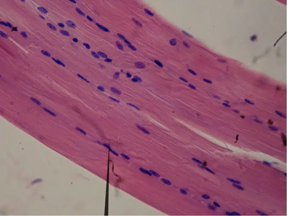
The smooth muscle fibres are small and spindle-shaped. They are broad in the middle and narrow at both ends. Each smooth muscle cell has a single centrally placed nucleus. The contractile proteins are not arranged in regular striated bands. They are arranged in a criss-cross manner, so the muscle looks smooth.
Smooth muscle is mainly present in the walls of hollow organs and internal passageways. It is found in stomach, intestine, blood vessels, respiratory tract, urinary bladder, reproductive organs, iris of eye and hair follicles of skin. In hair follicle it forms small muscle called arrector pili.
The main function of smooth muscle is to produce slow and sustained movement. It helps in movement of food through the alimentary canal by rhythmic waves. It also controls diameter of blood vessels and helps in regulation of blood pressure. In respiratory tract it controls the size of air passage. In urinary bladder and uterus it helps in contraction and relaxation.
The activity of smooth muscle is controlled by autonomic nervous system, hormones and local chemical substances. It is highly elastic and can stretch when organs like stomach and bladder become filled. It can maintain contraction for long time with less energy. This continuous partial contraction is called muscle tone. Smooth muscle does not fatigue easily and so it is suitable for long lasting internal functions.
Definition of Smooth Muscle
Smooth muscle is a type of involuntary and non-striated muscle tissue. It is found in the wall of internal organs like intestine, blood vessels and urinary bladder. It works automatically and helps in slow contraction and relaxation.
Characteristics of Smooth Muscle
The following are the important characteristics of smooth muscle–
- Involuntary control – Smooth muscle works without conscious control. It is controlled by autonomic nervous system, hormones and local chemical factors.
- Non-striated muscle – It does not show transverse bands or striations under microscope. The actin and myosin filaments are not arranged in regular sarcomere.
- Spindle-shaped cells – The cells of smooth muscle are fusiform in shape. They are broad at the middle and narrow at both ends.
- Single nucleus – Each smooth muscle cell contains only one nucleus. The nucleus is present at the centre of the cell.
- Small muscle fibre – The smooth muscle cells are small in size. They are much shorter than skeletal muscle fibres and usually about 30-200 µm long.
- Dense bodies present – The actin filaments are attached with dense bodies inside the cell. These dense bodies act like Z-disc of skeletal muscle.
- Troponin absent – Troponin is absent in smooth muscle. In this muscle contraction is regulated by calcium binding protein called calmodulin.
- Slow contraction – Smooth muscle contracts slowly. It can also remain contracted for long time.
- Energy efficient muscle – It uses very less energy during prolonged contraction. This long lasting contraction is called muscle tone.
- High elasticity – Smooth muscle can stretch very much without sudden rise of tension. This helps organs like stomach and urinary bladder to expand.
- Regeneration capacity – Smooth muscle cells can divide by mitosis. So it can increase in number, this is called hyperplasia.
- Two types – Smooth muscle is of two main types, single-unit smooth muscle and multi-unit smooth muscle. Single-unit type contracts together and multi-unit type contracts separately.
- Wide distribution – It is present in the wall of hollow organs like stomach, intestine, uterus, urinary bladder, blood vessels, respiratory tract, eye and skin hair follicles.
Location of Smooth Muscle
The following are the important location of smooth muscle–
- Gastrointestinal tract – Smooth muscle is present in the wall of oesophagus, stomach and intestine. It helps in movement of food.
- Blood vessels – It is found in the wall of arteries, veins and lymphatic vessels. It helps to control the diameter of vessels.
- Urinary system – Smooth muscle is present in urinary bladder and ureter. It helps in movement and removal of urine.
- Reproductive system – It is found in uterus and reproductive ducts. In male it is present in epididymis and vas deferens.
- Respiratory tract – Smooth muscle is present in the wall of bronchi and bronchioles. It controls the size of air passage.
- Eye – It is present in iris and ciliary body of eye. It controls pupil size and helps in changing the shape of lens.
- Skin – Smooth muscle is attached with hair follicle as arrector pili muscle. It helps in standing of hair and produces goosebumps.
Detail Structure or Anatomy of Smooth Muscle
The following are the detail structure of smooth muscle–
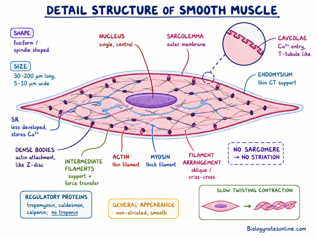
- Shape of cell – The smooth muscle cell is fusiform or spindle-shaped. It is broad in the middle and tapering at both ends.
- Size of cell – Smooth muscle fibres are small in size. They are about 30-200 µm long and 5-10 µm in diameter.
- Nucleus – Each smooth muscle cell has single nucleus. It is present at the centre of the cell.
- Sarcolemma – Sarcolemma is the outer cell membrane of smooth muscle cell. It surrounds the cytoplasm and helps in receiving stimulus.
- Caveolae – The sarcolemma has small pouch like depression called caveolae. These help in entry of calcium ion and acts like T-tubule of skeletal muscle.
- Sarcoplasmic reticulum – Sarcoplasmic reticulum is present inside the cell. It stores calcium ion, but it is less developed than skeletal muscle.
- Endomysium – Each smooth muscle cell is surrounded by thin connective tissue called endomysium. It gives support to the muscle cells.
- Dense bodies – Dense bodies are present in cytoplasm and also attached with inner side of sarcolemma. They act like Z-disc and give attachment to actin filaments.
- Intermediate filaments – Intermediate filaments connect one dense body with another dense body. They form internal supporting network and helps in transfer of contraction force.
- No sarcomere – Sarcomere is absent in smooth muscle. So it does not show striations and appears smooth under microscope.
- Actin filament – Actin is the thin filament present in large number. It is attached with dense bodies and helps in contraction of smooth muscle.
- Myosin filament – Myosin is the thick filament of smooth muscle. It pulls the actin filament and produce contraction.
- Arrangement of filaments – The actin and myosin filaments are not arranged in regular bands. They are arranged obliquely or criss-cross manner inside the cell.
- Regulatory proteins – Smooth muscle has tropomyosin, caldesmon and calponin. But troponin is absent in smooth muscle.
- General appearance – Due to absence of regular sarcomere and transverse band, smooth muscle looks non-striated. It contracts slowly and the whole cell becomes short in twisting manner.
Organ System Involved in Smooth Muscle
The following are the important organ system involved in smooth muscle–
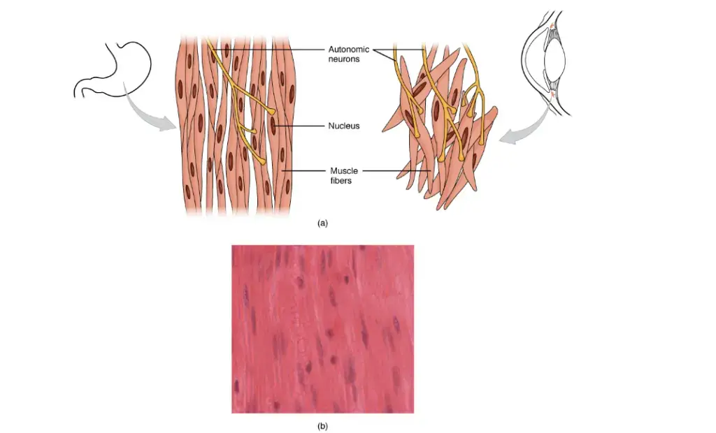
- Gastrointestinal system – Smooth muscle is present in the wall of stomach, intestine and other digestive organs. It helps in movement of food by peristalsis.
- Cardiovascular system – It is present in the wall of blood vessels. It controls blood flow and also helps in regulation of blood pressure.
- Lymphatic system – Smooth muscle is found in the wall of lymphatic vessels. It helps in movement of lymph through the vessels.
- Urinary system – It is present in urinary bladder, ureter and internal urethral sphincter. It helps in storage and removal of urine.
- Reproductive system – Smooth muscle is present in uterus and reproductive ducts. It helps in uterine contraction and movement of sperm in male reproductive tract.
- Respiratory system – It is present in bronchi and bronchioles. It helps to change the diameter of airway.
- Integumentary system – Smooth muscle is present in skin as arrector pili muscle. It is attached with hair follicle and causes standing of hair.
- Visual system – It is present in iris and ciliary muscle of eye. It controls pupil size and helps in changing the shape of lens.
Types of Smooth Muscle
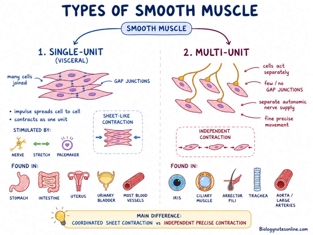
The smooth muscle is mainly of two types-
1. Single-unit smooth muscle
Single-unit smooth muscle is also called visceral smooth muscle. In this type, many smooth muscle cells are joined with each other by gap junctions. So the impulse passes from one cell to another cell and the whole muscle sheet contracts together as one unit. It may be stimulated by nerve impulse, stretching and pacemaker cells. It is found in wall of hollow organs like stomach, intestine, uterus, urinary bladder and most blood vessels.
2. Multi-unit smooth muscle
Multi-unit smooth muscle is the type of smooth muscle where each cell acts separately. The cells have very few or no gap junctions, so they do not contract as one common sheet. Each cell gets separate nerve supply from autonomic nervous system. So it gives fine and precise movement. It is found in iris and ciliary muscle of eye, arrector pili muscle of skin, trachea and large arteries like aorta.
Cross-Bridge Formation and Smooth Muscle Contraction
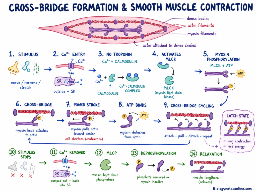
- First stimulus comes to smooth muscle cell. It may be nerve impulse, hormone or stretching.
- Then calcium ions (Ca²⁺) enter into the cell from outside fluid and also from sarcoplasmic reticulum.
- Smooth muscle has no troponin. So calcium binds with calmodulin protein.
- The calcium-calmodulin complex is formed inside the cell.
- This complex activates myosin light chain kinase (MLCK) enzyme.
- MLCK uses ATP and adds phosphate group to the myosin head.
- This addition of phosphate group is called phosphorylation of myosin.
- After phosphorylation, the myosin head becomes active and ready for contraction.
- The active myosin head attaches with actin filament.
- This attachment between actin and myosin is called cross-bridge formation.
- After cross-bridge formation, myosin head bends and pulls the actin filament.
- This pulling movement is called power stroke.
- The actin filaments are attached with dense bodies present inside the smooth muscle cell.
- So when actin is pulled, the whole cell becomes shortened and contracted.
- A new ATP binds with myosin head and myosin separates from actin.
- Then myosin again attaches with another site of actin and the cycle is repeated.
- This repeated attachment, pulling and detachment is called cross-bridge cycling.
- When stimulus stops, calcium ions are removed from cytoplasm.
- Myosin light chain phosphatase (MLCP) removes phosphate group from the myosin head.
- Due to removal of phosphate, myosin becomes inactive and smooth muscle relaxes.
- In some condition, few cross-bridges remain attached for long time. This is called latch state.
- Latch state helps smooth muscle to keep contraction for long time with very less energy.
Control of Contraction on Smooth Muscle
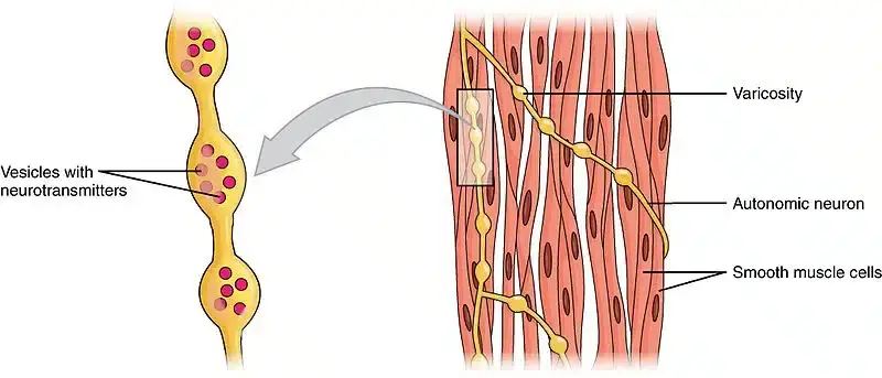
- Autonomic nervous system– Smooth muscle contraction is controlled by autonomic nerves and not by will. The nerve fibres have small swelling called varicosities, which release neurotransmitters directly into the muscle tissue. Acetylcholine usually stimulates contraction and helps in peristalsis, while norepinephrine and epinephrine may cause contraction or relaxation according to receptor type.
- Hormonal control– Smooth muscle cells have receptors for different hormones. Oxytocin causes contraction of uterus during labour. Vasopressin (ADH), angiotensin II, serotonin and histamine also control contraction or relaxation of smooth muscle in different organs.
- Local chemical factors– Smooth muscle is affected by chemical changes in surrounding tissue fluid. Low oxygen, high carbon dioxide, change in pH, lactic acid and adenosine can change contraction. In blood vessels, low oxygen and high carbon dioxide usually relax the smooth muscle and causes vasodilation.
- Mechanical stretch– Stretching of organ wall can start contraction of smooth muscle automatically. In intestine, food stretches the wall and causes peristaltic movement. In stomach and urinary bladder, stretching first increases tension and then muscle slowly relaxes, this is called stress-relaxation response.
- Pacemaker activity– Some smooth muscles can contract by their own without nerve impulse. This is called myogenic activity and is seen mainly in single-unit smooth muscle. Pacemaker cells produce slow electrical waves which spread through gap junctions and whole muscle sheet contracts rhythmically.

Mechanism of Action of Smooth Muscle
The following are the step by step mechanism of smooth muscle contraction–
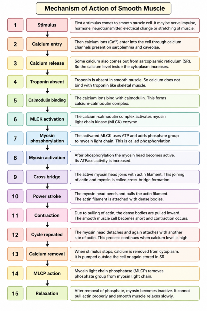
- Stimulus– First a stimulus comes to smooth muscle cell. It may be nerve impulse, hormone, neurotransmitter, electrical change or stretching of muscle.
- Calcium entry– Then calcium ions (Ca²⁺) enter into the cell through calcium channels present on sarcolemma and caveolae.
- Calcium release– Some calcium also comes out from sarcoplasmic reticulum (SR). So the calcium level inside the cytoplasm increases.
- Troponin absent– Troponin is absent in smooth muscle. So calcium does not bind with troponin like skeletal muscle.
- Calmodulin binding– The calcium ions bind with calmodulin. This forms calcium-calmodulin complex.
- MLCK activation– The calcium-calmodulin complex activates myosin light chain kinase (MLCK) enzyme.
- Myosin phosphorylation– The activated MLCK uses ATP and adds phosphate group to myosin light chain. This is called phosphorylation.
- Myosin activation– After phosphorylation the myosin head becomes active. Its ATPase activity is increased.
- Cross bridge– The active myosin head joins with actin filament. This joining of actin and myosin is called cross-bridge formation.
- Power stroke– The myosin head bends and pulls the actin filament. The actin filament is attached with dense bodies.
- Contraction– Due to pulling of actin, the dense bodies are pulled inward. The smooth muscle cell becomes short and contraction occurs.
- Cycle repeated– The myosin head detaches and again attaches with another site of actin. This process continues when calcium level is high.
- Calcium removal– When stimulus stops, calcium is removed from cytoplasm. It is pumped outside the cell or again stored in sarcoplasmic reticulum.
- MLCP action– Myosin light chain phosphatase (MLCP) removes phosphate group from myosin light chain.
- Relaxation– After removal of phosphate, myosin becomes inactive. It cannot pull actin properly and smooth muscle relaxes slowly.
Examples of Smooth Muscle
The following are the important examples of smooth muscle–
- Digestive organs– Smooth muscle is present in wall of stomach, small intestine, large intestine, pancreas, liver and gall bladder. It helps in movement of food and digestive materials.
- Blood vessels– It is present in wall of arteries, veins, aorta, meta-arterioles and precapillary sphincters. It helps in control of blood flow and blood pressure.
- Lymphatic vessels– Smooth muscle is present in the wall of lymphatic vessels. It helps in movement of lymph fluid.
- Urinary organs– It is found in urinary bladder, ureter and internal urethral sphincter. In urinary bladder it forms detrusor muscle and helps in passing of urine.
- Reproductive organs– Smooth muscle is present in uterus and male and female reproductive tract. It helps in uterine contraction and movement of reproductive materials.
- Respiratory tract– It is found in trachea, bronchi and bronchioles. It helps in changing the size of air passage.
- Skin– Smooth muscle is present as arrector pili muscle attached with hair follicle. It causes standing of hair and produces goosebumps.
- Eye– It is present in iris and ciliary muscle. Iris muscles control pupil size and ciliary muscle changes the shape of lens.
Functions of Smooth Muscle
The following are the important functions of smooth muscle–
- Transport– Smooth muscle produces rhythmic wave like contraction called peristalsis. It pushes food through digestive tract and moves lymph fluid through lymphatic vessels.
- Vascular control– It controls the diameter of blood vessels by vasoconstriction and vasodilation. It helps in blood flow, blood distribution and maintaining blood pressure.
- Sphincter control– Smooth muscle forms sphincters in different passages. It remains contracted to close the passage and relaxes when contents need to pass.
- Airway control– It is present in bronchi and bronchioles. It changes the diameter of air passage and controls flow of air into and outside the lungs.
- Mechanical digestion– Smooth muscle helps in mixing of food in gastrointestinal tract. This mixing movement is called segmentation and helps in mechanical digestion.
- Reproduction– It produces strong contraction of uterus during child birth. In male reproductive tract it helps in movement of sperm.
- Urine removal– Smooth muscle of urinary bladder forms detrusor muscle. It contracts to expel urine and peristalsis of ureter moves urine from kidney to bladder.
- Vision– Smooth muscle of ciliary muscle changes the shape of lens for focusing. Smooth muscle of iris controls dilation and constriction of pupil.
- Skin function– Smooth muscle forms arrector pili muscle attached with hair follicle. Its contraction causes hair to stand and produces goosebumps during cold or fear.
References
- Oregon State University. (n.d.). 10.7 Smooth muscle tissue – Anatomy & physiology 2e.
- Biology LibreTexts. (n.d.). 12.3 Smooth muscle.
- Mechanobiology Institute, National University of Singapore. (2023). Alpha-actinin. Cytoskeleton Dynamics.
- Hafen, B. B., Shook, M., & Burns, B. (2023). Anatomy, smooth muscle. In StatPearls. StatPearls Publishing.
- Hafen, B. B., Shook, M., & Burns, B. (2023). Anatomy, smooth muscle. In StatPearls. StatPearls Publishing.
- Calcium sensitization mechanisms in gastrointestinal smooth muscles. (n.d.). PubMed Central (PMC) – NIH.
- Caldesmon and calponin phosphorylation in regulation of smooth muscle contraction. (n.d.). PubMed.
- Caveolae and calcium handling, a review and a hypothesis. (n.d.). PubMed Central (PMC) – NIH.
- Caveolae in smooth muscles: Nanocontacts. (n.d.). PubMed Central (PMC) – NIH.
- Expression of CPI-17 and myosin phosphatase correlates with Ca2+ sensitivity of protein kinase C-induced contraction in rabbit smooth muscle. (n.d.). PubMed Central (PMC).
- Functional interrelationship between calponin and caldesmon. (n.d.). PubMed Central (PMC).
- Brookbush Institute. (n.d.). Length-tension relationship. Glossary.
- Schultz, M. L. C., Kachmar, L., Liu, C., Bai, A., Fletcher, S., & Lauzon, A.-M. (2026). Molecular mechanics of smooth muscle contraction and relaxation modulated by caldesmon. bioRxiv.
- Myosin light chain kinase activation and calcium sensitization in smooth muscle in vivo. (n.d.). PubMed.
- Wikipedia contributors. (2025). Myosin light-chain kinase. Wikipedia, The Free Encyclopedia.
- Gash, M. C., Kandle, P. F., Murray, I. V., & Varacallo, M. A. (2023). Physiology, muscle contraction. In StatPearls. StatPearls Publishing.
- Hafen, B. B., & Burns, B. (2023). Physiology, smooth muscle. In StatPearls. StatPearls Publishing.
- Randar, S., Silva-Velasco, D. L., Priviero, F., & Webb, R. C. (2025). RhoA/Rho-Kinase signaling in vascular smooth muscle and endothelium: Mechanistic insights and translational implications in hypertension. Biomolecules, 15(11), 1607.
- Role of caveolae and caveolin in vascular physiology and pathology. (n.d.). Journal of the American Heart Association.
- Signaling through myosin light chain kinase in smooth muscles. (n.d.). PubMed Central (PMC) – NIH.
- Slow cycling of unphosphorylated myosin is inhibited by calponin, thus keeping smooth muscle relaxed. (n.d.). Proceedings of the National Academy of Sciences.
- Champaneri, V. I. (n.d.). Smooth muscle [PDF]. Department of Physiology.
- Jack Westin. (n.d.). Smooth muscle – Biology.
- Smooth muscle contraction. (n.d.). In BIO 461 Principles of Physiology. EdTech Books.
- Cleveland Clinic. (2025). Smooth muscle function & anatomy. Health Library.
- OpenStax College. (2013). Smooth muscle. In Anatomy and Physiology I. Lumen Learning.
- OpenStax College. (2013). Smooth muscle. In Anatomy & Physiology. UH Pressbooks.
- Kamiya, A., & Rosenfeld, C. (n.d.). Smooth muscle: Structure & functions. Study.com.
- Agriculture Institute. (2025). Smooth muscle: Structure and role in the body. Agriculture Notes.
- Wikipedia contributors. (2025). Smooth muscle. Wikipedia, The Free Encyclopedia.
- Pearson. (n.d.). Smooth muscle has some unique properties, such as low energy. In Marieb & Hoehn (Eds.), Human Anatomy & Physiology (7th ed.).
- Structural dynamics of α-actinin-vinculin interactions. (n.d.). PubMed Central (PMC) – NIH.
- The latch-bridge hypothesis of smooth muscle contraction. (n.d.). PubMed Central (PMC) – NIH.
- The physiology and biomechanical architecture of smooth muscle: A comprehensive review. (n.d.).
- The importance of Rho-associated kinase-induced Ca2+ sensitization as a component of electromechanical and pharmacomechanical coupling in rat ureteric smooth muscle. (n.d.). PubMed Central (PMC).
- The role of caveolae and caveolin 1 in calcium handling in pacing and contraction of mouse intestine. (n.d.). PubMed Central (PMC).
- Bois, P. R. J., O’Hara, B. P., Nietlispach, D., Kirkpatrick, J., & Izard, T. (2006). The vinculin binding sites of talin and alpha-actinin are sufficient to activate vinculin. Journal of Biological Chemistry, 281(11), 7228–7236.
- Tonic and phasic smooth muscle contraction is not regulated by the PKCα–CPI-17 pathway in swine stomach antrum and fundus. (n.d.). PubMed Central (PMC).
- Vascular smooth muscle phenotypic diversity and function. (n.d.). PubMed Central (PMC) – NIH.
- Garay, R. P. (2000). [Cellular mechanisms of smooth muscle contraction]. Revue des Maladies Respiratoires, 17(2 Pt 2), 531–533.