What is Skeletal system?
- The skeletal system serves as the foundational structure of the human body, around which all other body parts are built. Comprised of bones, cartilages, and joints, it encompasses the rigid components that provide support and shape to the body. Without the skeletal system, the human body would lack the ability to perform various movements.
- One of the vital functions of the skeletal system is to offer support and protection to the internal organs. It acts as a framework that surrounds and safeguards organs such as the brain, spinal cord, heart, lungs, esophagus, and major sensory organs including the eyes, ears, nose, and tongue. By encasing these vital organs, the skeletal system plays a crucial role in preserving their integrity and preventing injury.
- Moreover, the skeletal system provides attachment points for muscles, enabling movement and locomotion. Muscles are connected to bones through tendons, which allow the contraction and relaxation of muscles to produce various types of movements. The skeletal system acts as an anchor for these muscles, giving them leverage and enabling coordinated motion.
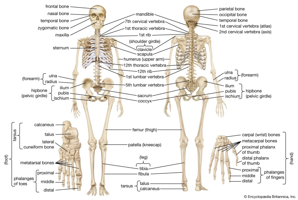
- In humans, the skeletal system consists of 206 bones, along with an assortment of joints and associated cartilages. These bones can be divided into two main components: the axial skeleton and the appendicular skeleton. The axial skeleton is centered around the body’s central axis and includes the skull, spine, and ribcage. It not only provides protection for vital organs like the brain and spinal cord but also supports the upper body and facilitates movements such as bending, twisting, and flexing.
- On the other hand, the appendicular skeleton is responsible for supporting the limbs. It comprises the bones of the arms and legs, along with the shoulder and hip girdles that connect the limbs to the axial skeleton. The appendicular skeleton enables a wide range of movements and activities, allowing humans to walk, run, grasp objects, and perform various complex tasks.
- It is worth noting that while humans possess an endoskeleton, where the bones lie internally beneath the skin and muscles, other organisms like insects possess an exoskeleton. In these organisms, the skeletal system is located on the outside of the body, providing protection and support in a different manner.
- In conclusion, the skeletal system serves as the fundamental framework of the human body, providing support, protection, and attachment points for muscles. It consists of bones, joints, and associated cartilages, with the axial skeleton protecting vital organs and the appendicular skeleton supporting the limbs. Without the skeletal system, human movement and the integrity of internal organs would be severely compromised.
Definition of Skeletal system
The skeletal system is the framework of bones, joints, and cartilages that provides support, protection, and facilitates movement in the human body.
Types of Skeletal Systems
There are three main types of skeletal systems found in different organisms: hydrostatic skeleton, exoskeleton, and endoskeleton.
- Hydrostatic skeleton: A hydrostatic skeleton is formed by a fluid-filled compartment called the coelom, which supports the organs of the organism and resists external compression. It is found in soft-bodied animals such as sea anemones, earthworms, and other invertebrates. Movement in a hydrostatic skeleton is achieved through muscular contractions that change the shape of the coelom, causing the fluid to produce movement.
- Exoskeleton: An exoskeleton is an external skeleton consisting of a hard encasement on the surface of an organism. It provides defense against predators, supports the body, and allows for movement through attached muscles. Examples of organisms with exoskeletons include crabs and insects. Arthropods like crabs and lobsters have exoskeletons made up of chitin, a strong and flexible material secreted by epidermal cells. Arthropods periodically shed their exoskeletons to accommodate growth.
- Endoskeleton: An endoskeleton is a skeleton composed of hard, mineralized structures located within the soft tissues of organisms. It provides support, protects internal organs, and allows for movement through muscle contractions. The human skeleton is an example of an endoskeleton, consisting of 206 bones in adults. The skeletal system in vertebrates is divided into the axial skeleton (skull, vertebral column, rib cage) and the appendicular skeleton (limb bones, shoulder and pelvic girdles).
Each type of skeletal system has its advantages and adaptations suited to the needs and environments of the organisms possessing them.
Anatomy of the Skeletal System
The skeleton is subdivided into two divisions:
- Axial Skeleton
- Appendicular skeleton
A. Axial Skeleton
The axial skeleton, which serves as the body’s longitudinal axis, is divided into three sections: the skull, the vertebral column, and the bony thorax.
1. Skull
The skull is formed by two sets of bones:
- the cranium
- the facial bones.
Cranium
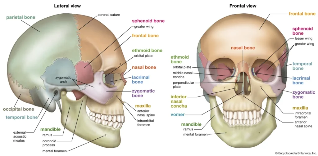
The cranium is an essential part of the skull that encloses and protects the delicate brain tissue. It is composed of eight large flat bones, each with its own specific features:
- Frontal bone: The frontal bone forms the forehead, the bony projections beneath the eyebrows, and the superior part of each eye’s orbits.
- Parietal bones: The paired parietal bones form the majority of the superior and lateral walls of the cranium. They meet at the midline of the skull at the sagittal suture and also form the coronal suture, where they join the frontal bone.
- Temporal bones: The temporal bones are situated below the parietal bones and connect to them at the squamous sutures. They have several notable structures:
- External acoustic meatus: This is a canal that leads to the eardrum and the middle ear. It serves as the pathway for sound to enter the ear.
- Styloid process: Located just inferior to the external acoustic meatus, the styloid process is a sharp, needle-like projection.
- Zygomatic process: The zygomatic process is a thin bridge of bone that articulates with the cheekbone (zygomatic bone) anteriorly.
- Mastoid process: Posterior and inferior to the external acoustic meatus, the mastoid process is a rough projection filled with air cavities (mastoid sinuses). It provides attachment sites for certain neck muscles.
- Jugular foramen: Situated at the junction of the temporal and occipital bones, the jugular foramen allows passage for the jugular vein, the largest vein in the head, which drains the brain. Just anterior to it in the cranial cavity is the internal acoustic meatus, through which cranial nerves VII and VIII pass.
- Occipital bone: The occipital bone joins with the parietal bones anteriorly at the lambdoid suture. It contains a large opening called the foramen magnum, which surrounds the lower part of the brain and allows the spinal cord to connect with the brain.
- Sphenoid bone: Shaped like a butterfly, the sphenoid bone spans the width of the skull and forms part of the cranial cavity floor. It features notable structures, including:
- Sella turcica: A small depression in the midline of the sphenoid bone, it serves as a snug enclosure for the pituitary gland.
- Foramen ovale: A large oval opening located in line with the posterior end of the sella turcica, allowing cranial nerve V fibers to pass to the chewing muscles of the lower jaw.
- Optic canal: This canal permits the passage of the optic nerve to the eye.
- Superior orbital fissure: A slit-like opening through which the cranial nerves controlling eye movements pass.
- Sphenoid sinuses: The central part of the sphenoid bone contains air cavities known as sphenoid sinuses.
- Ethmoid bone: The irregularly shaped ethmoid bone is positioned anterior to the sphenoid bone. It forms the roof of the nasal cavity and part of the medial walls of the orbits. Notable features include:
- Crista galli: This projection on the superior surface of the ethmoid bone serves as an attachment point for the outermost covering of the brain.
- Cribriform plates: These holey areas in the ethmoid bone allow nerve fibers carrying impulses from the olfactory receptors of the nose to reach the brain.
- Superior and middle nasal conchae: Extensions of the ethmoid bone, these structures contribute to the lateral walls of the nasal cavity and increase the turbulence of airflow through the nasal passages.
Together, these bones form the cranium, providing protection and structural support for the brain while also accommodating important sensory and functional structures.
Facial Bones
The face is composed of fourteen bones, with twelve of them existing in pairs, while the mandible and vomer are single bones. Let’s explore the different facial bones:
- Maxillae: The maxillae, or maxillary bones, fuse together to form the upper jaw. They are considered the “keystone” bones of the face, as all other facial bones (excluding the mandible) connect to them. The maxillae support the upper teeth in the alveolar margin.
- Palatine bones: Positioned behind the palatine processes of the maxillae, the paired palatine bones contribute to the posterior section of the hard palate.
- Zygomatic bones: Also known as the cheekbones, the zygomatic bones are responsible for forming a significant portion of the lateral walls of the eye sockets (or orbits).
- Lacrimal bones: The lacrimal bones are small finger-sized bones that form part of the medial walls of each orbit. They contain a groove that serves as a pathway for tears.
- Nasal bones: These small rectangular bones create the bridge of the nose, providing support and structure to this central facial feature.
- Vomer bone: The vomer is a single bone situated in the midline of the nasal cavity. It constitutes the majority of the bony nasal septum.
- Inferior nasal conchae: The inferior nasal conchae are thin and curved bones that project medially from the lateral walls of the nasal cavity. They help increase the surface area of the nasal passages.
- Mandible: The mandible, or lower jawbone, is the largest and strongest bone in the face. It articulates with the temporal bones on each side, forming the only freely movable joints in the skull. The horizontal part of the mandible forms the chin, while the vertical sections (rami) connect the mandible to the temporal bone.
These facial bones collectively contribute to the unique structure and appearance of the face, ensuring proper support, protection, and functionality.
2. The Hyoid Bone
The hyoid bone, while not considered part of the skull, is closely associated with the mandible (lower jawbone) and temporal bones. Here are some key points about the hyoid bone:
- Location: The hyoid bone is situated in the midneck region, approximately 2 cm (1 inch) above the larynx (voice box). It is suspended and anchored by ligaments to the styloid processes of the temporal bones.
- Structure: The hyoid bone has a horseshoe-shaped structure consisting of a body and two pairs of projections known as horns or cornua. The body forms the central part of the hyoid bone, while the horns extend outward from each end.
- Function: The hyoid bone serves important functions in the body. Firstly, it acts as a movable base for the tongue, providing support and attachment for the muscles involved in tongue movement and speech production. Additionally, the hyoid bone serves as an attachment point for neck muscles that are responsible for raising and lowering the larynx during swallowing and speaking.
Overall, the hyoid bone plays a crucial role in tongue movement, swallowing, and speech. Its unique position in the midneck region and its connection to the mandible and temporal bones make it an important component of the intricate musculoskeletal system in the head and neck.
3. Fetal Skull
The fetal skull, which includes the skull of a fetus or newborn infant, differs significantly from an adult skull in several ways:
- Size: The size of the fetal skull is distinct from that of an adult skull. In adults, the skull represents only about one-eighth of the total body length. However, in newborn infants, the skull is relatively larger, accounting for approximately one-fourth of the body’s total length. This discrepancy in size reflects the rapid growth and development that occurs during the fetal stage and early infancy.
- Fontanels: The newborn skull possesses fibrous regions known as fontanels, which are areas where the cranial bones have yet to fully convert into solid bone. These fontanels are essentially fibrous membranes that connect the cranial bones. They play a crucial role in accommodating the growth and development of the brain, as well as providing flexibility during childbirth.
- Anterior Fontanel: The largest and most prominent fontanel in the fetal skull is the anterior fontanel. It is diamond-shaped and is located at the junction of several cranial bones, including the frontal, parietal, and temporal bones. The anterior fontanel is easily recognizable as a soft spot on the baby’s head. This fontanel serves a vital purpose during childbirth by allowing the fetal skull to undergo slight compression, facilitating the passage of the baby through the birth canal.
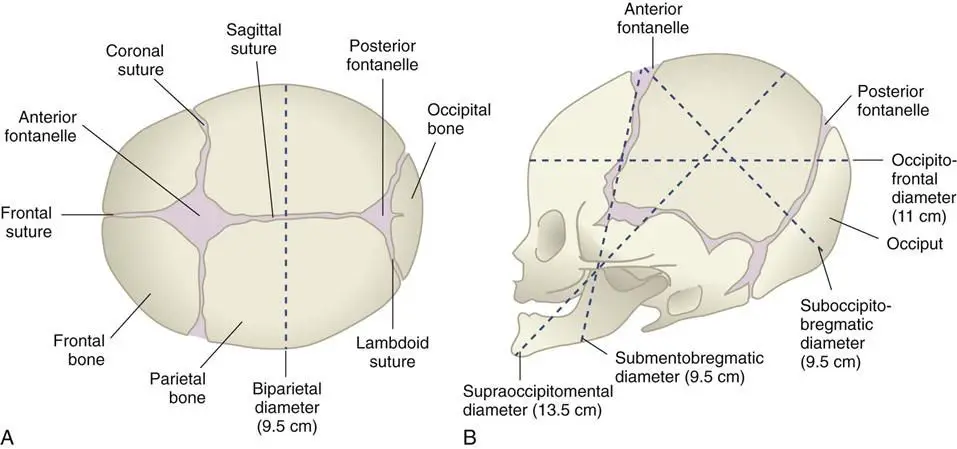
The fontanels found in the fetal skull are temporary structures that gradually close and convert into bone as the baby grows. The conversion of fontanels into solid bone is part of the normal process of skull development and typically occurs within the first two years of life.
In summary, the fetal skull differs from the adult skull in terms of size, the presence of fontanels, and the prominent anterior fontanel. These features are important adaptations that allow for the growth, flexibility, and safe passage of the baby during childbirth.
4. Vertebral Column (Spine)
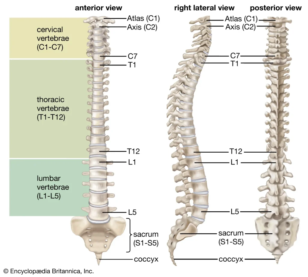
- The vertebral column, commonly known as the spine, serves as the central support structure of the body. It extends from the skull, providing support for it, all the way down to the pelvis, where it transmits the weight of the body to the lower limbs. Composed of 26 irregular bones, the spine forms a flexible and curved structure, reinforced by ligaments.
- Running through the central cavity of the vertebral column is the delicate spinal cord, which is surrounded and protected by the vertebral column. Before birth, the spine consists of 33 separate bones called vertebrae. However, as development progresses, nine of these vertebrae fuse together to form two composite bones known as the sacrum and the coccyx. The sacrum and coccyx construct the inferior portion of the vertebral column.
- Among the 24 single bones that remain, the first seven vertebrae in the neck are called cervical vertebrae. The next 12 vertebrae are known as thoracic vertebrae, and they are located in the chest region. The remaining five vertebrae, supporting the lower back, are called lumbar vertebrae.
- The individual vertebrae are separated by intervertebral discs, which are pads of flexible fibrocartilage. These discs cushion the vertebrae, absorb shock, and allow for flexibility in the spine. They play a crucial role in maintaining the structural integrity of the vertebral column.
- The vertebral column exhibits two types of curvatures: primary curvatures and secondary curvatures. The primary curvatures are present at birth and are found in the thoracic and sacral regions. The secondary curvatures, which develop after birth, are located in the cervical and lumbar regions.
- Each vertebra has distinct features. The body or centrum is a disc-like structure that bears weight and faces anteriorly in the vertebral column. The vertebral arch is formed by the joining of all the posterior extensions, known as the laminae and pedicles, from the vertebral body. The vertebral foramen is a canal through which the spinal cord passes.
- Additionally, each vertebra has transverse processes, which are two lateral projections from the vertebral arch. The spinous process is a single projection that arises from the posterior aspect of the vertebral arch. The superior and inferior articular processes are paired projections located laterally to the vertebral foramen. These processes enable a vertebra to form joints with adjacent vertebrae, allowing for movement and flexibility in the spine.
- Overall, the vertebral column, with its complex structure and various components, plays a vital role in supporting the body, protecting the spinal cord, and providing flexibility for movement.
Cervical Vertebrae
- The cervical vertebrae, consisting of seven individual bones labeled C1 to C7, form the region of the spine known as the neck. Each cervical vertebra possesses unique characteristics that contribute to its functions and structure.
- Starting with the first cervical vertebra, known as the atlas or C1, it differs from the other vertebrae in that it lacks a body. Instead, the superior surfaces of its transverse processes feature large depressions that receive the occipital condyles of the skull. This arrangement allows for the articulation between the skull and the vertebral column, facilitating the nodding motion of the head.
- The second cervical vertebra, called the axis or C2, plays a crucial role as a pivot point for the rotation of the atlas (and consequently, the skull) above it. The axis is characterized by a large upright process known as the dens or odontoid process. The dens protrudes superiorly from the body of the axis and acts as the pivotal structure around which the atlas and skull rotate. This arrangement enables the side-to-side movement of the head, such as shaking or rotating it.
- The transverse processes of the cervical vertebrae contain foramina, which are openings that serve important functions. These foramina provide passage for the vertebral arteries, which transport blood to the brain. The vertebral arteries pass through these foramina as they make their way to the brain, ensuring a sufficient blood supply for proper brain function.
- The cervical vertebrae as a whole contribute to the overall flexibility and movement of the neck region. Their unique structures, such as the atlas and axis, allow for a wide range of motions, including nodding, rotating, and tilting the head. Additionally, the presence of the foramina in the transverse processes ensures the necessary blood supply to the brain, highlighting the vital role of the cervical vertebrae in both structural support and physiological functions.
Thoracic Vertebrae
- The thoracic vertebrae, numbering twelve and labeled T1 to T12, are considered typical vertebrae due to their common characteristics and functions within the spine.
- One distinguishing feature of the thoracic vertebrae is their larger size compared to the cervical vertebrae. This increased size is necessary to support the weight and load-bearing functions associated with the thoracic region of the spine. Furthermore, the thoracic vertebrae are unique as they are the only vertebrae that articulate with the ribs.
- The body of each thoracic vertebra is somewhat heart-shaped and exhibits two costal facets on each side. These costal facets serve as points of articulation for the heads of the ribs. The presence of these facets enables the connection between the thoracic vertebrae and the rib cage, contributing to the structure and stability of the chest region.
- The transverse processes of the thoracic vertebrae have a specific function related to the ribs. They articulate with the nearby knob-like tubercles present on the ribs, forming a joint that allows for movements involved in breathing and overall rib cage mobility. This articulation contributes to the expansion and contraction of the rib cage during respiration.
- A distinctive feature of the thoracic vertebrae is the shape of their spinous process. The spinous process is elongated and curves sharply downward, resembling the shape of a giraffe’s head when viewed from the side. This unique appearance is notable and helps identify the thoracic vertebrae.
- Overall, the thoracic vertebrae play a crucial role in supporting the rib cage, protecting the vital organs within the thoracic cavity, and facilitating movements associated with breathing. Their size, shape, and specific articulations with the ribs are key characteristics that contribute to their function and position within the vertebral column.
Lumbar Vertebrae
- The lumbar vertebrae, consisting of five individual bones labeled L1 to L5, are characterized by their unique features and functions within the vertebral column.
- One notable characteristic of the lumbar vertebrae is the massive, blocklike shape of their bodies. This design provides increased strength and stability to withstand the substantial stress placed on the vertebral column, particularly in the lumbar region. The lumbar vertebrae are responsible for bearing most of the load and weight of the upper body, making them the sturdiest among the vertebrae.
- The spinous processes of the lumbar vertebrae contribute to their distinctive appearance. These processes are short and hatchet-shaped, resembling the silhouette of a moose head when viewed from the side. This feature helps in distinguishing the lumbar vertebrae from other vertebrae within the spine.
- Due to their robust structure and placement in the lower back, the lumbar vertebrae play a crucial role in supporting the weight of the upper body and maintaining stability during various activities such as walking, running, lifting, and bending. The lumbar region experiences significant stress and strain, and the lumbar vertebrae are specifically designed to withstand these forces.
- By providing strength and stability, the lumbar vertebrae contribute to the overall functionality of the vertebral column and the protection of the spinal cord. Their blocklike bodies, coupled with the unique appearance of their spinous processes, showcase their specialized adaptations for the demanding tasks and load-bearing functions associated with the lumbar region.
Sacrum
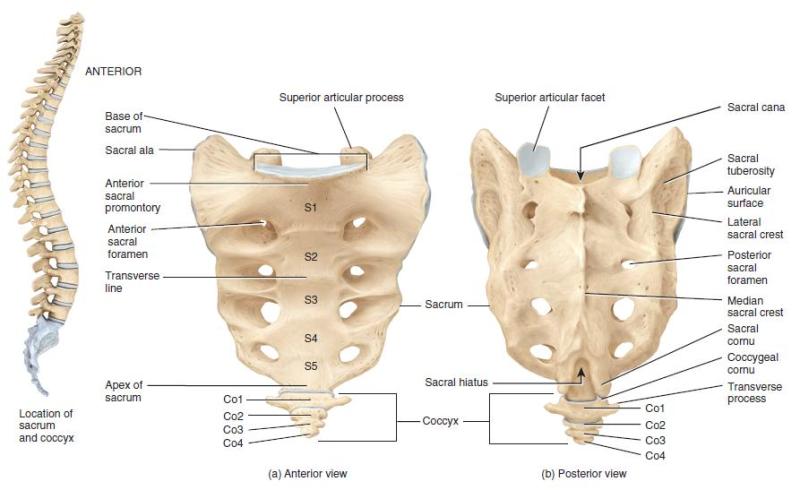
- The sacrum is a triangular bone located at the base of the vertebral column, formed by the fusion of five vertebrae known as the sacral vertebrae.
- The sacrum features winglike extensions called alae, which articulate laterally with the hip bones, forming the sacroiliac joints. These joints provide stability and support between the sacrum and the hip bones, contributing to the overall strength and structure of the pelvic region.
- On the posterior midline surface of the sacrum, there is a roughened area known as the median sacral crest. This crest is formed by the fused spinous processes of the sacral vertebrae. It serves as an attachment point for ligaments and muscles in the lower back region.
- The posterior sacral foramina are located on each side of the sacrum, flanking the median sacral crest. These foramina provide passageways for nerves and blood vessels, facilitating communication and transportation between the spinal cord and other parts of the body.
- Inside the sacrum, the vertebral canal continues as the sacral canal. This canal serves as a protective passage for the continuation of the spinal cord within the sacrum. At the bottom of the sacral canal, there is a large opening called the sacral hiatus. The sacral hiatus is the inferior termination of the sacral canal and is an important landmark for certain medical procedures and injections.
- The sacrum plays a crucial role in supporting the weight of the upper body, transmitting it to the pelvis and lower limbs. Its fusion of vertebrae provides strength and stability to the lower part of the vertebral column, enhancing posture and enabling movements such as walking and running.
- Overall, the sacrum, with its unique features such as the alae, median sacral crest, posterior sacral foramina, sacral canal, and sacral hiatus, serves as an integral component of the pelvic region, contributing to structural support, stability, and the protection of neural and vascular structures within the vertebral column.
Coccyx
- The coccyx, commonly referred to as the tailbone, is a small, triangular bone located at the base of the vertebral column. It is formed by the fusion of three to five tiny, irregular-shaped vertebrae.
- The coccyx is often considered a vestigial structure, meaning it is a remnant of a tail that our evolutionary ancestors possessed. While many other vertebrate animals have tails, the coccyx is the human equivalent, though significantly reduced in size and functionality. Its presence in humans serves as a reminder of our evolutionary history.
- The coccyx is positioned below the sacrum and curves slightly inward. It consists of fused vertebrae that gradually decrease in size from the base to the tip. The number of vertebrae that fuse to form the coccyx can vary from person to person, typically ranging from three to five.
- Although the coccyx is relatively small and has limited mobility, it serves several important functions. It provides attachment points for ligaments, tendons, and muscles in the pelvic region, contributing to stability and support during activities such as sitting and standing. It also acts as a weight-bearing structure, helping to distribute the weight and pressure exerted on the pelvis.
- The coccyx is susceptible to injury, particularly from falls or trauma to the lower back. These injuries can cause pain and discomfort in the tailbone area. However, in most cases, conservative treatment measures such as rest, pain management, and physical therapy can help alleviate coccyx-related symptoms.
- In summary, the coccyx is a small bone formed by the fusion of three to five vertebrae. It serves as the human “tailbone,” a vestigial structure inherited from our evolutionary past. While reduced in size and function compared to tails in other animals, the coccyx provides attachment points for muscles and contributes to stability and weight-bearing in the pelvic region.
5. Thoracic Cage
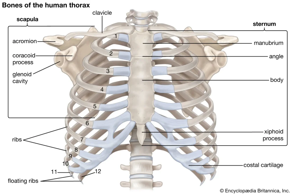
The bony thorax is made up of the sternum, ribs, and thoracic vertebrae; it is commonly referred to as the thoracic cage because it forms a protective, cone-shaped cage of slender bones around the organs of the thoracic cavity.
Sternum
- The sternum, also known as the breastbone, is a flat bone that serves as a central component of the rib cage. It is formed by the fusion of three distinct bones: the manubrium, body, and xiphoid process.
- The sternum features three significant bony landmarks that aid in its identification and serve as important anatomical references. The first landmark is the jugular notch, which is a concave depression located at the upper border of the manubrium. This notch can be easily palpated and is generally aligned with the level of the third thoracic vertebra.
- The second landmark is the sternal angle, which is formed by the meeting point of the manubrium and the body of the sternum. At this junction, there is a slight angle between the two components, creating a transverse ridge. The sternal angle is typically found at the level of the second ribs and serves as an essential reference point for counting ribs and locating other structures within the thoracic region.
- The third landmark is the xiphisternal joint. It is the point of fusion between the sternal body and the xiphoid process, the smallest and most inferior portion of the sternum. The xiphisternal joint is located at approximately the level of the ninth thoracic vertebra.
- The sternum plays a crucial role in protecting vital organs within the thoracic cavity, such as the heart and lungs. Additionally, it serves as an attachment point for various muscles and ligaments, including those involved in respiration and movement of the upper limbs.
- Understanding the landmarks of the sternum, such as the jugular notch, sternal angle, and xiphisternal joint, is important for accurate anatomical identification and clinical assessments. These landmarks aid in determining the level and position of other structures within the thoracic region, facilitating proper diagnosis and treatment.
Ribs
- The ribs are a set of twelve pairs of bones that form the structure of the bony thorax, also known as the rib cage. They play a crucial role in protecting the vital organs within the thoracic cavity, such as the heart and lungs, while also providing support and flexibility for breathing movements.
- The first seven pairs of ribs are called true ribs. They are directly attached to the sternum (breastbone) through their own costal cartilages, which are bands of hyaline cartilage that connect the ribs to the sternum. The true ribs not only contribute to the shape and structure of the rib cage but also provide stability and protection to the underlying organs.
- The next five pairs of ribs are known as false ribs. Unlike the true ribs, they either attach indirectly to the sternum or are not attached to the sternum at all. The eighth, ninth, and tenth pairs of ribs are attached to the sternum through their own costal cartilages, but they are indirectly connected to the sternum via the cartilage of the rib above. These are referred to as the vertebrochondral ribs.
- The eleventh and twelfth pairs of ribs are unique and are called floating ribs. They lack any direct or indirect attachment to the sternum. Instead, their free posterior ends do not articulate with any other bone, giving them a floating appearance. The floating ribs are often shorter and have a reduced attachment, serving a more minimal role in protecting the underlying organs.
- The ribs, along with the sternum and the thoracic vertebrae, form a protective cage around the thoracic organs, allowing for respiration and providing support to the upper body. Their arrangement and attachments contribute to the flexibility of the rib cage, enabling expansion and contraction during breathing movements.
- Understanding the classification of ribs into true ribs, false ribs, and floating ribs helps in comprehending their anatomical structure and their role in maintaining the integrity and functionality of the thoracic region.
B. Appendicular Skeleton
The appendicular skeleton is made up of 126 limb bones as well as the pectoral and pelvic girdles, which connect the limbs to the axial skeleton.
1. Bones of the Shoulder Girdle
- The bones of the shoulder girdle, also known as the pectoral girdle, consist of two bones: the clavicle (collarbone) and the scapula (shoulder blade).
- The clavicle is a slender, doubly curved bone that serves as a strut connecting the manubrium of the sternum medially to the scapula laterally. It helps to form the shoulder joint and acts as a brace to hold the arm away from the top of the thorax. The clavicle plays a crucial role in preventing shoulder dislocation.
- The scapula, or shoulder blade, is a triangular bone that flares when we move our arms posteriorly, resembling wings. It has a flattened body and two important processes: the acromion and the coracoid. The acromion is the enlarged end of the spine of the scapula and connects with the clavicle laterally at the acromioclavicular joint. The coracoid process, shaped like a beak, extends over the top of the shoulder and provides attachment points for some of the arm muscles. Just medial to the coracoid process is the suprascapular notch, which serves as a passageway for nerves.
- The scapula has three borders: the superior border, the medial (vertebral) border, and the lateral (axillary) border. It also has three angles: the superior angle, the inferior angle, and the lateral angle. The lateral angle contains the glenoid cavity, a shallow socket that receives the head of the humerus bone, forming the shoulder joint.
- The shoulder girdle allows for free movement due to several factors. It attaches to the axial skeleton at only one point, the sternoclavicular joint, which provides limited stability. The scapula has a loose attachment, allowing it to slide back and forth against the thorax as muscles act. The glenoid cavity, being shallow, is poorly reinforced by ligaments, contributing to the wide range of motion at the shoulder joint.
- Together, the clavicle and scapula form the shoulder girdle, providing stability, support, and attachment sites for muscles involved in shoulder movements. The shoulder girdle allows for the flexibility and mobility required for various arm and shoulder actions, such as reaching, lifting, and throwing.
2. Bones of the Upper Limb
The skeletal framework of each upper limb is made up of thirty distinct bones that serve as the foundations of the arm, forearm, and hand.
a. Arm
- The arm is primarily composed of a single bone known as the humerus, which is classified as a typical long bone.
- The humerus has several anatomical features that contribute to its structure and function. Just below the head of the humerus, there is a slight constriction called the anatomical neck. Anterolateral to the head, there are two bony projections known as the greater and lesser tubercles, which are separated by the intertubercular sulcus. These tubercles serve as attachment sites for muscles involved in arm movements.
- Located just distal to the tubercles is the surgical neck of the humerus. This particular region is called the surgical neck because it is a common site for fractures.
- In the midpoint of the humerus shaft, there is a roughened area known as the deltoid tuberosity. This is where the large, fleshy deltoid muscle of the shoulder attaches, playing a significant role in arm movements.
- Running obliquely down the posterior aspect of the humerus shaft is the radial groove. This groove marks the course of the radial nerve, which is an important nerve of the upper limb.
- At the distal end of the humerus, there are two prominent features: the medial trochlea and the lateral capitulum. The trochlea resembles a spool and articulates with the ulna bone, while the capitulum has a ball-like shape and articulates with the radius bone. Together, these processes form the elbow joint and allow for movements of the forearm.
- On the anterior surface above the trochlea, there is a depression called the coronoid fossa. This fossa, along with the posterior olecranon fossa, allows the corresponding processes of the ulna to move freely during bending and extending of the elbow joint. The medial and lateral epicondyles flank these depressions and provide attachment sites for ligaments and muscles associated with the elbow and forearm.
- Overall, the humerus bone forms the main structure of the arm and serves as a critical component for various movements and functions of the upper limb.
b. Forearm
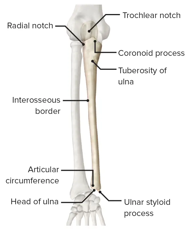
- The forearm consists of two bones, namely the radius and the ulna, which play a vital role in the structure and function of the arm.
- The radius is positioned on the lateral side of the forearm when the body is in the anatomical position, meaning it is located on the thumb side. However, when the hand is rotated so that the palm faces backward, the distal end of the radius crosses over and ends up medial to the ulna. This allows for specific movements and rotations of the forearm.
- Both the radius and the ulna articulate with each other at small radioulnar joints, both proximally and distally. These joints enable rotational movements of the forearm. Additionally, the two bones are connected along their entire length by the interosseous membrane, which provides flexibility and stability to the forearm.
- At the distal end of both the ulna and the radius, they possess styloid processes. These bony projections serve as attachment sites for ligaments and provide some support to the wrist joint.
- The disc-shaped head of the radius forms a joint with the capitulum of the humerus, allowing for pivotal movements of the forearm. Just below the head of the radius is the radial tuberosity, which serves as the attachment point for the tendon of the biceps muscle.
- The ulna, when the upper limb is in the anatomical position, is located on the medial side (little-finger side) of the forearm. Its proximal end features the coronoid process and the posterior olecranon process, which are separated by the trochlear notch. These structures, together with the trochlea of the humerus, form a pliers-like joint, providing stability and allowing for bending and extending motions of the elbow.
- Overall, the radius and the ulna work in conjunction to enable various movements of the forearm, including rotation, flexion, and extension. Their anatomical features and articulations with other bones in the arm contribute to the overall functionality of the upper limb.
c. Hand
- The hand is composed of three main groups of bones: the carpals, the metacarpals, and the phalanges. Together, they form the skeletal framework of the hand and allow for its intricate movements and dexterity.
- The carpal bones are a collection of eight small bones that are arranged in two irregular rows of four bones each. They are situated at the base of the hand, commonly referred to as the wrist or carpus. Ligaments connect and stabilize these carpal bones, limiting excessive movements between them.
- Next, we have the metacarpals, which are numbered from 1 to 5 starting from the thumb side of the hand to the little finger. These five metacarpal bones extend from the carpus to the base of each finger. When the hand is clenched into a fist, the heads of the metacarpals become prominent and are commonly referred to as the “knuckles.”
- Finally, the phalanges make up the bones of the fingers. Each hand consists of a total of 14 phalanges. Within each finger, except for the thumb, there are three phalanges: the proximal, middle, and distal phalanges. The thumb, on the other hand, has only two phalanges: the proximal and distal phalanges.
- The arrangement and articulation of these bones in the hand allow for a wide range of movements, precision, and versatility. The carpals provide stability to the wrist joint, while the metacarpals and phalanges facilitate gripping, grasping, and intricate movements of the fingers. The muscles, tendons, and ligaments surrounding these bones work together to enable the hand’s remarkable abilities in activities such as writing, typing, playing musical instruments, and countless other tasks requiring fine motor skills.
3. Bones of the Pelvic Girdle
- The pelvic girdle, also known as the hip bones, is formed by two coxal bones called ossa coxae. These bones are large, heavy, and securely attached to the axial skeleton, as their primary function is to bear weight and support the upper body.
- The sockets of the pelvic girdle, which receive the thigh bones, are deep and reinforced by strong ligaments, ensuring a firm attachment between the limbs and the girdle. Additionally, the bony pelvis provides protection to important organs such as the reproductive organs, urinary bladder, and a portion of the large intestine.
- The ilium is a large, flaring bone that forms the majority of the hip bone. It connects posteriorly with the sacrum at the sacroiliac joint. When placing your hands on your hips, you are resting them on the winglike portions of the ilia, known as the alae. The upper edge of an ala is called the iliac crest, which is a significant anatomical landmark. The iliac crest extends anteriorly to the anterior superior iliac spine and posteriorly to the posterior superior iliac spine.
- The ischium forms the most inferior part of the coxal bone and is often referred to as the “sit-down” bone. It features a roughened area called the ischial tuberosity, which bears weight when you are sitting. Superior to the tuberosity is the ischial spine, another important anatomical landmark, especially in pregnant women, as it narrows the outlet of the pelvis through which the baby must pass during childbirth.
- The ischium also includes the greater sciatic notch, which allows blood vessels and the large sciatic nerve to pass from the pelvis posteriorly into the thigh.
- The pubis, or pubic bone, is the most anterior part of the coxal bone. It features an opening called the obturator foramen, through which blood vessels and nerves pass into the anterior part of the thigh. The pubic bones of each hip bone fuse anteriorly at a cartilaginous joint called the pubic symphysis.
- The ilium, ischium, and pubis fuse together to form the acetabulum, a deep socket that receives the head of the thigh bone (femur). The term “acetabulum” translates to “vinegar cup” in Latin.
- The pelvic girdle has two important divisions: the false pelvis and the true pelvis. The false pelvis is located superiorly to the true pelvis and is medial to the flaring portions of the ilia. The true pelvis is surrounded by bone and lies inferiorly to the flaring parts of the ilia and the pelvic brim. The dimensions of the true pelvis, particularly the outlet (measured between the ischial spines) and the inlet (measured between the right and left sides of the pelvic brim), are crucial, especially during childbirth, as they must be large enough to allow the infant’s head to pass.
- Obstetricians carefully measure these pelvic dimensions to ensure a safe delivery.
4. Bones of the Lower Limbs
When we stand upright, our lower limbs support our entire body weight; thus, the bones that make up the three lower limb segments (thigh, leg, and foot) are more thicker and stronger than the corresponding upper limb bones.
a. Thigh
- The thigh is primarily composed of the femur, also known as the thigh bone, which is the only bone in this region and is recognized as the heaviest and strongest bone in the human body.
- The proximal end of the femur features several important structures. It has a ball-like head that articulates with the acetabulum of the hip bone, forming the hip joint. Adjacent to the head is the neck of the femur, which is a common site for fractures, particularly in older individuals. Additionally, the greater and lesser trochanters are present on the proximal end, with the intertrochanteric line anteriorly and the intertrochanteric crest posteriorly, serving as attachment points for muscles.
- Moving along the shaft of the femur, one can find the gluteal tuberosity and other markings that provide attachment sites for muscles.
- Distally, the femur exhibits the lateral and medial condyles, which articulate with the tibia below, forming the knee joint. These condyles are separated posteriorly by the deep intercondylar fossa, which provides stability and space for ligament attachments.
- On the anterior side of the distal femur, there is a smooth patellar surface. This surface forms a joint with the patella, also known as the kneecap, and aids in the proper functioning of the knee joint.
- The femur plays a crucial role in supporting body weight and facilitating movements of the lower limb, making it an essential bone for walking, running, and various activities that involve the thigh and knee.
b. Leg
- The leg is comprised of two bones, namely the tibia and fibula, which are connected by an interosseous membrane, forming the skeletal framework of the leg.
- The tibia, commonly referred to as the shinbone, is the larger and more medial of the two bones. At its proximal end, it articulates with the distal end of the femur, specifically with the medial and lateral condyles, contributing to the formation of the knee joint. On the anterior surface of the tibia, there is a roughened area known as the tibial tuberosity, to which the patellar (kneecap) ligament attaches.
- Moving distally, the tibia presents a prominent bony projection called the medial malleolus, which forms the inner bulge of the ankle. This serves as a distinctive landmark for the medial aspect of the ankle region. On the anterior surface of the tibia, there is a sharp ridge known as the anterior border, which is easily palpable beneath the skin as it is unprotected by muscles.
- In contrast, the fibula lies parallel to the tibia and forms joints with it at both its proximal and distal ends. The fibula is thinner and more stick-like compared to the tibia. It does not contribute to the formation of the knee joint. At the distal end of the fibula, there is another bony prominence called the lateral malleolus, which forms the outer part of the ankle. This serves as a recognizable feature on the lateral aspect of the ankle region.
- Together, the tibia and fibula provide structural support, stability, and mobility to the leg. While the tibia plays a major role in weight-bearing and forms part of the knee joint, the fibula contributes to the overall strength of the leg and plays a role in muscle attachment.
c. Foot
- The foot is a remarkable structure consisting of three main components: the tarsals, metatarsals, and phalanges. It serves two vital functions: supporting the body’s weight and acting as a lever for propulsion during walking and running.
- The tarsus is located in the posterior half of the foot and comprises seven tarsal bones. Among these, the calcaneus, commonly known as the heel bone, and the talus, situated between the tibia and the calcaneus, bear the majority of the body’s weight.
- The sole of the foot is formed by five metatarsals, which are long bones extending from the tarsus to the base of the toes. These metatarsals provide stability and flexibility to the foot during movement.
- The toes are composed of 14 phalanges, with each toe, except the great toe, possessing three phalanges. The great toe, also known as the hallux, has two phalanges. The phalanges contribute to the foot’s dexterity and provide balance during activities such as walking, running, and maintaining posture.
- A distinctive feature of the foot is the presence of three strong arches formed by the arrangement of its bones. These arches include two longitudinal arches, known as the medial and lateral arches, and one transverse arch. The arches enhance the foot’s flexibility, resilience, and shock-absorbing capabilities, distributing body weight evenly and minimizing the impact on joints and soft tissues.
- Overall, the foot’s intricate structure and arches work in harmony to support the body’s weight and facilitate efficient movement. Whether we are standing, walking, or engaging in more dynamic activities, the foot plays a crucial role in maintaining balance, stability, and propulsion.
5. Joints
Joints, also known as articulations, serve two primary functions: they provide stability by holding bones together securely, and they enable movement within the rigid skeleton.
Joints can be classified based on their function and structure. The functional classification focuses on the amount of movement allowed at the joint. There are three types of functional joints:
- Synarthroses: These are immovable joints where the bones are tightly joined together. Synarthroses joints provide stability and are found mainly in the axial skeleton, such as the sutures in the skull.
- Amphiarthroses: These joints are slightly movable, allowing limited mobility. Amphiarthroses joints are characterized by the presence of fibrous or cartilaginous connections between the bones. Examples include the intervertebral discs in the spinal column.
- Diarthroses: Also known as freely movable joints, diarthroses joints allow a wide range of motion. These joints are predominant in the limbs, where mobility is essential. Examples of diarthroses joints include the shoulder joint and the knee joint.
Joints can also be classified based on their structure. There are three main structural classifications:
- Fibrous joints: These joints are held together by fibrous connective tissue and have minimal or no movement. Fibrous joints can be further divided into three types: sutures (found in the skull), syndesmoses (such as the distal tibiofibular joint), and gomphoses (seen in the tooth sockets).
- Cartilaginous joints: These joints are connected by cartilage and allow limited movement. Cartilaginous joints can be further categorized into two types: synchondroses (where hyaline cartilage unites the bones, as seen in the epiphyseal plates of growing long bones) and symphyses (where fibrocartilage connects the bones, like the intervertebral discs and the pubic symphysis).
- Synovial joints: These joints are the most common type of joint in the body and allow a wide range of movement. Synovial joints are characterized by the presence of a joint cavity filled with synovial fluid. They are further classified into six types based on their shape and movement capabilities, including hinge joints (elbow joint), ball-and-socket joints (hip joint), and saddle joints (thumb joint).
In summary, joints are essential for both providing stability and enabling movement in the skeletal system. They can be classified based on their function, considering the degree of mobility, or based on their structure, taking into account the type of connective tissue or presence of a joint cavity. Understanding the different types of joints helps us appreciate the diverse capabilities of the human body in terms of mobility and flexibility.
a. Fibrous Joints
- Fibrous joints are characterized by the union of bones through fibrous tissue. These joints provide stability and have minimal or no movement between the bones involved.
- One prominent example of fibrous joints is seen in the sutures of the skull. Sutures are fibrous joints where the irregular edges of the cranial bones interlock and are tightly bound together by connective tissue fibers. This arrangement ensures a strong union between the bones, offering maximum protection to the underlying brain. Due to their immovable nature, sutures prevent any significant movement between the bones, promoting structural integrity.
- Another type of fibrous joint is known as syndesmosis. In syndesmoses, the fibers connecting the bones are longer compared to sutures, allowing for some degree of mobility or “give” at the joint. An example of a syndesmosis is the joint connecting the distal ends of the tibia and fibula. The fibrous tissue between these two bones provides stability while still permitting limited movement. This arrangement is important for maintaining proper alignment and coordination during movements of the lower leg.
- In both sutures and syndesmoses, fibrous tissue plays a crucial role in holding the bones together. The collagen fibers within the fibrous tissue provide strength and resistance, ensuring the stability of the joint and preventing excessive movement that could compromise the function and protection of surrounding structures.
- Fibrous joints, with their strong fibrous connections, are essential in areas where stability and protection are paramount. While they limit movement, they provide a solid foundation for the bones, enabling them to function harmoniously and safeguard vital structures within the body.
b. Cartilaginous Joints
- Cartilaginous joints are characterized by the connection of bone ends through cartilage. These joints provide some degree of movement while maintaining stability and support.
- One example of a slightly movable cartilaginous joint is the pubic symphysis of the pelvis. In this joint, the articulating bone surfaces are connected by a pad of fibrocartilage. This fibrocartilaginous disc allows for limited movement and flexibility between the bones while still providing stability and support to the pelvis.
- Another example of a cartilaginous joint is found in the intervertebral joints of the spinal column. These joints are formed by intervertebral discs, which are made of fibrocartilage. The intervertebral discs act as shock absorbers and allow for slight movement and flexibility between adjacent vertebrae, contributing to the overall mobility and function of the spine.
- Some cartilaginous joints are immovable and are classified as synarthrotic. An example of an immovable cartilaginous joint is the hyaline cartilage epiphyseal plates found in growing long bones. These plates consist of cartilage and are responsible for bone growth and development. They provide stability and serve as a site for longitudinal bone growth but do not allow for any movement between the bones.
- Another example of an immovable cartilaginous joint is seen between the first ribs and the sternum. These joints, known as the costal cartilages, are made of hyaline cartilage and provide stability to the rib cage. They allow for minimal movement during breathing but are primarily responsible for maintaining the structural integrity of the rib cage and protecting the underlying organs.
- Cartilaginous joints, with their cartilaginous connections, offer a balance between mobility and stability. They provide flexibility and shock absorption while ensuring structural support and protection. These joints play important roles in various parts of the body, contributing to overall movement, growth, and function.
c. Synovial Joints
Synovial joints are a type of joint characterized by the presence of a joint cavity filled with synovial fluid, separating the articulating bone ends. These joints are found in the limbs and account for the majority of movable joints in the body.
Articular cartilage covers the ends of the bones within synovial joints, providing a smooth surface that reduces friction and absorbs shock during movement. The joint surfaces are enclosed by a fibrous articular capsule, which consists of connective tissue. The inner lining of the capsule is called the synovial membrane, responsible for producing synovial fluid, a lubricating substance that nourishes and reduces friction within the joint cavity.
The joint cavity is the space enclosed by the articular capsule, containing synovial fluid. This fluid helps to lubricate the joint, reducing friction and facilitating smooth movement. The fibrous capsule of a synovial joint is often reinforced by ligaments, which provide additional stability and limit excessive movement.
Bursae are fluid-filled sacs lined with synovial membrane, located at areas where friction may occur, such as between ligaments, muscles, tendons, and bones. They act as cushions, reducing friction and allowing smooth movement.
Tendon sheaths are elongated bursae that wrap around tendons, protecting them from excessive friction. They provide a lubricated environment for tendons to glide easily as they pass through joints.
Synovial joints can be classified based on the shapes of their articulating bone surfaces, which determine the types of movements they allow:
- Plane joints: The articulating surfaces are flat, allowing short slipping or gliding movements. Plane joints are nonaxial, meaning they do not involve rotation around any axis. Examples include the intercarpal joints of the wrist.
- Hinge joints: These joints allow movement in only one plane, similar to a mechanical hinge. They are uniaxial joints. Examples include the elbow joint, ankle joint, and the joints between the phalanges of the fingers.
- Pivot joints: In pivot joints, one bone has a rounded end that fits into a ring of bone, allowing rotational movement around a single axis. Examples include the proximal radioulnar joint and the joint between the atlas and the dens of the axis.
- Condyloid joints: Condyloid joints have an egg-shaped articular surface that fits into an oval concavity in another bone. They allow movement from side to side and back and forth, but not rotation around the long axis. Condyloid joints are biaxial. An example is the metacarpophalangeal joints in the fingers.
- Saddle joints: Saddle joints have articular surfaces with both convex and concave areas, resembling a saddle. They are also biaxial joints and allow movements similar to condyloid joints. The carpometacarpal joint in the thumb is a prime example.
- Ball-and-socket joints: These joints consist of a spherical head of one bone fitting into a rounded socket of another bone. Ball-and-socket joints are multiaxial and allow movement in all directions, including rotation. The shoulder and hip joints are examples of ball-and-socket joints.
Synovial joints provide a wide range of motion and flexibility, enabling various movements essential for daily activities and physical mobility.
Functions of the Skeletal System
The skeletal system serves several crucial functions in the human body:
- Support: The bones form the internal framework that provides support to the body and cradles the soft organs. They act as the “steel girders” and “reinforced concrete” of the body. For instance, the bones of the legs act as pillars to support the body trunk when standing, and the rib cage supports the thoracic wall.
- Protection: Bones play a vital role in protecting soft body organs. The fused bones of the skull create a protective enclosure for the brain, the vertebrae surround and safeguard the spinal cord, and the rib cage provides protection to the vital organs in the thorax.
- Movement: Skeletal muscles, which are attached to bones via tendons, utilize the bones as levers to facilitate movement. When muscles contract, they pull on the bones, causing movement in the body and its various parts.
- Storage: Bones serve as storage sites for different substances. The internal cavities of bones store adipose tissue (fat). Additionally, bones act as a reservoir for essential minerals, with calcium and phosphorus being the most important ones. Calcium ions, crucial for various bodily functions, are stored in bones. When the body needs more calcium ions, they can be released from the bones into the bloodstream.
- Blood Cell Formation: Certain bones house marrow cavities where blood cell formation, known as hematopoiesis, takes place. These specialized bone marrow regions produce red blood cells, white blood cells, and platelets, contributing to the body’s immune response and oxygen transport.
Overall, the skeletal system provides structural support, protects vital organs, enables movement, stores substances like fat and minerals, and plays a role in blood cell production. These functions are essential for maintaining the body’s integrity, movement, and overall well-being.
What kinds of Conditions Affect the Skeletal System?
The skeletal system can be affected by various conditions that impact both the appendicular and axial skeleton. Some of the common conditions include:
- Fractures: Fractures occur when bones break due to accidents or traumatic events, such as falls or car accidents. They can also result from underlying conditions like osteoporosis, where bone density decreases and bones become fragile and prone to fractures.
- Metabolic bone diseases: Metabolic bone diseases affect the composition of bones, including proteins and minerals like calcium and phosphate. Conditions like hyperthyroidism, where there is an excess of thyroid hormone, can lead to the withdrawal of calcium from bones, weakening them. Insufficient levels of vitamin D can also affect bone health as it plays a crucial role in calcium absorption.
- Arthritis: Arthritis refers to inflammation of the joints. Osteoarthritis is the most common form of arthritis and is often associated with aging and wear and tear of the joints. Rheumatoid arthritis and gout are other significant forms of arthritis. Arthritis can be caused by factors like joint stress, repetitive use, joint inflammation, crystal deposition, or immune responses.
- Cancer: Bone cancer is rare but can be a serious condition. It primarily affects the pelvis and long bones of the arms and legs. Different types of bone cancers include chondrosarcoma, Ewing sarcoma, and osteosarcoma. Osteosarcoma is the most common type, typically found in long bones. Symptoms of bone cancer include pain, swelling, fatigue, and weakness.
- Spinal curvatures: Abnormalities in the curvature of the spine can lead to various pathological conditions. These curvatures include:
- Lordosis: This condition involves inward curves in the lower back, causing excessive arching.
- Kyphosis: Kyphosis refers to an abnormal rounding of the upper back, leading to a hunched posture.
- Scoliosis: Scoliosis is characterized by an S-shaped or C-shaped curve of the spine, resulting in a sideways curvature. It can cause asymmetry and uneven distribution of weight on the spine.
These are just a few examples of conditions that can affect the skeletal system. Proper diagnosis, treatment, and management of these conditions are crucial for maintaining skeletal health and overall well-being.
FAQ
What is the skeletal system?
The skeletal system refers to the framework of bones, joints, and cartilage that provides structure, support, and protection to the body. It also plays a vital role in movement, blood cell production, and mineral storage.
How many bones are in the human body?
The human body has a total of 206 bones. These bones come in different shapes and sizes, and they are grouped into the axial skeleton (skull, vertebral column, and ribcage) and the appendicular skeleton (bones of the limbs and their attachments).
What is the purpose of joints?
Joints are the points where two or more bones come together. They provide mobility and flexibility, allowing various types of movements like bending, rotating, and gliding. Joints are essential for activities such as walking, running, and performing everyday tasks.
How does the skeletal system support the body?
The skeletal system provides structural support to the body. It forms the rigid framework that holds the body upright and gives it shape. Without the skeletal system, the body would lack stability and the ability to maintain its form.
What is bone marrow, and what role does it play?
Bone marrow is a soft, spongy tissue found inside bones. It is responsible for the production of blood cells, including red blood cells, white blood cells, and platelets. Red bone marrow is involved in the production of new blood cells, while yellow bone marrow stores fat.
Can bones heal themselves if they are fractured?
Yes, bones have the ability to heal themselves after a fracture. When a bone breaks, the body initiates a process called bone healing or bone remodeling. Specialized cells called osteoblasts and osteoclasts work together to repair the broken bone by forming new bone tissue.
What is osteoporosis?
Osteoporosis is a medical condition characterized by a decrease in bone density and mass, making the bones brittle and more prone to fractures. It is most commonly seen in older adults, particularly women after menopause, but can also affect men. Lifestyle factors, hormonal changes, and genetics can contribute to the development of osteoporosis.
Can exercise help improve skeletal health?
Yes, regular exercise is beneficial for maintaining skeletal health. Weight-bearing exercises, such as walking, jogging, and weightlifting, help stimulate bone growth and increase bone density. Additionally, exercise promotes joint flexibility, muscle strength, and overall bone and muscle health.
How does nutrition affect the skeletal system?
Proper nutrition plays a crucial role in maintaining skeletal health. Calcium and vitamin D are essential nutrients for bone health. Calcium is necessary for bone strength, while vitamin D helps the body absorb calcium. Other nutrients like vitamin K, magnesium, and phosphorus also contribute to bone health.
What are some common skeletal system disorders?
Some common skeletal system disorders include osteoarthritis, rheumatoid arthritis, osteoporosis, scoliosis, fractures, and bone cancers. These disorders can cause pain, limited mobility, deformities, and other complications. Early detection, proper medical care, and lifestyle modifications can help manage these disorders effectively.
References
- https://training.seer.cancer.gov/anatomy/skeletal/
- https://www.biologyonline.com/dictionary/skeletal-system
- https://www.getbodysmart.com/skeletal-system/
- https://nurseslabs.com/skeletal-system/
- https://openstax.org/books/anatomy-and-physiology-2e/pages/6-1-the-functions-of-the-skeletal-system
- https://www.uc.edu/content/dam/uc/ce/images/OLLI/Page%20Content/THE%20SKELETAL%20SYSTEM.pdf
- https://biologydictionary.net/skeletal-system/
- https://flexbooks.ck12.org/cbook/ck-12-middle-school-life-science-2.0/section/11.6/primary/lesson/human-skeletal-system-ms-ls/
- https://courses.lumenlearning.com/suny-wmopen-biology2/chapter/skeletal-systems/
- https://www.biologycorner.com/anatomy/chap7.html