Contents
What is skeletal muscle?
- Skeletal muscle is a type of muscle tissue that plays a vital role in the human body. It is attached to the bones and is responsible for various movements and functions. Unlike visceral and cardiac muscles, skeletal muscles are voluntary muscles, meaning they are under the control of the somatic nervous system.
- The structure of skeletal muscle is unique. It consists of long, elongated cells called muscle fibers or myocytes. These muscle fibers are much longer than those found in other types of muscle tissue and are often referred to as myofibers. The muscle tissue of skeletal muscles appears striated or striped due to the arrangement of contractile units called sarcomeres.
- Each skeletal muscle is made up of multiple fascicles, which are bundles of muscle fibers. Surrounding each individual fiber and muscle is a connective tissue layer known as fascia. During development, myoblasts fuse together to form muscle fibers through a process called myogenesis, resulting in long multinucleated cells. The nuclei, called myonuclei, are located along the inside of the cell membrane. Muscle fibers also contain multiple mitochondria to meet their energy needs.
- Muscle fibers are further composed of myofibrils, which are composed of actin and myosin filaments called myofilaments. These myofilaments are repeated in units called sarcomeres, which are the basic functional units responsible for muscle contraction. The contraction of these sarcomeres enables skeletal muscles to produce movement and perform various functions.
- In terms of composition, skeletal muscle makes up approximately 35% of the body weight in humans. Its primary functions include generating movement, maintaining body posture, controlling body temperature, and stabilizing joints. Interestingly, skeletal muscle also acts as an endocrine organ, secreting various proteins, lipids, amino acids, metabolites, and small RNAs under different physiological conditions.
- While myocytes make up the majority of skeletal muscle in terms of volume, there are also significant numbers of mononuclear cells present within the muscle tissue. These mononuclear cells include endothelial cells, macrophages, and neutrophils. In terms of nuclei, myocyte nuclei account for about half of the nuclei present in skeletal muscle, with the remaining half belonging to resident and infiltrating mononuclear cells.
- Skeletal muscle research has primarily focused on the myocytes, studying their structure, function, and contractile properties. However, recent interest has also shifted towards studying the different types of mononuclear cells present in skeletal muscle and exploring the endocrine functions of muscle tissue.
- In summary, skeletal muscle is a vital component of the human body. It consists of long muscle fibers attached to the bones, enabling voluntary movement and various functions. Understanding the structure, properties, and functions of skeletal muscle is crucial for comprehending human anatomy, physiology, and movement.
Definition of skeletal muscle
Skeletal muscle is a type of voluntary muscle tissue that is attached to the bones and responsible for movement and various bodily functions.
Properties Of Skeletal Muscle
Skeletal muscles possess several important properties that contribute to their function and enable them to perform various tasks:
- Extensibility: Skeletal muscles exhibit extensibility, which means they have the ability to stretch and lengthen when an external force is applied. This property allows muscles to accommodate movements that require flexibility and elongation.
- Elasticity: The elasticity of skeletal muscles refers to their ability to return to their original shape and length after being stretched. Once the stretching force is removed, the muscles regain their resting position, enabling them to generate force and move efficiently.
- Excitability: Excitability, also known as irritability, is a fundamental property of skeletal muscles. It refers to the ability of muscle fibers to respond to a stimulus, usually an electrical signal from the nervous system. This electrical signal triggers a series of events that lead to muscle contraction.
- Contractility: Contractility is one of the most crucial properties of skeletal muscles. It refers to their ability to generate force and shorten in length when stimulated. When the muscle fibers receive a signal to contract, the actin and myosin filaments within the muscle fibers slide past each other, causing the muscle to contract and generate force.
These properties work together to allow skeletal muscles to perform a wide range of movements and functions. The extensibility and elasticity enable muscles to stretch and recoil, providing flexibility and resilience. Excitability allows muscles to respond to signals from the nervous system, while contractility allows them to generate force and perform tasks such as lifting, walking, and maintaining posture.
Understanding the properties of skeletal muscles is essential in fields such as sports science, rehabilitation, and anatomy, as it helps explain the mechanics of muscle function and how muscles interact with other structures in the body.
Structure of skeletal muscle
Gross anatomy
The gross anatomy of skeletal muscle encompasses several important aspects:
- Quantity and Distribution: The human body contains more than 600 skeletal muscles, accounting for approximately 40% of body weight in healthy young adults. These muscles are distributed throughout the body in bilateral pairs, serving both sides. They can be classified into different groups that work together to carry out specific actions.
- Muscle Compartments: Muscles are further categorized into compartments. For example, the arm has four compartments, and the leg also has four compartments. This compartmentalization helps organize and coordinate muscle actions within specific regions.
- Tendon Attachment: Muscles attach to bones through tendons. Tendons are dense fibrous connective tissues that join the non-contractile part of the muscle, known as the tendon, to the bones. Tendons are responsible for transmitting the force generated by the muscle to produce skeletal movement.
- Connective Tissues: Connective tissues, such as deep fascia, are present within muscles. Deep fascia surrounds and encloses individual muscle fibers as endomysium, muscle fascicles as perimysium, and entire muscles as epimysium. These layers collectively form the mysia. Deep fascia also separates muscle groups into compartments.
- Sensory Receptors: Skeletal muscles contain specialized sensory receptors that provide feedback and information about muscle tension and stretch. Muscle spindles, located in the muscle belly, act as stretch receptors. Golgi tendon organs, located at the myotendinous junction, inform about muscle tension.
- Muscle Fibers: Skeletal muscle cells, also known as muscle fibers, are the individual contractile units within a muscle. These fibers are multinucleated, meaning they contain multiple nuclei, which are often referred to as myonuclei. The myonuclei play a crucial role in protein synthesis and maintaining the muscle fiber’s functionality. Myosatellite cells, also called satellite cells, are muscle stem cells found between the basement membrane and the sarcolemma of muscle fibers. They can be activated to provide additional myonuclei for muscle growth or repair.
- Muscle Architecture: Muscle architecture refers to the arrangement of muscle fibers in relation to the axis of force generation. There are two main types: parallel muscles and pennate muscles. Parallel muscles have fascicles running parallel to the axis of force generation. Examples include fusiform, strap, and convergent muscles. Pennate muscles have fibers that run at an angle to the axis of force generation, allowing more fibers to be packed into a given muscle volume. Pennate muscles can be further classified into unipennate, bipennate, and multipennate types based on the arrangement of their fibers.
Ultrastructural of Skeletal Muscle
The ultrastructural appearance of skeletal muscle reveals the intricate organization of its contractile proteins and the functional units responsible for muscle contraction.
At the microscopic level, skeletal muscle fibers exhibit a striated appearance due to the arrangement of two main contractile proteins: actin and myosin. These proteins are organized within the functional unit of muscle contraction called the sarcomere.
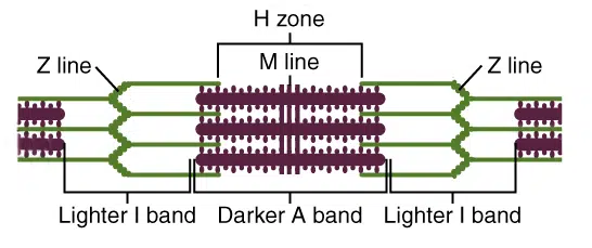
The sarcomere extends from one Z line to another Z line and is composed of several distinct sections. Let’s explore these sections:
- Z line: The Z line serves as an anchor for actin filaments. It marks the boundary between adjacent sarcomeres.
- M line: The M line is located in the center of the sarcomere and serves as an anchor for myosin filaments.
- I band: The I band is a light-colored region within the sarcomere that contains only actin filaments. It extends from the Z line to the edge of the overlapping myosin filaments.
- A band: The A band is a dark-colored region that represents the length of a myosin filament. It may contain overlapping actin filaments within its boundaries.
- H zone: The H zone is a region within the A band that contains only myosin filaments. It appears lighter due to the absence of overlapping actin filaments.
To remember the organization of these sections, a helpful acronym is MHAZI. It stands for M line, H zone, A band, Z line, and I band. Remember that the M line is inside the H zone, which is inside the A band, while the Z line is inside the I band.
The arrangement of actin and myosin filaments within the sarcomere allows for the sliding filament mechanism, which is responsible for muscle contraction. During contraction, the actin and myosin filaments slide past each other, causing the sarcomere to shorten.
Understanding the ultrastructural appearance of skeletal muscle provides valuable insights into the molecular mechanisms underlying muscle contraction. This knowledge is crucial for studying muscle function, muscle disorders, and the development of therapeutic interventions related to skeletal muscle.
Skeletal Muscle Contraction
Skeletal muscle contraction is initiated by the arrival of an action potential at the neuromuscular junction, a specialized synapse that connects an α-motor neuron and a skeletal muscle fiber.
When the action potential reaches the neuromuscular junction, voltage-gated calcium ion channels in the motor neuron terminal open, allowing calcium ions to enter. The influx of calcium ions triggers the release of vesicles containing a neurotransmitter called acetylcholine (ACh) into the synaptic cleft.
Acetylcholine diffuses across the synaptic cleft and binds to nicotinic acetylcholine receptors, which are ligand-gated ion channels located on the plasma membrane of the muscle fiber. Binding of acetylcholine to these receptors causes them to open, resulting in an influx of sodium ions into the muscle fiber. This influx of sodium ions depolarizes the muscle fiber membrane potential, leading to local depolarization.
The depolarization of the muscle fiber membrane activates voltage-sensitive sodium channels located in the membrane. This activation triggers the generation of an action potential that propagates along the entire length of the muscle fiber. The action potential travels deep into the muscle fiber through specialized invaginations of the plasma membrane called transverse tubules (T-tubules).
After acetylcholine has fulfilled its role in signal transmission, it is rapidly broken down (hydrolyzed) by the enzyme acetylcholinesterase present in the synaptic cleft. This enzymatic breakdown terminates the signal transmission by removing acetylcholine from the synaptic cleft.
The termination of the acetylcholine signal allows for repolarization of the muscle fiber membrane. Sodium ions are actively transported out of the cell, restoring the resting membrane potential and preparing the muscle fiber for subsequent contractions.
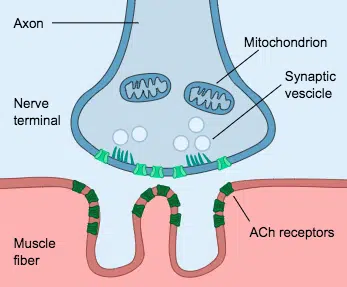
Overall, the process of skeletal muscle contraction involves the transmission of an action potential from the motor neuron to the muscle fiber via the neuromuscular junction. Acetylcholine plays a crucial role in initiating muscle fiber depolarization by binding to nicotinic acetylcholine receptors. The subsequent generation of an action potential leads to muscle contraction, while the breakdown of acetylcholine by acetylcholinesterase terminates the signal transmission and allows for membrane repolarization.
Excitation-Contraction Coupling
Excitation-contraction coupling is the process that links the electrical excitation of a skeletal muscle fiber with its subsequent contraction. It involves a series of events that allow the muscle fiber to generate force in response to an action potential.
The process begins with the action potential generated at the neuromuscular junction propagating along the sarcolemma, which is the cell membrane of the muscle fiber. The action potential then travels deep into the muscle fiber through a network of invaginations known as the transverse tubule (T-tubule) system.
As the action potential reaches the T-tubules, it causes the opening of voltage-gated L-type calcium channels, also called dihydropyridine receptors. These channels are located on the T-tubule membrane and allow calcium ions to enter the muscle fiber from the extracellular space.
The influx of calcium ions triggers a larger release of calcium from the sarcoplasmic reticulum, an intracellular calcium store. The calcium release is facilitated by the activation of ryanodine receptors located on the sarcoplasmic reticulum membrane. Calcium flows from the sarcoplasmic reticulum into the cytoplasm, leading to a significant increase in the intracellular calcium concentration.
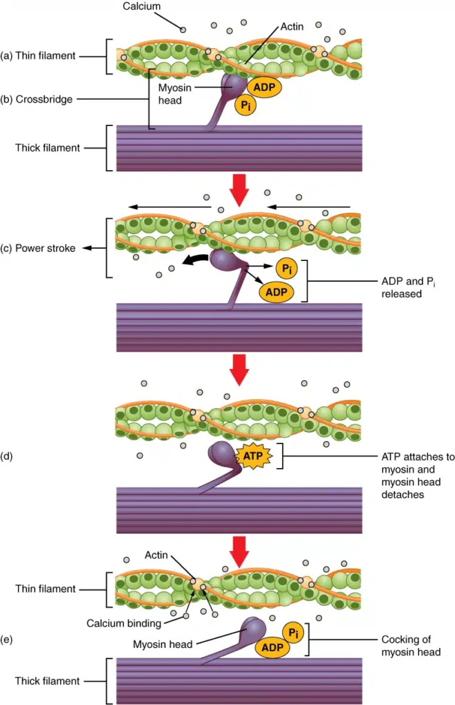
The increased concentration of calcium in the cytoplasm is essential for initiating muscle contraction. Calcium binds to troponin-C, a regulatory protein associated with the actin filaments within the muscle fiber. This binding induces a conformational change in the troponin-tropomyosin complex, exposing the binding sites on actin for the myosin heads.
The myosin heads, which are part of the myosin filaments, can then bind to the actin filaments. The binding of myosin to actin triggers ATP hydrolysis, providing energy for the myosin heads to undergo a conformational change. This change allows the actin and myosin filaments to slide past each other, shortening the length of the sarcomere—the basic functional unit of muscle contraction.
To initiate muscle relaxation, the concentration of calcium in the cytoplasm needs to be reduced. Calcium is actively pumped back into the sarcoplasmic reticulum by a calcium ATPase called sarco/endoplasmic reticulum Ca2+-ATPase (SERCA). As calcium is removed from the cytoplasm, the troponin-tropomyosin complex undergoes a conformational change, covering the binding sites on actin and preventing further interaction with myosin. This restores the sarcomere to its original length and allows the muscle fiber to relax.
Overall, excitation-contraction coupling ensures that the electrical excitation of a skeletal muscle fiber leads to a coordinated and synchronized contraction of the muscle. It relies on the precise regulation of calcium ions and the interaction between actin and myosin filaments to generate force and enable muscle movement.
Types Of Skeletal Muscle
Skeletal muscle, the type of muscle responsible for voluntary movement in our bodies, can be categorized into different types based on their characteristics and functions. The two main types of skeletal muscle fibers are Type I (slow-twitch) and Type II (fast-twitch) muscles.
- Type I: Type I, also known as slow-twitch or red muscle, is characterized by its dense capillary network, high mitochondrial content, and abundant myoglobin. These features give the muscle tissue its distinctive red color. Type I muscle fibers have a remarkable ability to sustain aerobic activity for extended periods. They are well-suited for endurance activities such as long-distance running or cycling. Due to their high oxidative capacity, Type I muscles can carry more oxygen, allowing for efficient energy production through aerobic metabolism.
- Type II: On the other hand, Type II muscle fibers are further classified into three subtypes: Type IIa, Type IIx, and Type IIb. These fibers are collectively known as fast-twitch muscles because they contract more rapidly than Type I fibers.
- Type IIa muscle fibers share some characteristics with Type I fibers. They possess a significant number of mitochondria and capillaries, and they also display a red color when deoxygenated. While they can still generate energy through aerobic metabolism, Type IIa fibers have a greater capacity for anaerobic activity compared to Type I fibers. This makes them suitable for activities that require a combination of endurance and strength, such as middle-distance running or swimming.
- Type IIx, also referred to as Type IId, is the fastest muscle type found in humans. These fibers have fewer mitochondria and myoglobin compared to Type I and IIa fibers. Consequently, Type IIx fibers rely more on anaerobic metabolism and are better suited for short, explosive bursts of activity. They can generate a significant amount of force but are less efficient in terms of endurance. Despite their name, Type IIx fibers are distinct from the previously mentioned Type IIB muscle fibers found in some older literature.
- Lastly, Type IIb muscle fibers, also known as white muscle, are primarily anaerobic and glycolytic. They have even fewer mitochondria and myoglobin compared to other muscle fiber types. Type IIb fibers are responsible for rapid, intense movements and are commonly found in small animals such as rodents. These muscles fatigue quickly and are not well-suited for prolonged activity.
It’s important to note that individuals possess a mixture of muscle fiber types in varying proportions. The specific composition of muscle fibers can affect an individual’s athletic performance, as certain activities may be better suited to the characteristics of a particular fiber type.
In summary, skeletal muscle can be classified into different types based on their contractile and metabolic properties. Type I muscles are slow-twitch, rich in mitochondria and myoglobin, and are suited for endurance activities. Type II muscles are fast-twitch and further divided into Type IIa, Type IIx, and Type IIb, each with different metabolic capacities and contractile speeds. Understanding the different types of skeletal muscle helps explain the varied abilities and functions of our muscles in different physical activities.
Functions Of Skeletal Muscle
Skeletal muscles serve a range of important functions in the human body. Here are some key functions of skeletal muscles:
- Body Movement: Skeletal muscles enable various voluntary movements in the body. When these muscles contract, they pull on tendons attached to bones, resulting in movement. Whether it’s typing on a keyboard, extending the arm, or engaging in activities like writing, skeletal muscles are responsible for these essential body movements.
- Posture Maintenance: The skeletal muscles play a crucial role in maintaining proper body posture. Muscles such as the gluteal muscles contribute to an erect posture by supporting the spine and pelvis. Other muscles, like the Sartorius muscle in the thighs, aid in body movement and help maintain balance and stability.
- Protection of Organs and Tissues: Skeletal muscles act as protective layers around internal organs and delicate tissues. They provide a cushioning effect, helping to safeguard these vital structures from potential injury and external impacts.
- Support for Entry and Exit Points: Skeletal muscles are involved in supporting the entry and exit points of the body. Sphincter muscles, found around the anus, mouth, and urinary tract, contract to reduce the size of the openings. This contraction facilitates essential functions like swallowing food, defecation, and urination, ensuring effective control over these processes.
- Regulation of Body Temperature: Skeletal muscles contribute to the regulation of body temperature. During strenuous exercise or physical activity, skeletal muscles contract vigorously, generating heat as a byproduct. This heat production aids in elevating the body’s temperature and helps maintain a stable internal environment.
In addition to these primary functions, skeletal muscles also assist in maintaining blood circulation, supporting joint stability, and participating in metabolic processes. Their coordinated actions are crucial for enabling a wide range of physical activities and ensuring overall body functionality.
It is important to recognize the multifaceted nature of skeletal muscle function, as their roles extend beyond movement and encompass vital aspects of protection, support, and thermoregulation in the human body.
FAQ
What is the ultrastructure of skeletal muscle?
The ultrastructure of skeletal muscle refers to its microscopic components and organization at a cellular level, including specialized structures such as sarcomeres, myofibrils, and individual muscle fibers.
What is a sarcomere?
A sarcomere is the basic functional unit of a skeletal muscle. It is the segment between two Z-discs and consists of overlapping thin actin filaments and thick myosin filaments.
What are myofibrils?
Myofibrils are cylindrical structures found within muscle fibers. They are made up of repeating units called sarcomeres and contain contractile proteins responsible for muscle contraction.
What are thin filaments and thick filaments?
Thin filaments are composed primarily of actin proteins, while thick filaments are made up of myosin proteins. These filaments slide past each other during muscle contraction, allowing the muscle to shorten or contract.
What is the role of the sarcoplasmic reticulum (SR)?
The sarcoplasmic reticulum is a specialized type of endoplasmic reticulum found in muscle cells. It plays a crucial role in regulating calcium ion concentration within the muscle cell, which is essential for muscle contraction.
What is the function of T-tubules?
T-tubules, or transverse tubules, are invaginations of the muscle cell membrane (sarcolemma) that extend deep into the muscle fiber. They help transmit electrical signals, known as action potentials, into the interior of the muscle fiber, allowing for synchronized muscle contraction.
What are motor units?
Motor units are composed of a motor neuron and the muscle fibers it innervates. When a motor neuron sends a signal to the muscle fibers it innervates, all the muscle fibers in that motor unit contract simultaneously.
What is the neuromuscular junction?
The neuromuscular junction is the point where the motor neuron meets the muscle fiber. It is responsible for transmitting the signal from the motor neuron to the muscle fiber, initiating muscle contraction.
What is the role of mitochondria in skeletal muscle?
Mitochondria are organelles responsible for energy production within cells. In skeletal muscle, mitochondria provide the necessary energy for muscle contraction by producing adenosine triphosphate (ATP) through aerobic metabolism.
How are muscle fibers organized within a skeletal muscle?
Muscle fibers are arranged in parallel bundles within a skeletal muscle. These bundles are surrounded by connective tissue layers, forming the overall structure of the muscle. The organization of muscle fibers allows for coordinated muscle contraction and efficient force generation.
References
- Gupta, R. C., Dettbarn, W.-D., & Milatovic, D. (2009). Skeletal Muscle. Handbook of Toxicology of Chemical Warfare Agents, 509–531. doi:10.1016/b978-012374484-5.00035-3
- Banks, R. (2014). Skeletal Muscle. Reference Module in Biomedical Sciences. doi:10.1016/b978-0-12-801238-3.00252-x
- Fossey, S. L., Greg Hall, D., & Leininger, J. R. (2018). Skeletal Muscle. Boorman’s Pathology of the Rat, 281–298. doi:10.1016/b978-0-12-391448-4.00017-4
- Damjanov, I. (2009). Skeletal Muscles. Pathology Secrets, 434–447. doi:10.1016/b978-0-323-05594-9.00021-0
- Eckel, J. (2018). Skeletal Muscle. The Cellular Secretome and Organ Crosstalk, 65–90. doi:10.1016/b978-0-12-809518-8.00003-9
- Franzini-Armstrong, C., & Engel, A. G. (2012). Skeletal Muscle. Muscle, 763–774. doi:10.1016/b978-0-12-381510-1.00053-3
- Rivas, D. A., & Fielding, R. A. (2013). Skeletal Muscle. Encyclopedia of Human Nutrition, 193–199. doi:10.1016/b978-0-12-375083-9.00188-4
- Gupta, R. C., Zaja-Milatovic, S., Dettbarn, W.-D., & Malik, J. K. (2015). Skeletal Muscle. Handbook of Toxicology of Chemical Warfare Agents, 577–597. doi:10.1016/b978-0-12-800159-2.00040-3


