

Intermediate filaments (IFs) are a diverse group of cytoskeletal protein polymers found mainly in animal cells. The cytoplasmic IFs are usually about 10 nm in diameter and unlike actin filaments and microtubules, they are non-polar. Different cells contain different IF proteins such as keratins, vimentin, desmin, and neurofilaments, while lamins form the intermediate filament system of the nucleus. Their major function is to provide mechanical strength and resilience to the cells and tissues. IFs are also involved in cell organization, adhesion, signaling and positioning of different cellular structures.
What are intermediate filaments?
Intermediate filaments (IFs) are protein filaments that form an important part of the cytoskeleton of animal cells. They are called intermediate filaments because their diameter is about 10 nm, which is intermediate between actin filaments and microtubules. These filaments are non-polar in nature and form strong rope-like structures within the cell.
Different types of cells contain different intermediate filament proteins. Keratins, vimentin, desmin and neurofilament proteins are some of the important cytoplasmic intermediate filaments. Lamins are present inside the nucleus and form the nuclear intermediate filament system. The major role of these filaments is providing mechanical strength to the cell and helping the cells to resist different mechanical stresses. They also take part in cell organization, cell adhesion and some cellular signaling processes.
Characteristics of Intermediate Filaments
The following are some of the important characteristics of intermediate filaments (IFs)–
- Intermediate filaments are fibrous protein polymers and form one of the major components of cytoskeleton in animal cells. They are mainly characteristic of metazoan cells.
- Cytoplasmic intermediate filaments are usually about 9–11 nm in diameter. Their size is intermediate between the actin filaments and microtubules, from which the name intermediate filament is derived.
- They are non-polar filaments. The two ends of an intermediate filament do not have distinct plus and minus polarity as found in actin filaments and microtubules.
- The basic IF proteins contain a central α-helical rod domain with variable amino-terminal and carboxy-terminal regions. Two protein chains coil around each other and form a coiled-coil dimer.
- The dimers associate in an antiparallel manner to form tetramers. Because of this antiparallel arrangement, polarity is lost during the formation of filament. These units further associate and form the rope-like filament structure.
- Their assembly does not require the hydrolysis of ATP or GTP. This is different from actin filaments and microtubules, whose assembly and dynamics are associated with nucleotide binding and hydrolysis.
- Intermediate filaments are strong and highly extensible structures. They can withstand large deformation and provide mechanical strength to cells, particularly the cells subjected to repeated mechanical stress.
- Different cell types contain different types of intermediate filament proteins. Keratins are mainly found in epithelial cells, vimentin in many mesenchymal cells, desmin in muscle cells and neurofilament proteins are abundant in neurons.
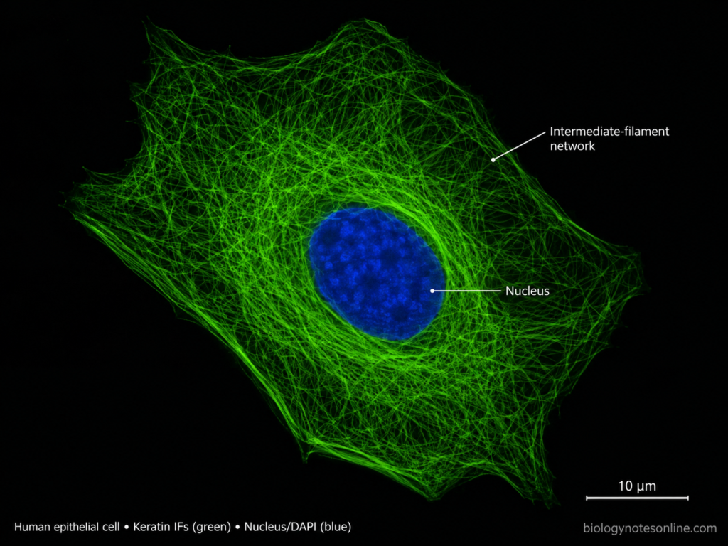
- Intermediate filaments form extensive networks within the cytoplasm. In many cells, the network extends around the nucleus towards the cell periphery and can be associated with junctions such as desmosomes and hemidesmosomes.
- Lamins are the nuclear members of the intermediate filament protein family. They form the nuclear lamina underlying the inner nuclear membrane and provide support to the nucleus.
- Intermediate filaments are comparatively stable structures, but they are not permanently fixed. Their networks undergo assembly, disassembly and reorganization according to the condition and activity of the cell.
- No conventional motor protein is known that moves along intermediate filaments as a track. Their major role is mechanical and structural, although IF proteins are also involved in cell organization and signaling processes.
Structure of Intermediate Filaments
The structure of intermediate filaments (IFs) is formed by the association of fibrous IF protein subunits. Most IF proteins have a similar basic molecular organization, although their size and amino acid sequence can differ.
- Monomer– Each IF protein contains a central α-helical rod domain. This rod region is present between a non-helical N-terminal head and C-terminal tail. The head and tail regions are more variable in different IF proteins.
- Dimer– Two IF protein molecules associate through their α-helical rod domains. They are arranged in parallel and coil around each other to form a coiled-coil dimer.
- Tetramer– Two dimers associate in an antiparallel arrangement to form a tetramer. Because of this arrangement, the structure does not have distinct plus and minus ends.
- ULF– Several tetramers associate laterally and form short structures called unit-length filaments (ULFs). These are important intermediate structures formed during IF assembly.
- Elongation– The unit-length filaments join longitudinally with one another. This results in increase in the length of the filament.
- Compaction– The elongated structures undergo radial compaction and rearrangement. A mature rope-like intermediate filament is then formed.
- Diameter– Mature cytoplasmic intermediate filaments are generally about 9–11 nm in diameter. This intermediate size is the basis for their name.
- Polarity– Intermediate filaments are non-polar in nature. They do not possess structurally different plus and minus ends like actin filaments and microtubules.
- Variation– The molecular organization is not exactly the same in all intermediate filaments. Keratin, vimentin, desmin, neurofilaments and lamins show differences in protein composition and filament organization.
Intermediate Filament Protein Structure
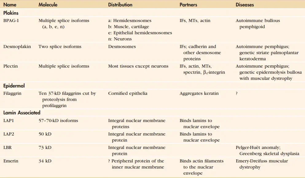
The proteins forming intermediate filaments (IFs) have a characteristic tripartite structure. Despite differences in their size and amino acid sequence, most IF proteins contain a central α-helical region with non-helical regions at both ends.
- Head domain– The N-terminal head domain is a non-helical region present at one end of the IF protein. Its length and amino acid sequence are variable among different intermediate filament proteins.
- Rod domain– A long α-helical rod domain forms the central region of the protein. It contains about 310 amino acids in most cytoplasmic IF proteins. In nuclear lamins, this region is about 350 amino acids long.
- Coil regions– The central rod is divided into four α-helical regions called 1A, 1B, 2A and 2B. These regions take part in coiled-coil formation between two IF protein molecules.
- Linker regions– The α-helical regions are separated by short non-helical linker sequences. These are called L1, L12 and L2.
- Tail domain– The C-terminal tail domain is present after the rod region. It is also non-helical and differs considerably in size and sequence between the different IF proteins.
- Coiled-coil structure– The rod domains of two protein molecules align parallel to each other and form a coiled-coil dimer. This is an important early structure during intermediate filament assembly.
- Variable ends– The head and tail domains show greater structural variation than the central rod domain. These variable regions contribute to the different properties and interactions of individual IF proteins.
- Common organization– Thus, the basic IF protein structure can be represented as head–rod–tail. This common organization is present in different members of the intermediate filament protein family, although the terminal regions are not identical.
Assembly of Intermediate Filaments
The assembly of intermediate filaments (IFs) takes place by association of IF protein molecules into dimers, tetramers and finally mature filaments. Unlike actin filaments and microtubules, this assembly does not depend on ATP or GTP hydrolysis. The major steps are as follows-
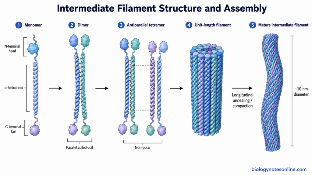
- Dimer formation– Two IF protein monomers associate through their central α-helical rod domains. They lie parallel to each other and form a coiled-coil dimer. The dimers can be homodimers, as in vimentin, or heterodimers as commonly found in keratins.
- Tetramer formation– Two dimers associate laterally in an antiparallel and staggered arrangement. This forms a tetramer containing four polypeptide chains. Due to antiparallel arrangement, the tetramer becomes non-polar.
- Lateral association– Several tetramers now associate side-by-side. This lateral association gives rise to short filamentous structures called unit-length filaments (ULFs). In in vitro studies of cytoplasmic IFs, ULF formation occurs very rapidly.
- ULF formation– The unit-length filament is an important intermediate during IF assembly. Its exact molecular packing can differ between various intermediate filament proteins. Thus, the number of polypeptide chains in the mature filament is not exactly same for all IF types.
- Longitudinal association– The ULFs join end-to-end with other ULFs. In this step, short filament units become progressively longer. This process is slower than the initial lateral association.
- Compaction– After elongation, the filament undergoes radial rearrangement and compaction. The diameter becomes smaller and a more compact filament structure is formed.
- Mature filament– A mature cytoplasmic intermediate filament is finally formed, generally about 9–11 nm in diameter. It is a rope-like and non-polar filament. The exact internal organization differs between IF proteins such as vimentin and keratin.
- Dynamic assembly– Intermediate filaments are relatively stable, but they are not permanent structures. Filaments can undergo assembly, disassembly and exchange of subunits according to cellular conditions. Their organization inside cells is therefore dynamic.
Why Intermediate Filaments Are Non-Polar
Intermediate filaments (IFs) do not show polarity because of the arrangement of their protein subunits. Two IF protein molecules first join in a parallel manner. A coiled-coil dimer is formed.
Two such dimers now associate in opposite directions. This arrangement is antiparallel and slightly staggered. Due to this arrangement, the tetramer has no separate directional ends.
The tetramers further associate to form the intermediate filament. So, the mature filament does not have distinct plus (+) and minus (−) ends like actin filaments and microtubules.
This non-polar nature mainly develops at the tetramer stage, when two dimers are arranged in opposite orientation.
Types of Intermediate Filaments
Intermediate filament proteins are classified into six different types based on their sequence, distribution and protein composition. Different types occur in different cells and tissues. They are as follows-
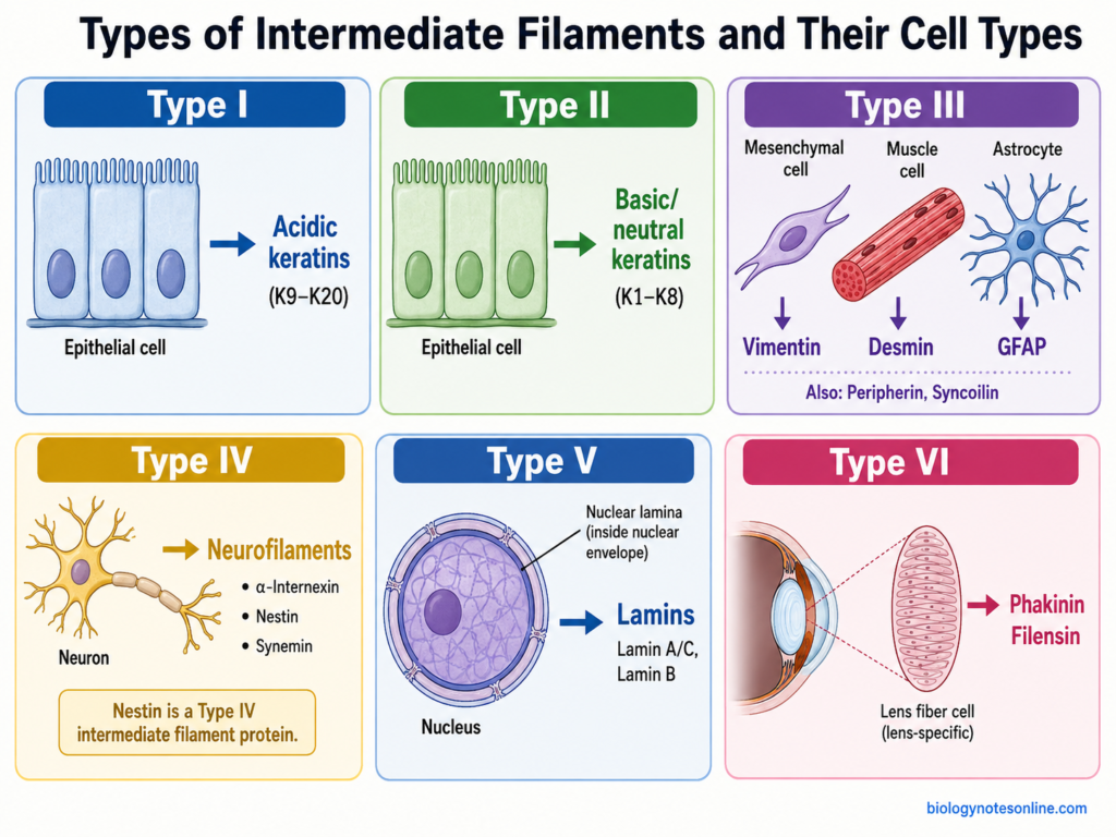
- Type I- Acidic keratins
These are the acidic keratin proteins mainly found in epithelial cells. Type I keratins associate with Type II keratins during filament formation. The keratin filaments give mechanical support to epithelial tissues and are attached with desmosomes and hemidesmosomes. - Type II- Basic keratins
Type II consists of basic or neutral-basic keratins. These are also present mainly in epithelial cells. A Type II keratin combines with a Type I keratin and forms the keratin heterodimer. They are important in maintaining strength of epithelial cells. - Type III- Vimentin group
This type includes vimentin, desmin, GFAP, peripherin and syncoilin. Vimentin is commonly present in mesenchymal cells. Desmin is characteristic of muscle cells, while GFAP (glial fibrillary acidic protein) occurs mainly in astrocytes. Peripherin is found largely in peripheral neurons. - Type IV- Neurofilament group
The major proteins of this type are neurofilament proteins, α-internexin, nestin and synemin. Neurofilaments are mainly present in neurons and are abundant in axons. They have an important role in maintaining the structure and diameter of axons. Nestin is mainly associated with developing and progenitor cells. - Type V- Lamins
These are the nuclear intermediate filament proteins. Lamin A/C and B-type lamins are the major members. They form the nuclear lamina below the inner nuclear membrane. Lamins support nuclear shape and are also associated with chromatin and nuclear organization. - Type VI- Beaded filament proteins
Type VI includes filensin (BFSP1) and phakinin (BFSP2/CP49). These proteins are mainly found in lens fiber cells of the eye. They associate to form the characteristic beaded filaments of lens cells.
Dynamics and Regulation of Intermediate Filaments
Intermediate filaments (IFs) are not permanently fixed structures inside the cell. Their filaments can be assembled, disassembled and reorganized according to cell condition. Different modifications of IF proteins take part in this regulation.
- Assembly and disassembly– IF proteins remain in equilibrium between soluble subunits and polymerized filaments. New subunits can be incorporated and filament networks can also be disassembled. This gives a dynamic nature to the IF network.
- Phosphorylation– It is one of the major mechanisms regulating intermediate filaments. Addition of phosphate groups at particular sites can change the assembly state of IF proteins. Increased phosphorylation commonly causes filament disassembly or reorganization.
- Dephosphorylation– Removal of phosphate groups is carried out by protein phosphatases. It can favor reassembly of soluble IF proteins into filaments. The balance between phosphorylation and dephosphorylation controls the organization of many IF networks.
- Mitotic regulation– During cell division, several IF proteins undergo extensive phosphorylation. Vimentin, for example, is phosphorylated by mitotic kinases such as Cdk1 and Plk1. This helps in rearrangement of the vimentin network during mitosis.
- Post-translational modifications– IF proteins also undergo modifications other than phosphorylation. These include O-GlcNAcylation, sumoylation, acetylation and ubiquitination, depending upon the IF protein and cell type. Such modifications can affect filament organization, interactions and stability.
- Subunit exchange– Intermediate filament proteins can exchange between soluble pools and already formed filaments. So, even apparently stable IF networks show continuous molecular turnover. The rate of this exchange is not same in all IF types.
- Severing and annealing– Some intermediate filaments can be broken into shorter filament pieces and these pieces can again join with each other. Such severing and re-annealing is well studied in vimentin and neurofilaments. It also helps in movement and remodeling of IFs within the cell.
- Protein interactions– Intermediate filaments interact with several cytoskeletal and regulatory proteins. These interactions affect their distribution, attachment and organization in the cytoplasm. IFs can also act as scaffolds for some signaling proteins.
- Cellular stress– Mechanical stress, heat stress and other cellular conditions can cause rearrangement of IF networks. Changes in phosphorylation and other modifications are often involved during these responses. The altered network helps the cell to adjust with changing conditions.
- Protein turnover– Damaged or excess IF proteins can be removed by cellular degradation systems. Proteasomal and other protein-clearing mechanisms are involved in maintaining normal IF protein levels. This process is especially important when abnormal IF proteins accumulate inside the cell.
Functions of Intermediate Filaments
The following are some of the important functions of intermediate filaments (IFs)–
- Mechanical strength– Intermediate filaments provide mechanical strength to cells. They help the cells to withstand stretching, compression and other mechanical stress. This function is especially important in epithelial and muscle tissues.
- Cell shape– IF networks help in maintaining the normal shape and structural integrity of cells. They form a supporting network extending throughout the cytoplasm.
- Tissue integrity– Intermediate filaments connect with cell junctions and help in maintaining the strength of tissues. Keratin filaments are attached with desmosomes and hemidesmosomes in epithelial cells.
- Nuclear support– Lamins form the nuclear lamina below the inner nuclear membrane. They support nuclear shape and provide mechanical stability to the nucleus.
- Organelle positioning– Intermediate filament networks take part in positioning and distribution of different organelles inside the cell. Mitochondria, Golgi apparatus and other cellular structures can associate with these networks.
- Cytoskeleton organization– IFs interact with actin filaments and microtubules through different linker proteins. These interactions help in organization of the cytoskeleton and cellular architecture.
- Cell adhesion– Intermediate filaments are associated with cell-cell and cell-matrix adhesion. Their attachment with junctional complexes helps in transferring mechanical forces between cells and tissues.
- Cell migration– Some intermediate filament proteins take part in cell migration. Their networks undergo reorganization during movement of cells and influence cellular mechanical properties.
- Signaling– Intermediate filaments also participate in different cellular signaling pathways. They can influence cell growth, survival, stress responses and other cellular activities.
- Chromatin organization– Nuclear lamins interact with chromatin and several nuclear proteins. They are involved in chromatin organization and are associated with regulation of nuclear functions.
- Neuronal support– Neurofilaments provide structural support to neurons. They are especially important in axons and help in maintaining axonal caliber.
- Muscle organization– Desmin filaments form an important structural network in muscle cells. They help in maintaining organization and mechanical integrity of muscle tissue.
Intermediate Filaments and Disease
Defects in intermediate filament (IF) proteins are associated with different human diseases. Mutation in these proteins can affect mechanical strength, cell structure and normal tissue function. In some diseases, abnormal intermediate filament proteins are also accumulated inside the cells.
The following are some of the important diseases associated with intermediate filaments-
- Epidermolysis bullosa simplex– This disease is mainly associated with mutation in keratin 5 (K5) and keratin 14 (K14). These keratins are present in basal epithelial cells of skin. Mutation makes the keratin network weak and the cells become more sensitive to mechanical stress. Blister formation takes place even after minor friction or injury.
- Keratin disorders– Mutations in different keratin genes can produce different epithelial disorders. Some of them affect skin, hair, nails and other epithelial tissues. The disease mainly depends on the type of keratin affected and the tissue where it is normally expressed.
- Desminopathies– Desmin is an important intermediate filament protein of muscle cells. Mutation in desmin can result in different myopathies and cardiomyopathies. Abnormal desmin proteins may form aggregates inside muscle cells. Muscle weakness is commonly observed and cardiac muscles can also be affected.
- Laminopathies– These are a group of disorders mainly caused by mutation in lamin A/C and other nuclear lamina-associated proteins. It includes muscular dystrophy, cardiomyopathy, lipodystrophy and some premature aging disorders. The nucleus often shows abnormal shape and altered mechanical stability.
- Progeria– Hutchinson-Gilford progeria syndrome is caused by a mutation in the LMNA gene in most cases. It produces an abnormal form of lamin A called progerin. This affects normal structure of the nuclear lamina and results in premature aging characteristics.
- Alexander disease– It is mainly associated with mutation in GFAP (glial fibrillary acidic protein). GFAP is an intermediate filament protein of astrocytes. Abnormal GFAP accumulates inside these cells and characteristic protein aggregates are formed. The disease mainly affects the central nervous system.
- Neurodegenerative diseases– Abnormal changes in neurofilaments are found in several neurological disorders. Accumulation of neurofilament proteins is seen in damaged neurons and axons. Changes in proteins such as peripherin and α-internexin have also been associated with some neurodegenerative conditions including amyotrophic lateral sclerosis (ALS).
- Cancer– Intermediate filament expression is also changed in many cancers. Keratins are commonly used as markers for tumors of epithelial origin. Vimentin expression is often increased in tumor cells showing mesenchymal characteristics. These proteins are therefore also useful in identification and study of different tumors.
Intermediate Filaments vs Microfilaments vs Microtubules
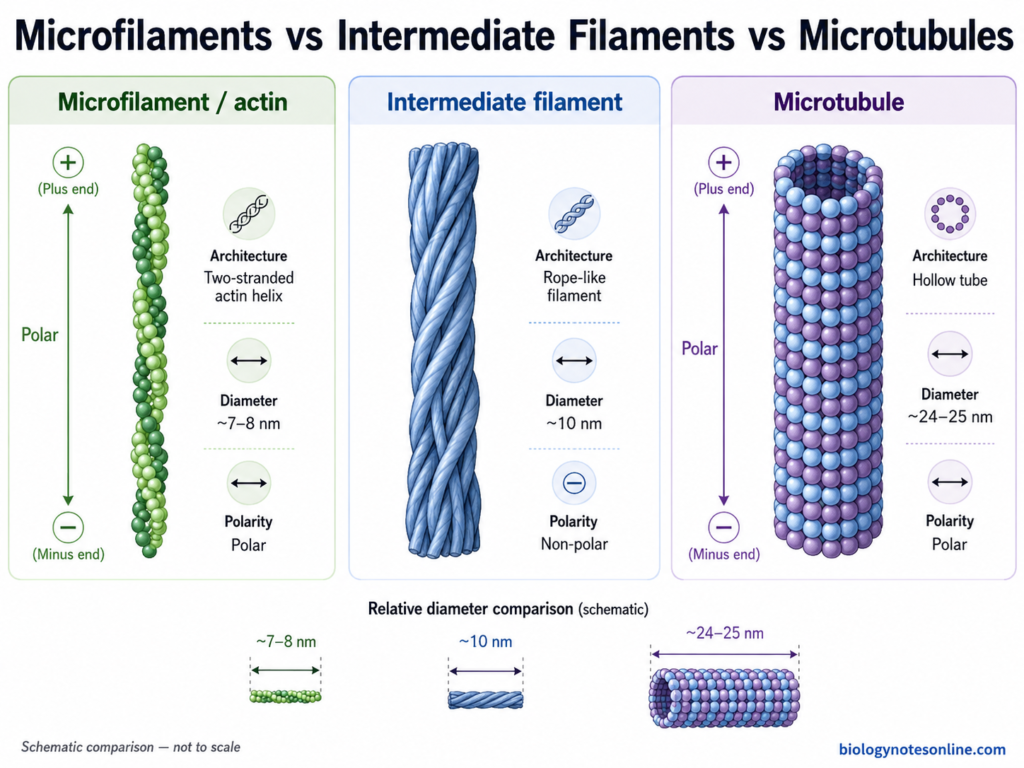
The three major components of the cytoskeleton are intermediate filaments, microfilaments and microtubules. They differ in their protein composition, diameter, polarity and functions.
| Features | Intermediate Filaments | Microfilaments | Microtubules |
|---|---|---|---|
| Basic protein | Made up of different IF proteins such as keratin, vimentin, desmin and neurofilament proteins. | Mainly formed of actin protein. | Made up of α-tubulin and β-tubulin heterodimers. |
| Diameter | Usually about 10 nm. | About 7–8 nm. | About 24–25 nm. |
| Structure | Rope-like solid filaments. | Thin, two-stranded helical filaments. | Hollow cylindrical structures. |
| Polarity | Non-polar. They do not have distinct plus and minus ends. | Polar and contain plus (+) and minus (−) ends. | Polar, with distinct plus (+) and minus (−) ends. |
| Assembly unit | IF proteins first form dimers, tetramers and then larger filament units. | G-actin monomers polymerize to form F-actin. | α/β-tubulin heterodimers form protofilaments, which associate to form the microtubule. |
| Nucleotide requirement | Assembly does not require ATP or GTP hydrolysis. | Actin polymerization is associated with ATP binding and hydrolysis. | Microtubule assembly and dynamics involve GTP bound to tubulin. |
| Stability | Comparatively more stable and mechanically resistant. | Highly dynamic. Assembly and disassembly takes place rapidly in many cellular regions. | Highly dynamic structures and show rapid polymerization and depolymerization. |
| Mechanical nature | Strong and highly extensible. They provide resistance against mechanical stress. | Mainly helps in tension generation and changes in cell shape. | More rigid and resist compression inside the cell. |
| Motor proteins | No conventional motor protein uses IFs as tracks. | Myosin moves along actin filaments. | Kinesin and dynein move along microtubules. |
| Major location | Distributed throughout cytoplasm. Lamins form the nuclear IF system. | Commonly present below plasma membrane and in structures such as stress fibers and microvilli. | Extend throughout cytoplasm, commonly from microtubule-organizing centres. |
| Major functions | Mechanical strength, tissue integrity, nuclear support and cellular organization. | Cell movement, muscle contraction, cytokinesis, cell cortex formation and changes in cell shape. | Intracellular transport, chromosome movement, mitotic spindle formation, cilia and flagella and maintenance of cell organization. |
| Examples | Keratins, vimentin, desmin, neurofilaments and lamins. | Actin filaments of microvilli, stress fibers and contractile ring. | Microtubules of mitotic spindle, cilia, flagella and cytoplasmic network. |
The diameter and polarity differences are particularly characteristic. Intermediate filaments are around 10 nm and non-polar, microfilaments are thinner at about 8 nm, while microtubules are hollow polar polymers about 24 nm in diameter.
References
- Broussard, J. A., Jaiganesh, A., Zarkoob, H., Conway, D. E., Dunn, A. R., Espinosa, H. D., Janmey, P. A., & Green, K. J. (2020). Scaling up single-cell mechanics to multicellular tissues: The role of the intermediate filament–desmosome network. Journal of Cell Science, 133(6), jcs228031. https://doi.org/10.1242/jcs.228031
- Coelho-Rato, L. S., Parvanian, S., Salajkova, S. A., Medalia, O., & Eriksson, J. E. (2024). Intermediate filaments at a glance. Journal of Cell Science, 137(16), jcs261386. https://doi.org/10.1242/jcs.261386
- Cooper, G. M. (2000). The cell: A molecular approach (2nd ed.). Sinauer Associates. https://www.ncbi.nlm.nih.gov/books/NBK9947/
- Eriksson, J. E., Dechat, T., Grin, B., Helfand, B., Mendez, M., Pallari, H.-M., & Goldman, R. D. (2009). Introducing intermediate filaments: From discovery to disease. Journal of Clinical Investigation, 119(7), 1763–1771. https://doi.org/10.1172/JCI38339
- Foisner, R. (n.d.). Dynamic connections of nuclear envelope proteins to chromatin and the nuclear matrix. In Madame Curie Bioscience Database. Landes Bioscience. https://www.ncbi.nlm.nih.gov/books/NBK6125/
- Hyder, C. L., Isoniemi, K. O., Torvaldson, E. S., & Eriksson, J. E. (2011). Insights into intermediate filament regulation from development to ageing. Journal of Cell Science, 124(9), 1363–1372. https://doi.org/10.1242/jcs.041244
- Izawa, I., & Inagaki, M. (2006). Regulatory mechanisms and functions of intermediate filaments: A study using site- and phosphorylation state-specific antibodies. Cancer Science, 97(3), 167–174. https://doi.org/10.1111/j.1349-7006.2006.00161.x
- Lane, E. B. (n.d.). Keratin intermediate filaments and diseases of the skin. In Madame Curie Bioscience Database. Landes Bioscience. https://www.ncbi.nlm.nih.gov/books/NBK6247/
- Lee, C.-H., & Coulombe, P. A. (2009). Self-organization of keratin intermediate filaments into cross-linked networks. Journal of Cell Biology, 186(3), 409–421. https://doi.org/10.1083/jcb.200810196
- Liem, R. K. H., & Messing, A. (2009). Dysfunctions of neuronal and glial intermediate filaments in disease. Journal of Clinical Investigation, 119(7), 1814–1824. https://doi.org/10.1172/JCI38003
- Phillips, C. L., Faridounnia, M., Armao, D., & Snider, N. T. (2023). Stability dynamics of neurofilament and GFAP networks and protein fragments. Current Opinion in Cell Biology, 85, 102266. https://doi.org/10.1016/j.ceb.2023.102266
- Sapra, K. T., & Medalia, O. (2021). Bend, push, stretch: Remarkable structure and mechanics of single intermediate filaments and meshworks. Cells, 10(8), 1960. https://doi.org/10.3390/cells10081960
- Smith, F. J. D., Hansen, C. D., Hull, P. R., Kaspar, R. L., McLean, W. H. I., O’Toole, E., & Sprecher, E. (2017). Pachyonychia congenita. In M. P. Adam et al. (Eds.), GeneReviews®. University of Washington, Seattle. https://www.ncbi.nlm.nih.gov/books/NBK1280/
- Vermeire, P.-J., Stalmans, G., Lilina, A. V., Fiala, J., Novak, P., Herrmann, H., & Strelkov, S. V. (2021). Molecular interactions driving intermediate filament assembly. Cells, 10(9), 2457. https://doi.org/10.3390/cells10092457