

Prophase is the first stage of mitosis and takes place after interphase. In this stage, the cell prepares for chromosome separation during nuclear division. The long chromatin fibres begin to condense into short, thick chromosomes.
DNA replication is completed before prophase, during the S phase of interphase. It involves copying the DNA. Each chromosome now consists of two sister chromatids. During chromosome condensation, the already replicated material is packed more tightly, no new copy of DNA is formed.
Prophase also occurs in both meiotic divisions, called prophase I in meiosis I and prophase II in meiosis II. These are the first stages of their respective divisions. Before prophase I, the cell passes through interphase where DNA is replicated. No further DNA replication takes place between meiosis I and meiosis II.
In mitosis, the stages following prophase are prometaphase, metaphase, anaphase and telophase, in this order.
Events During Mitotic Prophase
During mitotic prophase, several changes take place in the nucleus and cytoplasm of the cell. The chromosomes undergo condensation, while the cell starts forming the mitotic spindle for chromosome segregation.
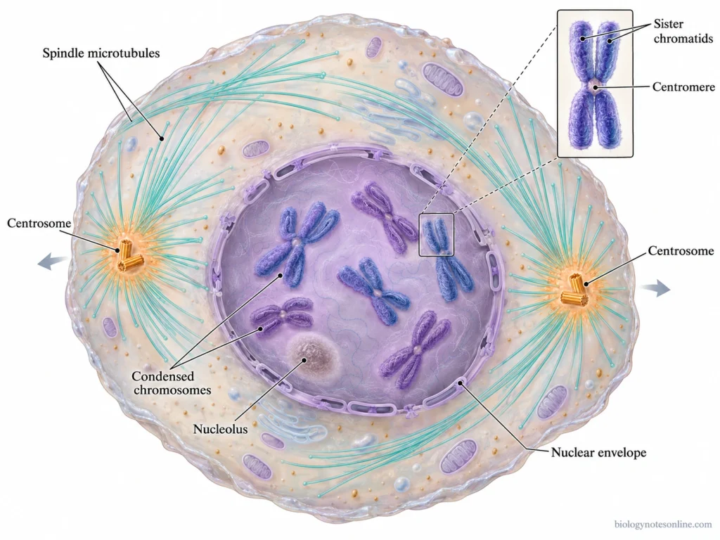
The major events occurring during mitotic prophase are as follows-
- Chromosome Condensation- The long chromatin fibres gradually condense to form short, thick and distinct chromosomes with the help of condensin proteins. Each chromosome consists of two identical sister chromatids formed during the S phase of interphase. These chromatids are held together mainly at the centromeric region by cohesin proteins.
- Disappearance of Nucleolus- During prophase, the nucleolus gradually disappears as the chromosomes undergo condensation.
- Centrosome Separation- In animal cells, the two centrosomes formed during interphase begin to move towards opposite sides of the nucleus. Each centrosome acts as a microtubule-organizing centre and starts forming an array of microtubules around it.
- Formation of Mitotic Spindle- The cytoplasmic microtubules are reorganized to form the mitotic spindle. Microtubules grow out from the two separating centrosomes and interact with each other. The spindle starts forming outside the nucleus. In higher plant cells, spindle formation occurs without centrioles.
- Kinetochore Formation- During late prophase, kinetochores are assembled at the centromeric region of each sister chromatid. These are protein complexes which provide the attachment sites for spindle microtubules during prometaphase.
- Nuclear Envelope Disassembly- The nuclear envelope begins to break down as prophase progresses. In cells undergoing open mitosis, the complete breakdown of the nuclear envelope marks the beginning of prometaphase, when the spindle microtubules gain access to the chromosomes.
Prophase I of Meiosis
Prophase I is the first and longest phase of meiosis I, which involves the pairing of homologous chromosomes and genetic recombination. The DNA replication takes place before prophase I. Each chromosome already contains two sister chromatids.
During this phase, the homologous chromosomes of maternal and paternal origin pair up and exchange their genetic material. The pairing of homologous chromosomes is called synapsis, whereas the exchange of genetic material is referred to as crossing-over.
There are five stages of prophase I, which are as follows-
Stages of Prophase I
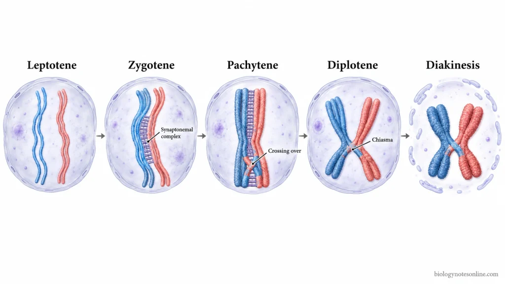
1. Leptotene
Leptotene is the initial stage of prophase I. The duplicated chromosomes begin to condense, appearing as long and thin thread-like structures.
Each chromosome is made up of two sister chromatids held together by cohesin proteins. These chromatids cannot be distinguished clearly at this stage.
The homologous chromosomes also start recognizing their corresponding partners. They come closer to each other during early prophase.
2. Zygotene
The pairing of homologous chromosomes begins during zygotene. This process is referred to as synapsis, in which the homologous chromosomes pair along their corresponding regions.
A specialized ladder-like protein structure called the synaptonemal complex (SC) starts forming between the paired homologous chromosomes.
The synaptonemal complex holds the chromosomes together and maintains their alignment. Pairing gradually progresses along the entire length of the homologous chromosomes and is completed by the beginning of pachytene.
3. Pachytene
In this stage, synapsis is completed, and the synaptonemal complex is fully developed. Each homologous chromosome consists of two sister chromatids. The paired homologous chromosomes contain four chromatids and form a structure known as bivalent or tetrad.
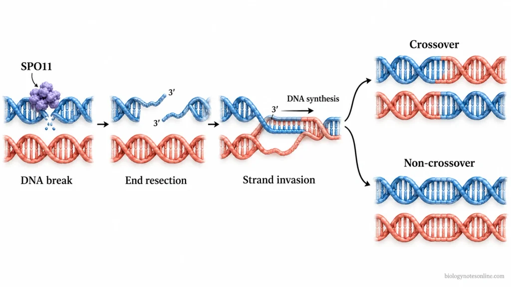
Crossing-over is one of the characteristic events associated with pachytene. It involves the exchange of corresponding DNA segments between the non-sister chromatids of homologous chromosomes.
The process of genetic recombination, however, begins earlier during prophase I.
An enzyme called SPO11 produces programmed double-strand breaks (DSBs) in the DNA. The broken DNA ends are then processed. One of the resulting single-stranded DNA ends enters the homologous DNA sequence, generally present on the non-sister chromatid of the homologous chromosome.
In this process, the homologous DNA is used as a template for DNA synthesis and repair. Some of the recombination events result in reciprocal exchange of DNA segments. Other events are completed without crossing-over.
The crossover events are generally completed during pachytene, producing chromatids with new combinations of maternal and paternal genetic material.
4. Diplotene
The synaptonemal complex breaks down during diplotene. The homologous chromosomes begin to move apart but remain attached at certain points where crossing-over has occurred.
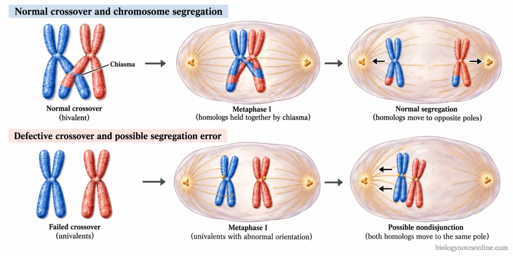
These attachment points are known as chiasmata (singular: chiasma). They become clearly visible during this stage and represent the sites of earlier crossover events between the non-sister chromatids.
The connections between homologous chromosomes are maintained by sister chromatid cohesion. Chiasmata hold the homologous chromosomes together for their proper alignment and subsequent separation during meiosis I.
5. Diakinesis
Diakinesis is the last stage of prophase I. The chromosomes undergo further condensation and become shorter and thicker.
The homologous chromosomes remain connected through chiasmata. During the transition towards metaphase I, the nuclear envelope breaks down, and the chromosomes become ready for attachment to the meiotic spindle.
Prophase II of Meiosis
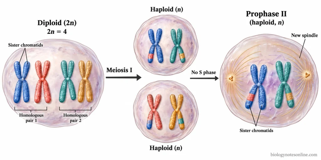
Prophase II is the first stage of meiosis II, which takes place in the haploid cells formed after the first meiotic division. Each cell contains a single set of chromosomes. The chromosomes, however, still consist of two sister chromatids, which remain attached after meiosis I.
In some organisms, a short interval called interkinesis occurs between the two meiotic divisions. No DNA replication takes place during this period. Some cells enter the second meiotic division almost directly without any considerable interval.
During prophase II, the chromosomes undergo condensation again if they were decondensed during telophase I. They become visible as condensed chromosomes.
The nuclear envelope, if formed after the first meiotic division, begins to break down. A new meiotic spindle is formed. In cells having centrosomes, these move towards opposite poles during spindle formation.
Unlike prophase I, the homologous chromosomes do not undergo extensive pairing or synapsis during prophase II. The homologous chromosomes have already separated during the first meiotic division. Crossing-over between homologous chromosomes also does not take place at this stage.
Prophase II is comparatively short, whereas prophase I involves the prolonged pairing of homologous chromosomes and genetic recombination.
Identifying Prophase in Diagrams and Microscopy
Prophase can be identified by observing the condensation of chromosomes, their arrangement and the changes occurring in the nucleus. Labelled diagrams and stained microscopic slides are used to study these features. The stained root-tip cells of onion (Allium cepa) can also be used to observe different stages of cell division.
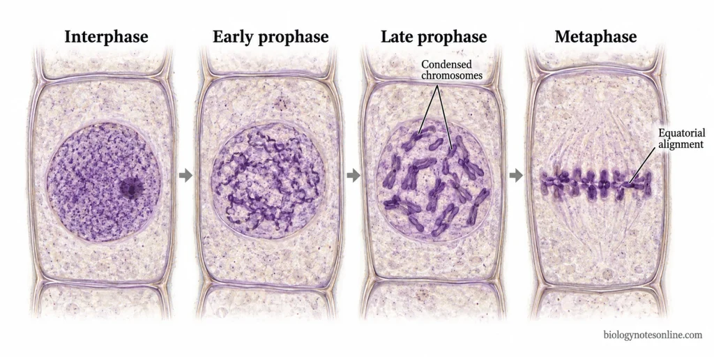
The following are the characteristic features used for identifying prophase-
- The chromatin starts condensing during prophase and appears as thick, darkly stained thread-like chromosomes under the microscope. In early prophase, the chromosomes are comparatively less condensed. They become more distinct during late prophase.
- The chromosomes are distributed within the nuclear region without any definite arrangement. They have not yet aligned at the equatorial plane (metaphase plate).
- In early prophase, the nuclear envelope may still be visible around the condensing chromosomes, while the nucleolus gradually disappears. The nuclear envelope starts breaking down as the cell progresses towards the later stage. Its complete breakdown is generally associated with prometaphase. However, some labelled diagrams include this event under late prophase.
- The spindle fibres also begin to form during prophase. In animal cells, the centrosomes move towards the opposite poles, and spindle microtubules start developing. These structures are represented in labelled diagrams, but they may not be clearly visible in an ordinary stained microscopic slide. Fluorescence microscopy with suitable staining is used for observing the developing spindle.
- In labelled diagrams, the duplicated chromosomes are generally represented as structures consisting of two sister chromatids joined at the centromere. The individual sister chromatids, however, may not be clearly distinguished under an ordinary light microscope.
- During interphase, the chromatin is present in a comparatively dispersed form. The nucleus is generally distinct, and the nucleolus may also be observed. Individual condensed chromosomes are not normally distinguishable at this stage.
- In metaphase, the chromosomes are highly condensed and arranged at the equatorial region of the cell, unlike prophase where the chromosomes have no definite equatorial arrangement. When viewed from the poles, the metaphase chromosomes may appear as a group instead of a straight line.
- Mitotic prophase differs from meiotic prophase I in the pairing of homologous chromosomes. In mitotic prophase, the chromosomes undergo condensation without extensive homologous chromosome pairing. During meiotic prophase I, the homologous chromosomes pair up by a process called synapsis, forming bivalents or tetrads. In suitable preparations of late meiotic prophase I, certain attachment points between homologous chromosomes, known as chiasmata, may be observed.
- An ordinary light microscope is used to observe the visible arrangement and condensation of chromosomes. However, all the molecular events occurring during prophase cannot be identified by this method. The actual exchange of DNA segments during crossing-over cannot be directly observed in routine microscopic preparations. Chiasmata represent the visible consequences of earlier crossover events and can be examined in suitable meiotic chromosome preparations.
Significance of Prophase
Prophase is involved in several important events required for chromosome separation during cell division. Some of the major functions of prophase are as follows-
- Chromosome condensation- During prophase, the long chromatin fibres are condensed into short and compact chromosomes. This condensation allows the chromosomes to move along the spindle without becoming tangled or broken during their separation.
- Formation of spindle apparatus- The spindle microtubules begin to form, and the centrosomes move towards the opposite poles in animal cells. The spindle is responsible for the movement and separation of chromosomes during the subsequent stages of cell division.
- Changes in the nucleus- The nucleolus disappears, and the nuclear envelope starts breaking down during prophase in cells undergoing open mitosis. After the breakdown of the nuclear envelope, spindle microtubules can reach the chromosomes and attach to their kinetochores during prometaphase.
- Homologous chromosome pairing- In meiotic prophase I, the maternal and paternal homologous chromosomes come together and undergo pairing by a process called “synapsis”. The synaptonemal complex holds the paired homologous chromosomes along their length, forming bivalents containing four chromatids.
- Genetic recombination- Crossing-over takes place during meiotic prophase I between the non-sister chromatids of homologous chromosomes. In this process, corresponding segments of DNA are exchanged, producing new combinations of maternal and paternal genetic material. It is one of the major sources of genetic variation during sexual reproduction.
- Proper chromosome segregation- The chiasmata formed following crossing-over help in holding the homologous chromosomes together until their separation during anaphase I. In most sexually reproducing organisms, these connections are required for the proper distribution of homologous chromosomes into the daughter cells.
- Preparation for meiosis II- During prophase II, the chromosomes undergo condensation again if they were decondensed after meiosis I. A new spindle is formed in the haploid cells for the separation of sister chromatids during the second meiotic division.
Consequences of Prophase Dysfunction
Abnormalities during prophase can affect the condensation of chromosomes, spindle formation and the genetic events occurring during meiosis. Some of the important consequences of prophase dysfunction are as follows-
- Defective chromosome condensation- Improper functioning of condensin proteins can affect the normal condensation and organisation of chromosomes during prophase. The chromosomes may not acquire their proper compact structure. Unresolved DNA connections between sister chromatids can lead to the formation of chromosome bridges during anaphase, sometimes resulting in DNA breakage.
- Abnormal spindle formation- In animal cells, the failure of centrosomes to separate properly can affect the formation of the mitotic spindle. Abnormal spindle organisation may produce incorrect attachments between chromosomes and spindle microtubules during the subsequent stages. Some chromosomes may lag behind during anaphase. These errors can produce daughter cells with an abnormal number of chromosomes, a condition referred to as aneuploidy.
- Nuclear envelope abnormalities- In cells undergoing open mitosis, defects in nuclear envelope breakdown can interfere with the proper interaction between chromosomes and spindle microtubules. Failure of chromosomes to become properly separated from nuclear envelope structures may also cause chromosome segregation defects.
- Failure of homologous chromosome pairing- During meiotic prophase I, abnormalities in the formation of synaptonemal complex can affect the pairing of maternal and paternal homologous chromosomes. This may prevent the completion of synapsis and interfere with meiotic recombination. Cells containing improperly paired chromosomes can activate the meiotic prophase checkpoint, resulting in cell cycle arrest and sometimes cell death. Such abnormalities are associated with infertility, particularly when sperm development is affected.
- Defective genetic recombination- The abnormal exchange of DNA segments between non-sister chromatids during prophase I can affect the formation and distribution of crossovers. When sufficient crossovers are not formed, the homologous chromosomes may fail to maintain their connections through chiasmata. This increases the possibility of improper chromosome separation during meiosis I. Gametes containing extra or missing chromosomes may be produced, which can contribute to pregnancy loss or chromosomal abnormalities in the offspring.
- Accumulation of DNA damage- The programmed double-strand breaks (DSBs) produced during meiotic prophase I must be repaired through the recombination process. Defects in DNA repair can leave these breaks unresolved. The persistent DNA damage may activate meiotic checkpoints and prevent further cell division. In some cases, the affected germ cells undergo apoptosis, resulting in reduced gamete production and infertility.
Prophase at a Glance
The important features of prophase in mitosis and meiosis are given below-
| Features | Mitotic Prophase | Meiotic Prophase I | Meiotic Prophase II |
|---|---|---|---|
| Occurrence | First stage of mitosis. | First stage of meiosis I. | First stage of meiosis II, following the first meiotic division. |
| Chromosome condensation | Chromatin condenses into visible chromosomes. | Chromosomes condense, followed by pairing of homologous chromosomes. | Chromosomes condense again if they were decondensed after meiosis I. |
| Chromosome number | Chromosome number remains unchanged. | The cell is diploid (2n). | The cell is haploid (n). |
| Sister chromatids | Each duplicated chromosome consists of two sister chromatids. | Each chromosome contains two sister chromatids. Paired homologous chromosomes form tetrads. | Each chromosome ordinarily still contains two sister chromatids. |
| Homologous pairing | Extensive pairing is absent. | Homologous chromosomes pair up through synapsis. | No extensive homologous pairing. |
| Crossing-over | Absent as a normal feature. | Takes place between non-sister chromatids of homologous chromosomes. | Absent as a normal feature. |
| Nuclear changes | Nucleolus disappears, and the nuclear envelope breaks down as division progresses. | Nuclear envelope breaks down towards the end of prophase I. | The nuclear envelope breaks down if it was reformed after meiosis I. |
| Spindle formation | Mitotic spindle starts developing. | Meiotic spindle develops for homologous chromosome separation. | A new spindle is formed for sister chromatid separation. |
| Duration | Comparatively short. | Longest and most complex meiotic prophase. | Generally short. |
| DNA replication | Already completed before mitosis. | Takes place before meiosis I. | No additional DNA replication between meiosis I and II. |
| Identification | Condensed chromosomes without equatorial alignment. | Paired homologous chromosomes, with chiasmata visible during suitable late-stage preparations. | Condensed chromosomes in haploid cells without extensive homologous pairing. |
Five stages of prophase I: Leptotene (chromosome condensation), Zygotene (synapsis), Pachytene (completion of synapsis and crossing-over), Diplotene (separation of homologous chromosomes with visible chiasmata), and Diakinesis (further chromosome condensation and preparation for metaphase I).
References
- Alberts, B., Johnson, A., Lewis, J., Raff, M., Roberts, K., & Walter, P. (2002a). Meiosis. In Molecular biology of the cell (4th ed.). Garland Science. https://www.ncbi.nlm.nih.gov/books/NBK26840/
- Alberts, B., Johnson, A., Lewis, J., Raff, M., Roberts, K., & Walter, P. (2002b). Mitosis. In Molecular biology of the cell (4th ed.). Garland Science. https://www.ncbi.nlm.nih.gov/books/NBK26934/
- Clark, M. A., Douglas, M., & Choi, J. (2018a). The cell cycle. In Biology 2e (Section 10.2). OpenStax. https://openstax.org/books/biology-2e/pages/10-2-the-cell-cycle
- Clark, M. A., Douglas, M., & Choi, J. (2018b). The process of meiosis. In Biology 2e (Section 11.1). OpenStax. https://openstax.org/books/biology-2e/pages/11-1-the-process-of-meiosis
- Cooper, G. M. (2000a). The events of M phase. In The cell: A molecular approach (2nd ed.). Sinauer Associates. https://www.ncbi.nlm.nih.gov/books/NBK9958/
- Cooper, G. M. (2000b). Meiosis and fertilization. In The cell: A molecular approach (2nd ed.). Sinauer Associates. https://www.ncbi.nlm.nih.gov/books/NBK9901/
- Dey, G., & Baum, B. (2021). Nuclear envelope remodelling during mitosis. Current Opinion in Cell Biology, 70, 67–74. https://doi.org/10.1016/j.ceb.2020.12.004
- Hann, M. C., Lau, P. E., & Tempest, H. G. (2011). Meiotic recombination and male infertility: From basic science to clinical reality? Asian Journal of Andrology, 13(2), 212–218. https://doi.org/10.1038/aja.2011.1
- Hillers, K. J., Jantsch, V., Martinez-Perez, E., & Yanowitz, J. L. (2017). Meiosis. In A. Villeneuve & D. Greenstein (Eds.), WormBook: The online review of C. elegans biology. WormBook. https://doi.org/10.1895/wormbook.1.178.1
- Llano, E., & Pendás, A. M. (2023). Synaptonemal complex in human biology and disease. Cells, 12(13), Article 1718. https://doi.org/10.3390/cells12131718
- Silkworth, W. T., & Cimini, D. (2012). Transient defects of mitotic spindle geometry and chromosome segregation errors. Cell Division, 7, Article 19. https://doi.org/10.1186/1747-1028-7-19
- Talibova, G., Bilmez, Y., & Ozturk, S. (2022). DNA double-strand break repair in male germ cells during spermatogenesis and its association with male infertility development. DNA Repair, 118, Article 103386. https://doi.org/10.1016/j.dnarep.2022.103386
- Wood, A. J., Severson, A. F., & Meyer, B. J. (2010). Condensin and cohesin complexity: The expanding repertoire of functions. Nature Reviews Genetics, 11(6), 391–404. https://doi.org/10.1038/nrg2794