

Chromosomes are thread-like structures that contain the genetic material of a cell. They are mainly made up of DNA and associated proteins. In eukaryotic cells, DNA is associated with histone proteins which helps in the proper packaging of long DNA molecules inside the nucleus.
The DNA present in chromosomes contains many genes. Genes are the units of heredity that carry genetic information from parents to offspring. These genes control different inherited characters and cellular functions.
Before cell division, the DNA of chromosomes is replicated and later distributed into the daughter cells. In this way, chromosomes help in the storage and transfer of genetic information from one cell to another and from one generation to the next.
What is a Chromosome?
A chromosome is a structure containing DNA and proteins, which carries genetic information in the cell. It bears the genes. These genes are arranged along the DNA molecule.
Chromosomes are made up of DNA with different associated proteins. In the eukaryotic cell, DNA remains associated mainly with histone proteins and forms chromatin, which becomes highly packed during cell division. The chromosome is formed from this condensed chromatin.
The information required for heredity is present in the DNA. Genes are particular regions of DNA present on chromosomes. They contain the information for formation of functional RNA or proteins and are involved in different inherited characters.
In eukaryotes, the chromosomes are usually linear. They are present within the nucleus. A chromosome contains a long DNA molecule together with its associated proteins, and during division it becomes more condensed and easily visible.
The chromosome in prokaryotes is not enclosed within a nucleus. It lies in the nucleoid region. Most bacterial cells contain a circular chromosome, although some bacteria contain linear chromosomes and some contain more than one chromosome.
Why Are They Called Chromosomes?
The term chromosome is derived from the Greek words chroma meaning color and soma meaning body. Literally, it means “colored body”.
During the early cytological studies, these structures were found to stain strongly with certain dyes. Because of this strong staining, the bodies could be seen clearly under microscope, particularly during cell division.
The term chromosome was introduced by Heinrich Wilhelm Waldeyer in 1888. He used the name for these strongly stained nuclear bodies. The name later became commonly used for the DNA-containing structures carrying genes.
What Are Chromosomes Made Of?
Chromosomes are mainly made up of DNA and proteins. The DNA forms its genetic part. In eukaryotic cells, the long DNA molecule remains associated with different proteins, making a DNA-protein material called chromatin.
The major proteins are the histones. DNA wraps around these small proteins and forms bead-like structures known as nucleosomes. Each nucleosome core is made up of eight histone proteins, two each of H2A, H2B, H3 and H4, around which about 146 base pairs of DNA is wrapped. Histone H1 is present with the linker DNA and takes part in further packing of the chromatin.
Some other proteins are also present in chromosomes, which are commonly called non-histone proteins. They are not all of same type and perform different functions. Some take part in DNA replication and repair, whereas others are involved with gene activity, chromosome organization and its separation during cell division.
The chromosomal DNA of bacteria does not have the same type of packing as that found in eukaryotic chromosomes. It is associated with different nucleoid-associated proteins (NAPs). Typical eukaryotic nucleosomes are not found in bacteria. In many Archaea, however, DNA remains associated with histone or histone-like proteins.
Where Are Chromosomes Found?
Chromosomes are present in both eukaryotic and prokaryotic cells. In eukaryotes, they are mainly found inside the nucleus. Prokaryotes have no true nucleus, here the chromosome remains in a region of cytoplasm called nucleoid.
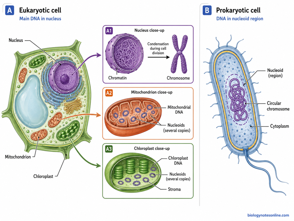
The following are the locations-
- Nucleus – It is the major location of chromosomes in eukaryotic cells. Plant, animal and fungal cells have their main genetic material enclosed within the nucleus. The DNA together with proteins forms chromatin. During cell division, this chromatin becomes more compact, forming the visible chromosomes.
- Nucleoid – In prokaryotic cells, membrane-bound nucleus is absent. The chromosome is present within the cytoplasm in an irregular region, called nucleoid. Bacteria generally contain a circular chromosome. It is folded and compacted within this region.
- Mitochondria – Mitochondria also have DNA of their own. It is different from the DNA present in nucleus. Several copies of mitochondrial DNA can be present, arranged in small groups called nucleoids. In most eukaryotic cells, the major amount of DNA still remains in nucleus.
- Chloroplasts – These are another DNA-containing organelles found in plants and algae. Chloroplast DNA is present in the stroma. It occurs in several copies and is grouped into nucleoids. Thus, plant cells have genetic material in nucleus, mitochondria and chloroplasts.
Structure of a Chromosome
A chromosome is formed of DNA associated with histone and other proteins. During cell division, it becomes condensed and the different structural regions can be seen more clearly. A replicated chromosome has two chromatids joined with each other. The following are the main parts-
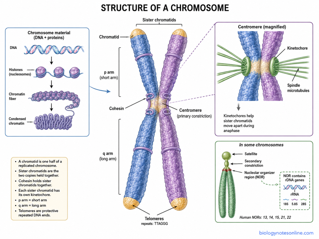
- Chromatid – Chromatid is one of the two halves of a replicated chromosome. It has one continuous double-stranded DNA molecule with associated proteins. After the two chromatids separate during cell division, each one becomes a chromosome of the daughter cell.
- Sister Chromatids – These are the two copies formed from the same chromosome after DNA replication. They remain joined with each other before separation. Cohesin proteins help in holding the sister chromatids together. During anaphase, these chromatids move apart.
- Centromere – This is the primary constricted region of chromosome. It holds an important position during chromosome separation and divides the chromosome into two arms. Depending on its position, the two arms may be equal or unequal in length. Kinetochore is formed in association with this region.
- Kinetochore – A kinetochore is a protein structure assembled on the centromere. The spindle microtubules are attached here during cell division. Each sister chromatid has its own kinetochore, which helps the chromatids to move toward opposite poles.
- p Arm – The shorter arm of chromosome is called p arm. The letter p is taken from the French word petit, meaning small. It extends between the centromere and telomere on the shorter side.
- q Arm – This is generally the longer arm of chromosome. It occurs on the other side of the centromere and the letter q follows p in chromosome nomenclature. Its terminal end also contains a telomere.
- Telomeres – Telomeres are present at both terminal ends of a linear chromosome. These are made up of repeated DNA sequences with associated proteins and protect the chromosome ends. In vertebrates, the repeated sequence is TTAGGG. Telomeres also prevent normal chromosome ends from being recognized as broken DNA.
- Secondary Constriction – Besides the centromere, another constricted region can be found in some chromosomes. It is called the secondary constriction. This region does not act as the primary site for spindle attachment. In some chromosomes, the secondary constriction is associated with the nucleolar organizer region.
- Nucleolar Organizer Region (NOR) – The NOR is a chromosomal region having repeated ribosomal DNA genes. These genes produce the precursor rRNA from which 18S, 5.8S and 28S rRNAs are formed. It is also involved in the formation of nucleolus. In humans, NORs are present on the short arms of acrocentric chromosomes 13, 14, 15, 21 and 22.
- Satellite – A small chromosome segment found beyond a secondary constriction is called a satellite. It remains connected to the main chromosome by a narrow stalk-like region. In human acrocentric chromosomes, satellites are found toward the ends of the short arms.
DNA Packaging into Chromosomes
In eukaryotic cells, a long DNA molecule is packed inside the nucleus with the help of histone and other proteins. DNA together with its associated proteins forms chromatin. The packaging takes place at different levels, which are as follows-
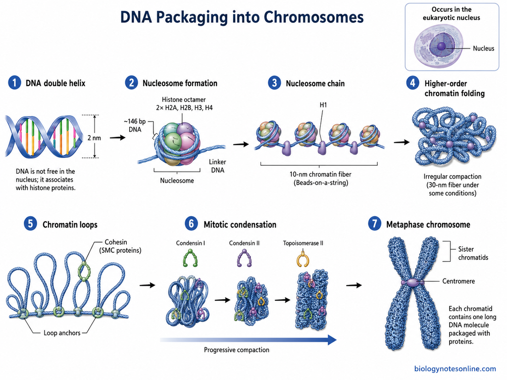
- DNA double helix – The first level is the double-stranded DNA molecule, about 2 nm in diameter. It is not present as free DNA inside the nucleus. The long DNA becomes associated with histone proteins for its packaging.
- Formation of nucleosomes – In this step, DNA wraps around a group of histone proteins called the histone octamer. It contains two molecules each of H2A, H2B, H3 and H4. About 146 base pairs of DNA is wrapped around this histone core, forming a nucleosome. A short region of linker DNA remains between two nucleosomes.
- Nucleosome chain – The nucleosomes connected by linker DNA form a bead-like chromatin structure. This is commonly referred to as the 10-nm chromatin fiber or “beads-on-a-string” structure. Histone H1 binds near the DNA entering and leaving the nucleosome and helps in further chromatin compaction.
- Higher-order chromatin folding – The nucleosome chain is further packed inside the nucleus. It does not generally form one uniform regular fiber. Instead, nucleosomes show irregular folding and interactions, producing more compact chromatin structures. The classical 30-nm fiber can be formed under some conditions, but it is not considered a regular basic structure in most cells.
- Formation of chromatin loops – The chromatin is organized into loops and different chromosomal domains. Proteins of the SMC (structural maintenance of chromosomes) family are involved in this organization. Cohesin has an important role in the organization of chromatin loops during interphase.
- Condensation during cell division – During mitosis, the chromatin undergoes extensive condensation. Condensin I and condensin II take part in formation and compaction of chromosome loops, producing a shorter and thicker chromosome structure. Topoisomerase II also helps in resolving intertwined DNA molecules during chromosome formation and segregation.
- Formation of the metaphase chromosome – The chromosome reaches a highly condensed condition at metaphase. After DNA replication, each chromosome is formed of two compact sister chromatids. Each chromatid contains one long DNA molecule packed with its associated proteins.
Types of Chromosomes
Chromosomes are classified into different types based on their structure and function. The position of centromere is one common basis. They can also be grouped as autosomes and sex chromosomes. According to the number or distribution of functional centromeric regions, acentric, monocentric, dicentric and holocentric forms are recognized.
A. Types Based on Centromere Position
Based on the location of centromere, four types of chromosomes are found-
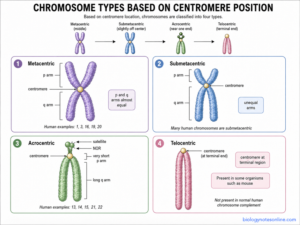
1. Metacentric Chromosome
In metacentric chromosome, the centromere lies nearly in the middle region. As a result, p and q arms are almost equal. It appears V-shaped during chromosome movement when the chromatids are considered. Human chromosomes 1, 3, 16, 19 and 20 are metacentric chromosomes.
2. Submetacentric Chromosome
The centromere is little away from the middle. Here, two arms are unequal, one is shorter and another is longer. The short arm is called p arm while the long one is q arm. Many human chromosomes are submetacentric.
3. Acrocentric Chromosome
These chromosomes contain the centromere near one end, giving a very short p arm and a long q arm. The difference in arm length is very prominent. Human chromosomes 13, 14, 15, 21 and 22 belong to this group. Their short arms contain stalks with nucleolar organizer regions (NORs) and satellites.
4. Telocentric Chromosome
Telocentric chromosomes have the centromere at the terminal region of chromosome. Practically one arm is therefore present. They occur in some organisms such as mouse. Normal human chromosome complement does not contain a telocentric chromosome.
B. Types Based on Biological Function
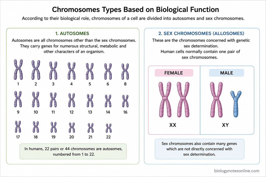
According to their biological role, chromosomes of a cell are divided into autosomes and sex chromosomes.
1. Autosomes
Autosomes are all chromosomes other than the sex chromosomes. They carry genes for numerous structural, metabolic and other characters of an organism. In humans, 22 pairs or 44 chromosomes are autosomes, numbered from 1 to 22.
2. Sex Chromosomes
These are the chromosomes concerned with genetic sex determination. They are also known as allosomes. Human cells normally contain one pair of sex chromosomes. XX is the usual female chromosome constitution and XY is the usual male chromosome constitution.
Sex chromosomes also contain many genes which are not directly concerned with sex determination.
C. Based on Number or Distribution of Centromeres
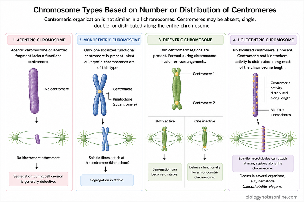
Centromeric organization is not similar in all chromosomes. Some have a single localized centromere, while in some organisms kinetochore activity occurs along most of chromosome length. Acentric and dicentric chromosomes can also arise, commonly due to chromosome rearrangements.
1. Acentric Chromosome
An acentric chromosome or acentric fragment lacks a functional centromere. Such fragments are commonly produced due to chromosome breakage or an abnormal rearrangement. As normal kinetochore attachment is absent, their segregation during cell division is generally defective.
2. Monocentric Chromosome
Only one localized functional centromere is found in a monocentric chromosome. Most of the commonly studied eukaryotic chromosomes are of this type. The kinetochore develops at this centromeric region and spindle fibres are attached here.
3. Dicentric Chromosome
Dicentric chromosome has two centromeric regions. It may be formed during chromosome fusion or different rearrangement processes. When both centromeres remain active, segregation can become unstable.
In many stable dicentric chromosomes, however, one centromere becomes inactive. The chromosome then behaves functionally like a monocentric chromosome.
4. Holocentric Chromosome
In holocentric chromosomes, one localized centromere is absent. Instead, centromeric and kinetochore activity is distributed along much of the chromosome length. Spindle microtubules can therefore interact at many regions along it.
These chromosomes occur in several groups of organisms. The nematode Caenorhabditis elegans is a well-known example.
Metacentric vs Submetacentric vs Acrocentric vs Telocentric Chromosomes
| Type | Centromere position | Relative arm length | Typical appearance | Human example |
|---|---|---|---|---|
| Metacentric | Centromere is present near the middle of chromosome. | p and q arms are almost equal in length. | Usually appears V-shaped during chromosome movement. | Chromosomes 1, 3, 16, 19 and 20 |
| Submetacentric | Centromere is slightly away from the middle. | One arm is shorter (p arm) and the other is longer (q arm). Difference is moderate. | Generally L-shaped. | Chromosome 2, and several others including 4–12, 17, 18 and X |
| Acrocentric | Centromere occurs close to one end. | p arm is very short, while q arm is much longer. | Usually J-shaped. The short arm may contain satellite and NOR in some chromosomes. | Chromosomes 13, 14, 15, 21 and 22 |
| Telocentric | Centromere is located at or very close to the terminal end. | Practically only one chromosome arm is visible. | Usually appears I-shaped. | Not normally present in humans |
Note: The V, L, J and I appearances are traditional morphological descriptions and can vary with chromosome condensation and orientation. Human chromosomes are classified mainly by centromere position and arm ratio.
Special Types of Chromosomes
Some chromosomes show unusual size, structure or behaviour in particular cells and organisms. The following are some of the important special types of chromosomes-
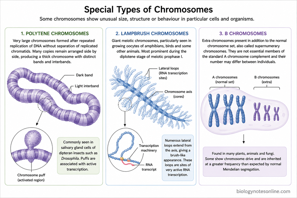
- Polytene chromosomes– These are very large chromosomes formed after repeated replication of DNA without separation of the replicated chromatids. Many copies therefore remain arranged side by side, producing a thick chromosome with distinct bands and interbands. Polytene chromosomes are commonly studied in the salivary gland cells of dipteran insects such as Drosophila. Some regions become expanded and form chromosome puffs, which are associated with active transcription.
- Lampbrush chromosomes– Lampbrush chromosomes are giant meiotic chromosomes, particularly seen in growing oocytes of amphibians, birds and some other animals. They are most prominent during the diplotene stage of meiotic prophase I. Numerous lateral loops extend from the chromosome axis, giving it a brush-like appearance. These loops are sites of very active RNA transcription.
- B chromosomes– These are extra chromosomes present in addition to the normal chromosome set, which are also called supernumerary chromosomes. They are not essential members of the standard A chromosome complement and their number may differ between individuals of the same species. B chromosomes are found in many plants, animals and fungi. Some also show chromosome drive and are inherited at a greater frequency than expected by normal Mendelian segregation.
Chromosome Number
Chromosome number is the number of chromosomes present in a cell. In most organisms, a particular species possesses a characteristic number of chromosomes. It is expressed in terms of n, 2n or more, depending on the number of chromosome sets present.
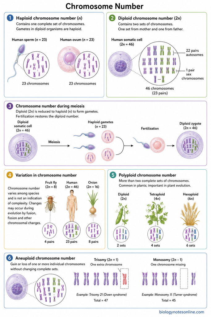
The different forms of chromosome number are as follows-
- Haploid chromosome number (n)– The chromosome number consisting of only one complete set is called haploid chromosome number. It is represented by n. Gametes in diploid organisms are haploid. In humans, the sperm and ovum contain 23 chromosomes, hence n = 23.
- Diploid chromosome number (2n)– It contains two sets of chromosomes and is represented by 2n. One set generally comes from the mother while another set comes from the father. Most human somatic cells have 46 chromosomes, arranged into 23 pairs. Thus, the diploid number of humans is 2n = 46. Of these, 22 pairs are autosomes and one pair is sex chromosomes.
- Chromosome number during meiosis– During meiosis, the diploid chromosome number is reduced to haploid number. The cells with two chromosome sets therefore form gametes having one set. At fertilization, the two haploid gametes unite. The diploid number is restored again.
- Variation in chromosome number– Chromosome number varies considerably among different species. Some organisms have a small number while others possess many chromosomes, and this number is not an indication of the complexity of an organism. Changes may also take place during evolution by chromosome fusion, fission and other chromosomal changes.
- Polyploid chromosome number– More than two complete sets of chromosomes in an organism is referred to as polyploidy. It may be 3n, 4n or still higher. This condition is particularly common in plants and has an important role in plant evolution.
- Aneuploid chromosome number– In aneuploidy, one or more individual chromosomes are gained or lost from the normal chromosome number, without the gain or loss of a complete set. Presence of three copies of one chromosome is called trisomy. When one chromosome of a pair is absent, the condition is referred to as monosomy.
How Many Chromosomes Do Humans Have?
Humans have 46 chromosomes in a normal somatic cell. These 46 chromosomes are arranged into 23 pairs. Of these, 22 pairs are autosomes. One pair is the sex chromosomes.
The chromosome numbers in humans are as follows-
- 46 chromosomes – Most of the nucleated body cells have 46 chromosomes. This number is called diploid number and represented as 2n = 46. The chromosomes are present inside nucleus. However, mature red blood cells have no nucleus and chromosomes are absent in them.
- 23 pairs – The total 46 chromosomes occur as 23 pairs. One chromosome of a pair is from mother, while another comes from father. Thus, two sets are present in somatic cells.
- 22 autosomal pairs – 22 pairs are autosomes, making total 44 autosomal chromosomes. These are numbered chromosome 1 to chromosome 22. Autosomes carry large number of genes for different body characters and cellular functions.
- One sex chromosome pair – The remaining one pair is sex chromosomes. Usually XX is present in females and XY in males. So, humans contain two sex chromosomes along with 44 autosomes.
- 23 chromosomes in gametes – The chromosome number becomes half in sperm and egg. Each gamete contains only 23 chromosomes and no pairs are present like somatic cells. It is the haploid number, written as n = 23.
During fertilization, 23 chromosomes from sperm and 23 chromosomes of egg come together. The zygote again contains 46 chromosomes.
Human Chromosome Quick Table
| Feature | Number |
|---|---|
| Total chromosomes in somatic cell | 46 |
| Total chromosome pairs | 23 pairs |
| Autosomal pairs | 22 pairs |
| Total autosomes | 44 |
| Sex chromosome pair | 1 pair |
| Total sex chromosomes | 2 |
| Chromosomes in sperm | 23 |
| Chromosomes in egg | 23 |
| Diploid number | 2n = 46 |
| Haploid number | n = 23 |
What are Homologous Chromosomes?
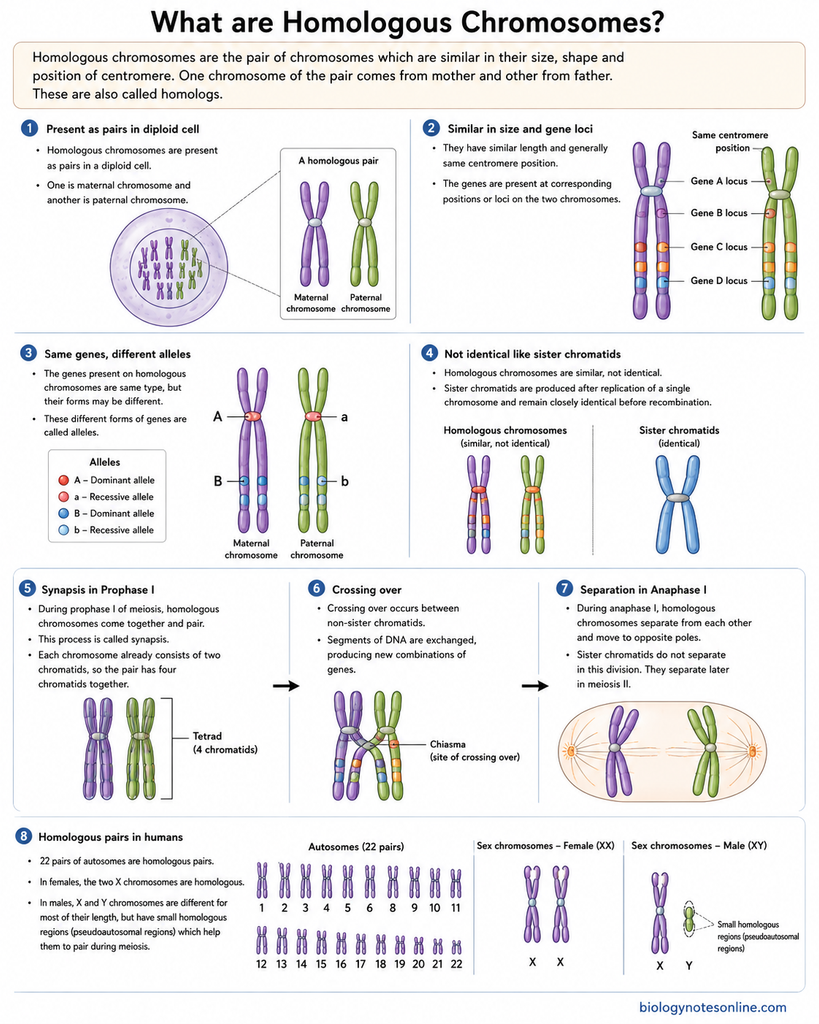
Homologous chromosomes are the pair of chromosomes which are similar in their size, shape and position of centromere. One chromosome of the pair comes from mother and other from father. These are also called homologs.
The following are some of the important characteristics of homologous chromosomes-
- Homologous chromosomes are present as pairs in a diploid cell. One is maternal chromosome and another is paternal chromosome.
- They have similar length and generally same centromere position. The genes are also present at corresponding positions or loci on the two chromosomes.
- The genes present on homologous chromosomes are same type, but their forms may be different. These different forms of genes are called alleles. For example, one chromosome may contain a dominant allele and its homolog can contain recessive allele.
- Homologous chromosomes are not identical copies of each other. They are similar chromosomes. Sister chromatids, on the other hand, are produced after replication of a single chromosome and remain closely identical before recombination.
- During prophase I of meiosis, the homologous chromosomes come together and pair. This process is called synapsis. At this time each chromosome already consists of two chromatids, therefore the paired homologous chromosomes have four chromatids together.
- Crossing over takes place between the non-sister chromatids of homologous chromosomes. Here, segments of DNA are exchanged. This results in new combinations of genes in the chromosomes.
- During anaphase I, homologous chromosomes separate from each other and move to opposite poles. The sister chromatids do not separate in this division. They separate later during meiosis II.
- In humans, the 22 pairs of autosomes are homologous pairs. In females, the two X chromosomes are also homologous. The X and Y chromosomes in males are different for most of their length, but have small homologous regions which help them to pair during meiosis.
What Is a Karyotype?
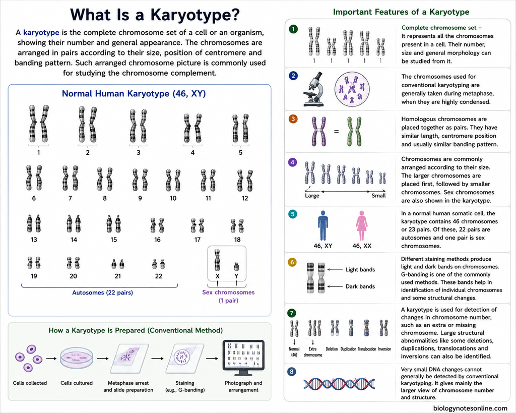
A karyotype is the complete chromosome set of a cell or an organism, showing their number and general appearance. The chromosomes can be arranged in pairs according to their size, position of centromere and banding pattern. Such arranged chromosome picture is commonly used for studying the chromosome complement.
The following are some of the important features of a karyotype-
- Complete chromosome set – It represents all the chromosomes present in a cell. Their number, size and general morphology can be studied from it.
- The chromosomes used for conventional karyotyping are generally taken during metaphase, when they are highly condensed. At this stage, individual chromosomes can be distinguished more easily.
- Homologous chromosomes are placed together as pairs. They have similar length, centromere position and usually similar banding pattern.
- Chromosomes are commonly arranged according to their size. The larger chromosomes are placed first, followed by smaller chromosomes. Sex chromosomes are also shown in the karyotype.
- In a normal human somatic cell, the karyotype contains 46 chromosomes or 23 pairs. Of these, 22 pairs are autosomes and one pair is sex chromosomes. A usual male karyotype is written as 46,XY, while usual female karyotype as 46,XX.
- Different staining methods produce light and dark bands on chromosomes. G-banding is one of the commonly used methods. These bands help in identification of individual chromosomes and some structural changes.
- A karyotype is used for detection of changes in chromosome number, such as an extra or missing chromosome. Large structural abnormalities like some deletions, duplications, translocations and inversions can also be identified.
- Very small DNA changes cannot generally be detected by conventional karyotyping. It gives mainly the larger view of chromosome number and structure.
How Chromosomes Change During the Cell Cycle
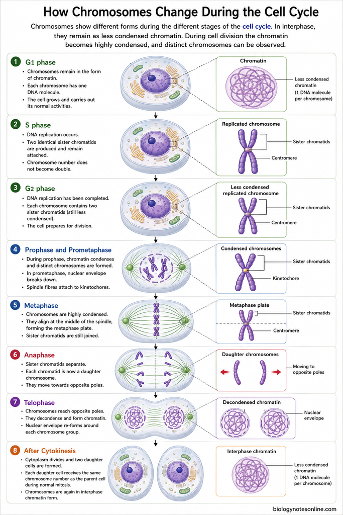
Chromosomes show different forms during the different stages of the cell cycle. In interphase, they remain as less condensed chromatin. During cell division the chromatin becomes highly condensed, and distinct chromosomes can be observed.
The changes of chromosomes during the cell cycle are as follows-
- G1 phase– Chromosomes remain in the form of chromatin during this phase. Each chromosome has one DNA molecule. The cell grows and carries out its normal activities.
- S phase– This is the phase of DNA replication. DNA of each chromosome is copied and two identical sister chromatids are produced. They remain attached with each other. However, chromosome number does not become double.
- G2 phase– DNA replication has already been completed. Each chromosome now contains two sister chromatids, although they are still present in less condensed form. The cell prepares for the following division.
- Prophase and prometaphase– During prophase, the chromatin becomes shorter and thicker due to condensation. Distinct chromosomes are now formed. In prometaphase, the nuclear envelope breaks down and spindle fibres become attached to the kinetochores.
- Metaphase– The chromosomes are highly condensed at this stage. They become arranged at the middle region of the spindle, forming the metaphase plate. Sister chromatids are still joined.
- Anaphase– In this step, the sister chromatids separate. Each separated chromatid is now referred to as a daughter chromosome and moves towards the opposite pole.
- Telophase– Chromosomes reach the two opposite poles. Their compact structure now starts disappearing and chromosomes again form less condensed chromatin. Nuclear envelope is also formed around each chromosome group.
- After cytokinesis– Cytoplasm divides and two daughter cells are formed. Each daughter cell receives the same chromosome number as the parent cell during normal mitosis. The chromosomes again remain in their interphase chromatin form.
Chromosomal Abnormalities
Chromosomal abnormalities are changes in the normal number or structure of chromosomes. These may involve whole chromosomes or only a part of chromosome. They are mainly divided into numerical and structural abnormalities.
Numerical Chromosome Abnormalities
Numerical abnormalities involve a change in chromosome number. The following are the major types-
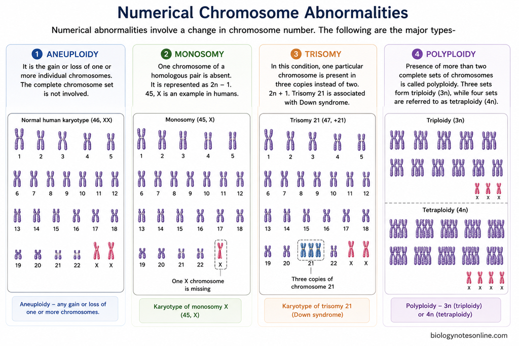
- Aneuploidy– It is the gain or loss of one or more individual chromosomes. The complete chromosome set is not involved.
- Monosomy– One chromosome of a homologous pair is absent. It is represented as 2n − 1. 45,X is an example in humans.
- Trisomy– In this condition, one particular chromosome is present in three copies instead of two. 2n + 1. Trisomy 21 is associated with Down syndrome.
- Polyploidy– Presence of more than two complete sets of chromosomes is called polyploidy. Three sets form triploidy (3n), while four sets are referred to as tetraploidy (4n).
Structural Chromosome Abnormalities
Structural abnormalities develop due to breakage and rearrangement of chromosome segments. A chromosome part may be lost, repeated or joined again in a different arrangement.
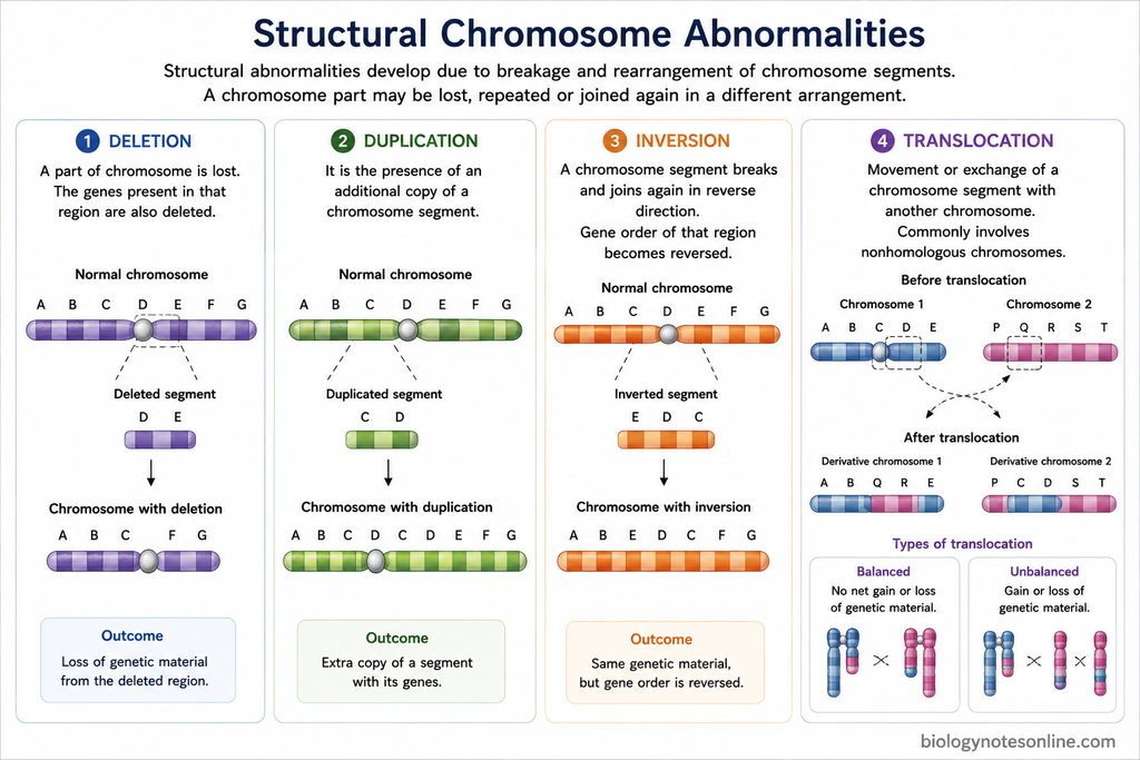
- Deletion– A part of chromosome is lost. The genes present in that region are also deleted.
- Duplication– It is the presence of an additional copy of a chromosome segment.
- Inversion– A chromosome segment breaks and joins again in reverse direction. Thus, the gene order of that region becomes reversed.
- Translocation– Movement or exchange of a chromosome segment with another chromosome is called translocation. It commonly involves nonhomologous chromosomes. The rearrangement may be balanced or unbalanced.
Examples of Chromosomal Disorders
Different chromosomal disorders occur due to change in chromosome number or chromosome structure. Some of the important examples are-
- Down syndrome– An example of autosomal trisomy is Down syndrome, which is caused due to an extra chromosome 21. Thus, the affected individual usually contains three copies of chromosome 21.
- Edwards syndrome– It is caused by the presence of an extra chromosome 18, and the condition is therefore referred to as trisomy 18. It causes severe developmental and congenital abnormalities.
- Patau syndrome– Patau syndrome occurs due to an extra copy of chromosome 13. This is also known as trisomy 13. Complete trisomy is the common form, but mosaic and translocation forms are also present.
- Turner syndrome– Loss of all or part of one X chromosome results in Turner syndrome. 45,X is the common chromosome constitution, although mosaic forms can also occur.
- Klinefelter syndrome– It is a sex chromosome disorder in males having an additional X chromosome, commonly 47,XXY. Other chromosome variants and mosaic forms are also found.
- Cri du Chat syndrome– Another example of structural chromosomal disorder is Cri du Chat syndrome. It results from deletion of chromosome material from the short arm of chromosome 5 (5p). A high-pitched cry during infancy is one of its characteristic features, along with developmental delay.
Functions of Chromosomes
The following are the major functions of chromosomes-
- Chromosomes carry genes, which contain the genetic information of a cell.
- They help in compact packing of the long DNA molecule inside the nucleus.
- During cell division, chromosomes are equally distributed into the newly formed daughter cells.
- Chromosomes also take part in control of gene expression. Some regions remain active while others are less active or inactive.
- They carry hereditary characters from one generation to another. This is the basis of heredity.
- During meiosis, chromosomes undergo crossing over and recombination, resulting in new gene combinations.
- Centromere, telomeres and origins of replication present in chromosomes help in their replication, protection and proper separation.
- The sex chromosomes are involved in determination of sex in organisms having chromosomal sex determination.
Importance of Chromosomes
Chromosomes have an important role in heredity, development, reproduction and different genetic studies. Some of the important roles of chromosomes are as follows-
- Chromosomes are the carriers of genes. The genetic information present in them is passed from parents to offspring, forming the basis of heredity.
- Genes present on chromosomes provide the information required during growth and development. Their proper expression is necessary for formation and normal functioning of different cells and tissues.
- During reproduction, chromosomes are distributed through meiosis and fertilization. Gametes receive one chromosome set and the diploid chromosome number is again restored after fertilization.
- Chromosomal changes are also involved in evolution, where fusion, fission, inversion and other rearrangements can change the organization of genome and some of these changes become associated with adaptation or formation of species.
- Crossing over takes place between homologous chromosomes during meiosis. It produces new combinations of alleles, therefore increasing genetic variation among the offspring.
- The study of chromosome number, structure, banding pattern and their behaviour is the basis of cytogenetics. Karyotyping and FISH are some of the methods used for chromosome analysis.
- In medical genetics, chromosomes are examined for numerical and structural abnormalities. Such studies are used in congenital disorders, prenatal diagnosis and also in different cancers.
- Chromosomes are also important in genome research. Chromosome mapping, chromosome-scale genome sequencing and comparison of chromosomes between species have provided information about genome structure and evolution. Modern studies also examine how chromosomes are organized inside the nucleus.
Chromosome Quick Revision Table
| Feature | Description |
|---|---|
| Definition | A chromosome is a DNA-protein structure that carries genetic information of a cell. |
| Location | In eukaryotes, chromosomes are present inside the nucleus. Prokaryotic chromosome is present in the nucleoid region. |
| Composition | Mainly made up of DNA and associated proteins, particularly histone proteins in eukaryotes. |
| Structural unit | Nucleosome is the basic structural unit of eukaryotic chromatin. |
| Chromatid | Each of the two identical copies of a replicated chromosome is called a chromatid. |
| Centromere | The specialized region of chromosome where sister chromatids remain associated and kinetochore is formed. |
| Kinetochore | A protein complex formed at the centromere. Spindle microtubules attach with chromosome through it. |
| Telomere | Specialized DNA-protein structure present at the ends of linear chromosomes. It protects chromosome ends. |
| p arm | The short arm of a chromosome. |
| q arm | The long arm of a chromosome. |
| Human diploid number | 46 chromosomes (2n = 46) or 23 pairs. |
| Human haploid number | 23 chromosomes (n = 23), present in sperm and ovum. |
| Autosomes | Humans contain 22 pairs of autosomes, making 44 autosomal chromosomes. |
| Sex chromosomes | One pair of sex chromosomes is present. Usually XX in females and XY in males. |
| Main chromosome types | Based on centromere position- metacentric, submetacentric, acrocentric and telocentric. |
| Primary functions | Storage and transmission of genetic information, carrying genes, DNA packaging, chromosome segregation, heredity and genetic variation. |
Prokaryotic vs Eukaryotic Chromosomes
| Feature | Prokaryotic Chromosomes | Eukaryotic Chromosomes |
|---|---|---|
| Location | Chromosome is present in the nucleoid region of cytoplasm. No true nucleus is present. | Chromosomes are mainly present inside the nucleus. |
| Number | Generally one main chromosome is present, although more than one chromosome occurs in some prokaryotes. | Usually many chromosomes are present. The number varies among different organisms. |
| Shape | Chromosome is generally circular in bacteria. Linear chromosomes are also found in some prokaryotes. | Nuclear chromosomes are linear. |
| DNA molecule | Usually contains one double-stranded DNA molecule forming the main chromosome. | Each chromosome contains one long double-stranded DNA molecule before replication. |
| Histone proteins | Typical bacterial chromosomes do not contain true histones. Histone-like proteins help in DNA organization. Archaeal chromosomes may contain histones. | DNA is closely associated with histone proteins, forming chromatin. |
| Chromatin formation | Typical nucleosomal chromatin like eukaryotes is absent in bacteria. | DNA and histones together form chromatin. |
| Centromere | Typical eukaryotic-type centromere is absent. | Centromere is generally present and has an important role during chromosome segregation. |
| Telomeres | Generally absent in circular chromosomes because no free chromosome ends are present. | Linear chromosomes contain telomeres at their ends. |
| Replication origins | Bacterial chromosomes commonly have a single major origin of replication. | Each chromosome generally contains many origins of replication. |
| Introns | Introns are less common in bacterial protein-coding genes. | Introns are common in many eukaryotic genes. |
| Gene density | Gene density is generally high, with comparatively less non-coding DNA. | Large amount of non-coding DNA can be present between and within genes. |
| Organization of genes | Genes are often arranged into operons in bacteria. | Operons are uncommon in most eukaryotes, and genes are generally regulated individually. |
| Separation during cell division | Chromosome segregation occurs during binary fission and does not involve a typical mitotic spindle. | Chromosomes are separated by the spindle apparatus during mitosis and meiosis. |
| Example | Bacterial chromosome of Escherichia coli. | Human nuclear chromosomes, with 46 chromosomes in 23 pairs in most nucleated somatic cells. |
Homologous Chromosomes vs Sister Chromatids
| Feature | Homologous Chromosomes | Sister Chromatids |
|---|---|---|
| Definition | A pair of chromosomes having similar size, shape and genes at corresponding loci. | Two copies of the same chromosome formed after DNA replication. |
| Origin | One chromosome is inherited from mother and another from father. | Both are produced from a single chromosome during S phase. |
| Genetic similarity | They contain the same types of genes, but may have different alleles. | They are usually nearly identical in DNA sequence before recombination or mutation. |
| Number of chromosomes involved | Two separate homologous chromosomes are involved. | Two chromatids of one replicated chromosome are involved. |
| Centromere | Each homolog has its own centromere. | Sister chromatids remain joined at the centromeric region after replication. |
| Pairing | Homologous chromosomes pair during prophase I of meiosis. | Sister chromatids remain attached after DNA replication. |
| Synapsis | Synapsis occurs between homologous chromosomes in meiosis I. | Synapsis does not occur between sister chromatids. |
| Crossing over | Crossing over occurs between non-sister chromatids of homologous chromosomes. | Sister chromatids normally do not exchange corresponding segments with each other during meiotic crossing over. |
| Separation | Homologous chromosomes separate during anaphase I of meiosis. | Sister chromatids separate during anaphase of mitosis and anaphase II of meiosis. |
| Alleles | Different alleles of the same gene may be present on the two homologs. | Same alleles are generally present because they are replicated copies of one chromosome. |
| Example | Maternal chromosome 1 and paternal chromosome 1 form a homologous pair. | The two chromatids of one replicated chromosome 1 are sister chromatids. |
Chromosome vs Chromatid
| Feature | Chromosome | Chromatid |
|---|---|---|
| Definition | Chromosome is a DNA-protein structure carrying genetic material of a cell. | Chromatid is one of the two copies of a replicated chromosome. |
| Formation | Chromosomes are formed by condensation of chromatin. | Chromatids are formed after DNA replication of a chromosome. |
| DNA molecule | An unreplicated chromosome contains one DNA molecule. After replication, it contains two DNA molecules as two sister chromatids. | Each chromatid contains one double-stranded DNA molecule. |
| Number of chromatids | Before DNA replication, one chromosome has one chromatid. After replication, it has two chromatids. | It represents a single chromatid of a chromosome. |
| Centromere | A replicated chromosome has two sister chromatids joined in the centromeric region. | Each chromatid has its own centromeric DNA and kinetochore region. |
| Occurrence | Chromosomes are present throughout the cell cycle, although their condensation changes. | Two clearly recognized sister chromatids are present after DNA replication and before their separation. |
| Genetic material | It carries genes arranged along DNA. | It contains one replicated copy of the genetic information of the chromosome. |
| Separation | Chromosome number is counted by the number of centromeres. | During anaphase, sister chromatids separate and each one is then considered an individual chromosome. |
| Structure | It may consist of one chromatid or two sister chromatids, depending on stage of cell cycle. | It is one longitudinal unit of a replicated chromosome. |
| Example | Human chromosome 1 before replication is one chromosome with one chromatid. | After chromosome 1 replicates, its two copies are called sister chromatids. |
Chromosome vs Chromatin
| Feature | Chromosome | Chromatin |
|---|---|---|
| Definition | Chromosome is a condensed DNA-protein structure carrying genetic material. | Chromatin is the DNA associated with histone and other proteins present inside the nucleus. |
| Condensation | It is highly condensed, especially during cell division. | It is comparatively less condensed during most part of interphase. |
| Appearance | Individual chromosomes can be seen clearly during mitosis and meiosis. | Chromatin appears as a more diffuse material and individual chromosomes are not clearly distinguished. |
| Basic composition | It is made up of DNA and chromosomal proteins. | It also contains DNA, histones and other associated proteins. |
| Structural relation | A chromosome is formed when chromatin becomes highly compacted. | Chromatin is the material from which chromosomes are formed. |
| Occurrence | Chromosomes remain as distinct condensed structures mainly during cell division. | Chromatin is present throughout the nucleus and becomes more prominent as an extended form during interphase. |
| Organization | Each chromosome contains one long DNA molecule before replication and two sister chromatids after replication. | Chromatin is organized into nucleosomes and higher levels of DNA packing. |
| Function | It helps in proper distribution of genetic material during cell division. | It helps in DNA packaging and also allows regulation of gene expression, replication and DNA repair. |
| Types | Chromosomes may be metacentric, submetacentric, acrocentric or telocentric based on centromere position. | Chromatin is commonly described as euchromatin and heterochromatin. |
| Example | Human chromosome 1, chromosome 2, X chromosome. | Euchromatin and heterochromatin present in a human cell nucleus. |
References
- Alberts, B., Johnson, A., Lewis, J., Raff, M., Roberts, K., & Walter, P. (2002). Molecular biology of the cell (4th ed.). Garland Science. https://www.ncbi.nlm.nih.gov/books/NBK21054/
- Chauhan, P. R., & Tanaka, J. (2026). Genetics, chromosome abnormalities. In StatPearls. StatPearls Publishing. https://www.ncbi.nlm.nih.gov/books/NBK557691/
- Cooper, G. M. (2000). The cell: A molecular approach (2nd ed.). Sinauer Associates. https://www.ncbi.nlm.nih.gov/books/NBK9839/
- Dame, R. T., Rashid, F.-Z. M., & Grainger, D. C. (2020). Chromosome organization in bacteria: Mechanistic insights into genome structure and function. Nature Reviews Genetics, 21(4), 227–242. https://doi.org/10.1038/s41576-019-0185-4
- Genetic Alliance, & The New York-Mid-Atlantic Consortium for Genetic and Newborn Screening Services. (2009). Understanding genetics: A New York, Mid-Atlantic guide for patients and health professionals. Genetic Alliance. https://www.ncbi.nlm.nih.gov/books/NBK115508/
- Gerton, J. L., & Hawley, R. S. (2005). Homologous chromosome interactions in meiosis: Diversity amidst conservation. Nature Reviews Genetics, 6(6), 477–487. https://doi.org/10.1038/nrg1614
- Jackson, M., Marks, L., May, G. H. W., & Wilson, J. B. (2018). The genetic basis of disease. Essays in Biochemistry, 62(5), 643–723. https://doi.org/10.1042/EBC20170053
- Monen, J., Maddox, P. S., Hyndman, F., Oegema, K., & Desai, A. (2005). Differential role of CENP-A in the segregation of holocentric C. elegans chromosomes during meiosis and mitosis. Nature Cell Biology, 7(12), 1248–1255. https://doi.org/10.1038/ncb1331
- Morrison, P. J., & Spence, R. A. J. (2005). Genetics for surgeons. Remedica. https://www.ncbi.nlm.nih.gov/books/NBK7524/
- National Institutes of Health, & Biological Sciences Curriculum Study. (2007). NIH curriculum supplement series. National Institutes of Health. https://www.ncbi.nlm.nih.gov/books/NBK20362/
- National Research Council, Committee on DNA Forensic Science: An Update. (1996). The evaluation of forensic DNA evidence. National Academies Press. https://www.ncbi.nlm.nih.gov/books/NBK232615/
- Olson, S. (1989). Shaping the future: Biology and human values. National Academies Press. https://www.ncbi.nlm.nih.gov/books/NBK218591/
- Rose, R. J. (2019). Sustaining life: Maintaining chloroplasts and mitochondria and their genomes in plants. Yale Journal of Biology and Medicine, 92(3), 499–510. https://pmc.ncbi.nlm.nih.gov/articles/PMC6747931/