

Prokaryotic cells are relatively simple and small cells, which do not possess a true membrane-bound nucleus and other membrane-bound cell organelles that are commonly present in the eukaryotic cells. Bacteria and Archaea are made up of prokaryotic type of cells. They have their genetic material inside the cell but it is not enclosed within a nuclear membrane. This region containing the major cellular DNA is referred to as the nucleoid.
Generally, prokaryotic cells are much smaller than most of the eukaryotic cells. Their internal cellular organization is also simpler. The major difference between prokaryotic and eukaryotic cell is the presence of a membrane-bound nucleus and different membrane-bound organelles in eukaryotic cells, while a true nucleus is absent in the prokaryotic cells.
What Are Prokaryotic Cells?
The term prokaryote is derived from pro meaning “before” and karyon meaning “nucleus”. It is used for organisms having a prokaryotic type of cell organization. Prokaryotic cell is a type of cell which does not contain a true membrane-bound nucleus. These cells are generally present as unicellular organisms.
The organisms of Bacteria and Archaea have prokaryotic cells. They are predominantly unicellular. The DNA is present inside the cell, but it is not enclosed within a nuclear envelope. Instead, the major cellular DNA remains in a region called the nucleoid. This is one of the main features of prokaryotic cellular organization.
The terms prokaryotic cell and prokaryote are related but do not indicate exactly the same thing. Prokaryotic cell refers to the type of cell. A prokaryote is an organism having this type of cellular organization, which includes members of Bacteria and Archaea.
Characteristics of Prokaryotic Cells
Some of the important characteristics of prokaryotic cells are-
- Cell type- Members are unicellular in general. Some bacteria occur together in chains, colonies or filament-like forms.
- Occurrence- Bacteria and Archaea consist of prokaryotic type of cells.
- Cell size- Most of the prokaryotic cells are very small, generally occurring in micrometre range. The size however varies greatly among different members.
- Nucleus- True membrane-bound nucleus is absent. No nuclear envelope is formed around the genetic material.
- Nucleoid- The major DNA of cell is located in an irregular cytoplasmic region known as nucleoid. It is not an organelle.
- Chromosome- In many members, the major chromosome is circular DNA. However, this is not same for all prokaryotes, as linear chromosomes and more than one chromosome are also found in some forms. Small DNA molecules called plasmids may be present.
- Cell organelles- Membrane-bound cell organelles are absent, such as mitochondria, Golgi bodies, and endoplasmic reticulum (ER). Thus, the internal organization is less compartmentalized than eukaryotic cells.
- Ribosomes- 70S ribosomes occur freely in the cytoplasm. Protein synthesis takes place on these ribosomes.
- Plasma membrane- It forms the boundary around cytoplasm and controls movement of different materials. In bacteria, several energy-producing reactions are also associated with this membrane.
- Cell wall- Most bacteria possess a rigid cell wall containing peptidoglycan, but it is not a feature of every bacterial cell. Mycoplasma does not contain cell wall. Archaea have walls of different chemical nature and typical bacterial peptidoglycan is absent.
- Cell division- Binary fission is the common method of multiplication in bacteria. Mitosis does not occur.
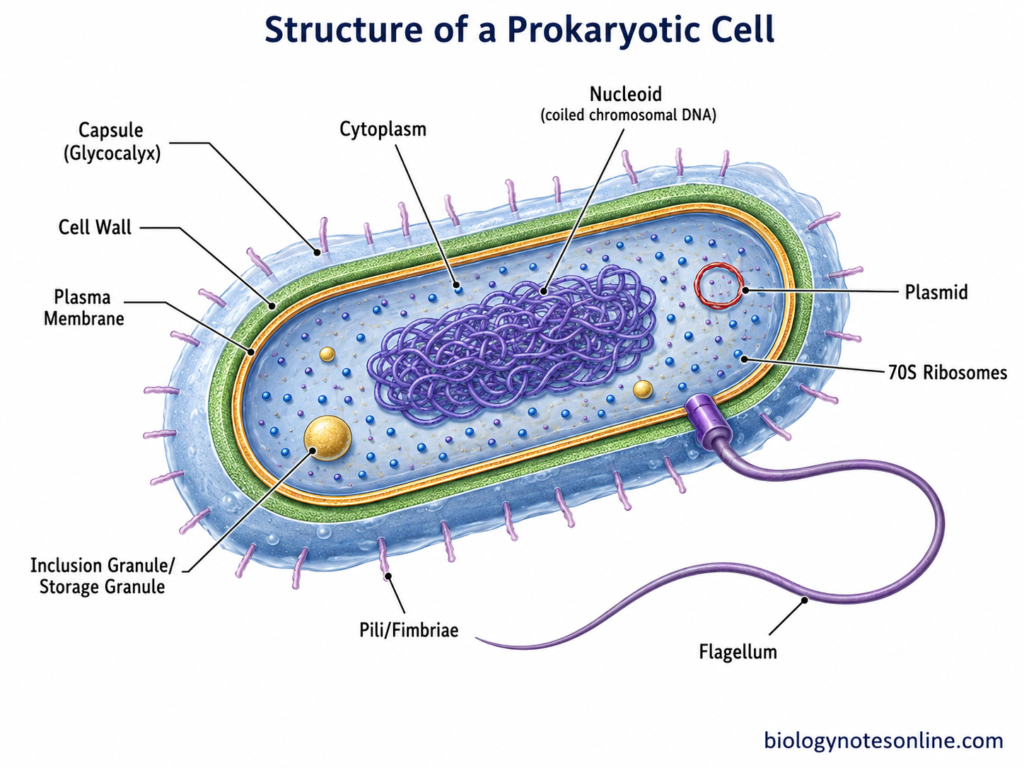
- External structures- Flagella, pili, fimbriae and capsule may occur in different prokaryotic cells. These are not present in every member. Flagella are mainly associated with movement, while pili and fimbriae have roles mainly in attachment and other functions.
Size and Shape of Prokaryotic Cells
Cell Size
Some of the important points about the size of prokaryotic cells are-
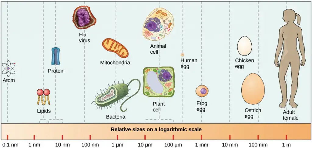
- Microscopic size- Prokaryotic cells are generally microscopic and cannot be viewed clearly with unaided eye. Most of the bacterial cells are only a few micrometres in size.
- General range- A commonly given size range for prokaryotic cells is about 0.1–5.0 µm, particularly in introductory descriptions. However, this is not the actual limit for all prokaryotes and much greater variation occurs among different bacterial and archaeal cells.
- Typical bacteria- Cells of many common bacterial species are around 1–2 µm in their major dimension. The size is not fixed for all bacteria. Some are much smaller, whereas others attain unusually large sizes.
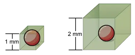
- Surface area- Small cells have a high surface-area-to-volume ratio. As the cell size increases, volume increases more rapidly than its surface area and the surface-area-to-volume ratio becomes lower.
- Advantages- The small size allows nutrients and other molecules to move through short distances within the cell. Removal of wastes is also rapid. A greater amount of cell surface is available relative to the cytoplasmic volume for exchange with surrounding environment.
- Limitations- Very small size also has a lower biological limit, as the cell must contain its DNA, ribosomes and different molecular components required for metabolism and reproduction. Thus, bacterial cells cannot simply become indefinitely smaller.
- Large bacteria- Some bacteria are exceptionally large and do not follow the usual microscopic size range. Candidatus Thiomargarita magnifica has an average cell length greater than 9,000 µm (9 mm) and the cell can be seen by unaided eye. Some specimens occur around centimetre scale. Such giant bacteria are unusual exceptions rather than the normal condition.
Common Cell Shapes
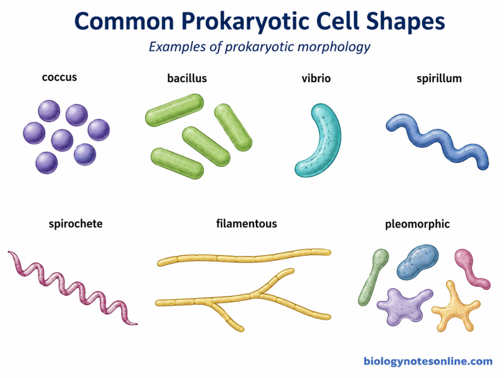
Prokaryotic cells show considerable variation in their shape. The commonly used morphological terms are particularly common for bacteria, while Archaea also have a wide spectrum of cell shapes and some show considerable morphological plasticity.
- Coccus- These are round or nearly spherical cells. The plural form is cocci.
- Bacillus- A bacillus is rod-shaped cell. Bacilli may differ greatly in their length and width, with some forms appearing as short rods.
- Vibrio- These cells are curved rods, generally having a comma-like appearance. Vibrio cholerae is a well-known example.
- Spirillum- Spirilla are spiral or corkscrew-like bacterial cells. They generally have a more rigid spiral form.
- Spirochete- These are thin and flexible helical cells. The spiral is more flexible than that of typical spirilla. Treponema is one of the common examples.
- Filamentous forms- Some bacteria grow as long filamentous cells and the filaments may also show branching. Members such as Actinomyces show this type of morphology.
- Pleomorphic forms- A fixed cell shape is not present in some prokaryotes. These are referred to as pleomorphic forms. Mycoplasma, which lacks a cell wall, shows pleomorphic morphology, and pleomorphism is also present in some archaeal groups.
Cell Arrangements
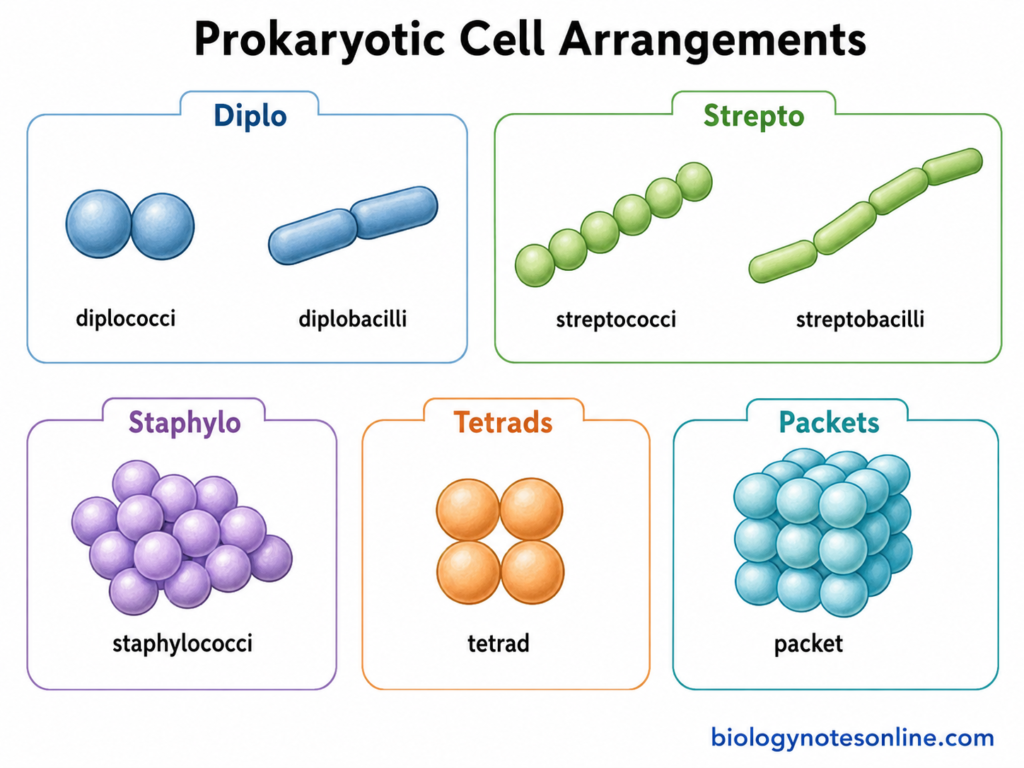
After cell division, some bacterial cells remain attached with one another and produce characteristic arrangements. These terms are used mainly in describing bacterial morphology.
- Diplo- Cells remain in pairs after division. Two attached cocci are called diplococci, while paired rods can be referred to as diplobacilli.
- Strepto- The cells are arranged in chains. Thus, chains of cocci are described as streptococci and chains of bacilli as streptobacilli.
- Staphylo- Cocci occur in irregular, grape-like clusters. This arrangement is characteristic of many members of Staphylococcus.
- Tetrads- Four coccal cells remain together, generally forming a square-like group. This arrangement results from a particular pattern of cell division.
- Packets- Some cocci form regular cubic packets after repeated divisions in different planes. Such packet-like groups are found in certain bacterial forms and are different from the irregular clusters of staphylococci.
Structure of a Prokaryotic Cell
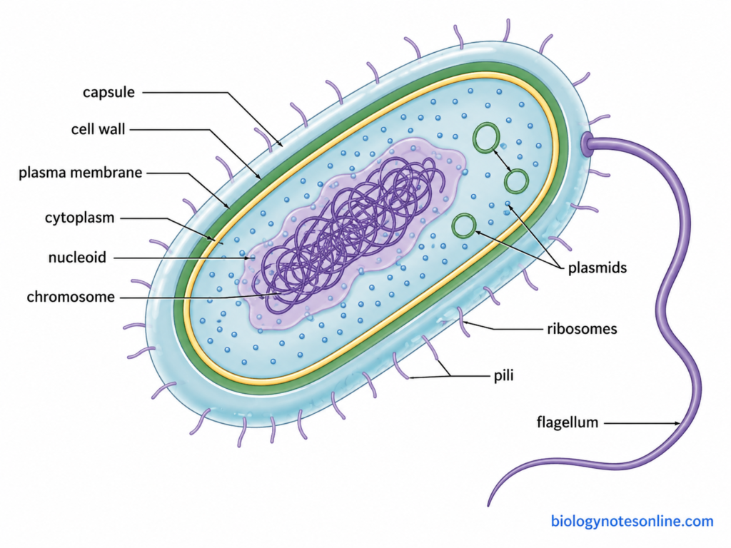
1. Plasma Membrane
The plasma membrane forms the boundary between cytoplasm and the external region. It is present in both Bacteria and Archaea, but their membrane chemistry is not same.
- In bacteria, it is mainly a phospholipid bilayer with different proteins present within the membrane. It surrounds the entire cytoplasm.
- The membrane is selectively permeable. Movement of different substances therefore does not occur freely in the same manner.
- Nutrients are transported into the cell through different membrane transport systems, while waste products and other substances are moved outside. Many of these processes need specific membrane proteins.
- Different proteins are embedded within the membrane. They have role in transport, enzymatic reactions, receptors and several other cellular processes.
- In bacteria, the membrane also has an important role in energy production. Components of respiratory electron transport and ATP synthase are located here. Thus, many functions performed by mitochondria in eukaryotic cells occur at the bacterial plasma membrane.
- Archaeal membrane lipids are different. Bacterial membrane lipids generally contain fatty acids attached to glycerol by ester bonds, whereas Archaea mainly possess isoprenoid chains attached through ether bonds. Some archaeal tetraether lipids can also extend across the complete membrane and form a monolayer.
2. Cell Wall
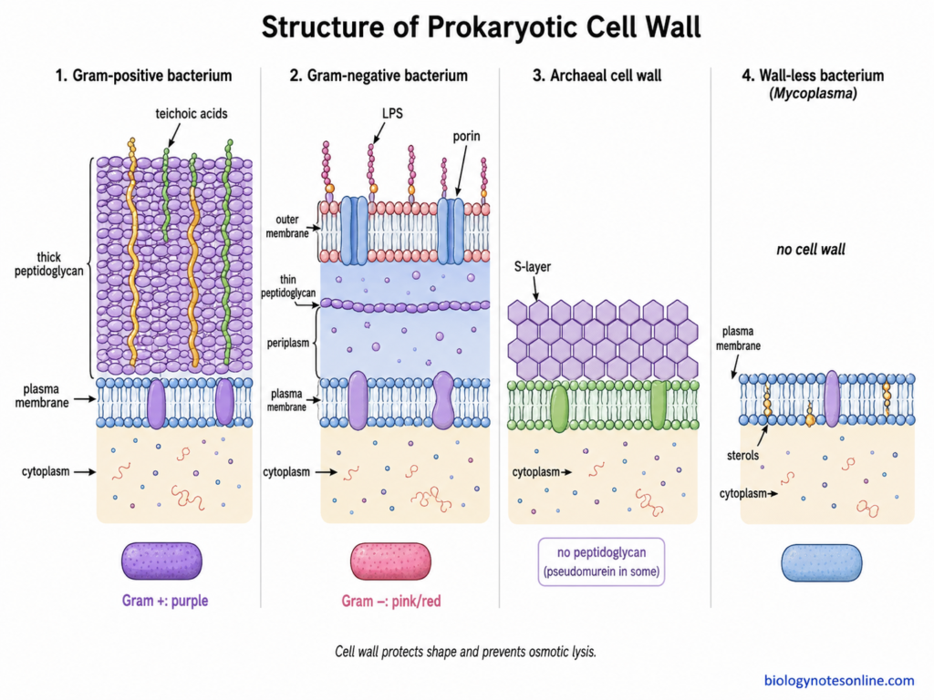
The cell wall is present outside the plasma membrane in most prokaryotic cells. Its composition however is not universal.
- It gives mechanical protection to the cell. The wall also helps in maintaining the characteristic cellular shape.
- Bacterial cells normally contain a high concentration of dissolved substances inside the cytoplasm. The rigid wall protects the cell from excessive expansion and osmotic lysis, especially when present in a hypotonic surrounding.
- Peptidoglycan (murein) is the characteristic wall material of bacteria. It forms a strong network around the bacterial cell. The amount and arrangement is different among bacterial groups.
- Archaeal walls do not contain bacterial peptidoglycan. Many members possess a protein or glycoprotein S-layer as the major wall structure. Pseudomurein and some other wall materials are also found in particular archaeal groups.
- Cell wall is absent in some bacteria. Mycoplasma is an important wall-less bacterial form and lacks the usual peptidoglycan layer.
Gram-Positive and Gram-Negative Cell Envelopes
The Gram-positive and Gram-negative arrangement is mainly used for bacterial cells. Their cell envelope is different in structure.
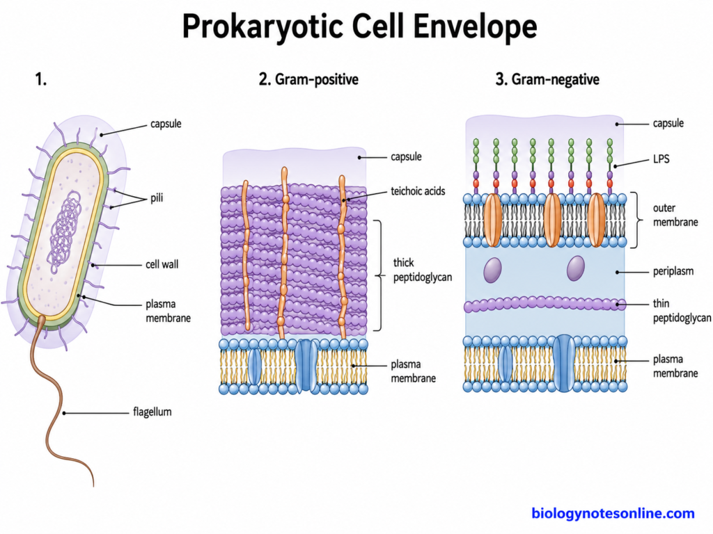
- Gram-positive bacteria generally possess a thick peptidoglycan layer outside the plasma membrane. No typical Gram-negative outer membrane is present.
- In Gram-negative bacteria, the peptidoglycan layer is much thinner and occurs between an inner and an outer membrane. This space forms part of the periplasmic region.
- The outer membrane is a characteristic component of the typical Gram-negative envelope. Its outer leaflet contains lipopolysaccharide (LPS), while phospholipids mainly occur in the inner leaflet.
- During Gram staining, Gram-positive bacteria retain the crystal violet-iodine complex after decolorization and appear purple. Gram-negative cells are decolorized and then take the counterstain, generally producing pink or red cells. This staining difference is mainly related with their different cell envelope organization.
3. Capsule and Glycocalyx
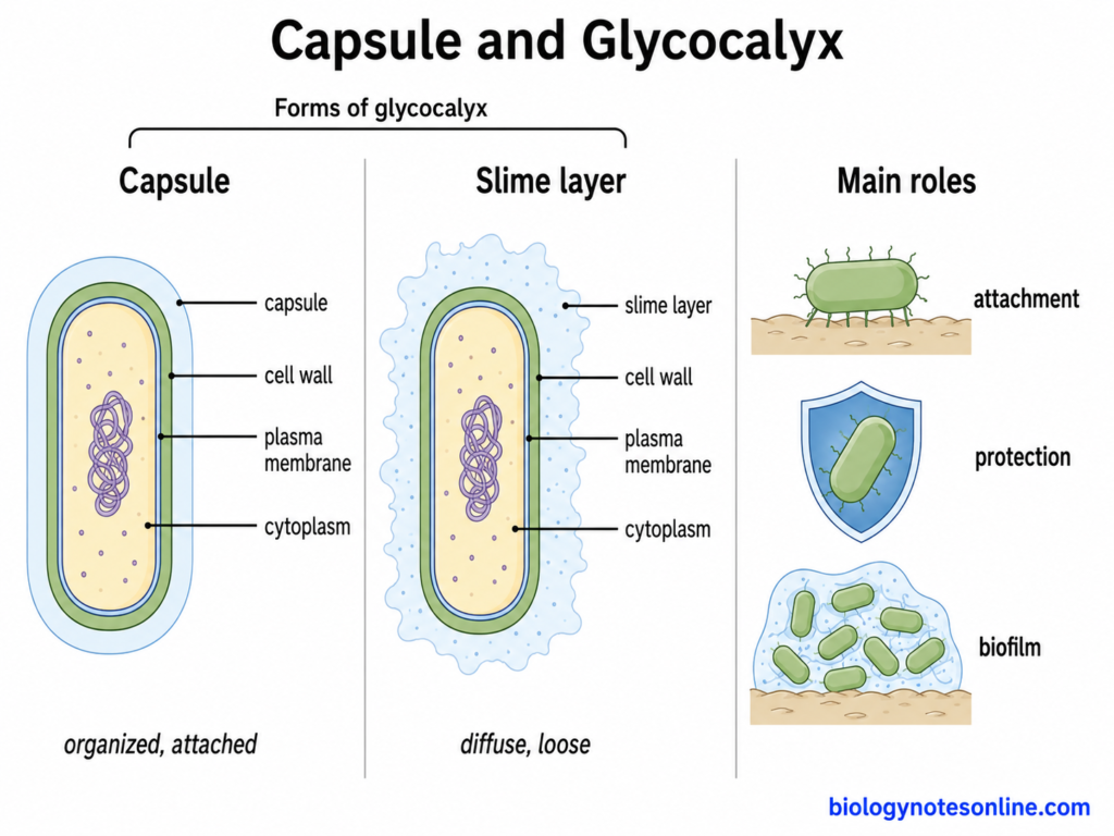
A glycocalyx is an extracellular covering found around many bacterial cells. It is not present in all prokaryotes.
- When this outer material remains well organized and closely associated with cell surface, it is generally called a capsule. Bacterial capsules are commonly polysaccharide in composition, although this is not same for every capsule.
- A more diffuse and loosely attached extracellular material is commonly referred to as the slime layer.
- These surface materials can help the bacterial cell to become attached with host tissues, other cells and non-living surfaces.
- Capsule also provides protection. In several pathogenic bacteria it helps the cell to resist different host defence mechanisms, while extracellular coverings can also protect against environmental stress.
- Attachment and production of extracellular material are important during formation of biofilms. The cells now remain together on a surface within an extracellular matrix.
4. Cytoplasm
The cytoplasm is present within the plasma membrane. It contains different cellular materials but lacks the typical membrane-bound organelles of eukaryotic cells.
- The fluid portion of cytoplasm is the cytosol. Water, ions, soluble molecules and other components remain in it.
- A large number of enzymes required for different metabolic reactions occur in the cytoplasm. Different metabolites are also present.
- Ribosomes are distributed within the cytoplasmic region and are involved in protein synthesis.
- The nucleoid containing the major chromosome is also present within cytoplasm. It is not enclosed inside a nuclear membrane.
- Different reserve substances may remain as granules or inclusion materials. The type of stored material is different among organisms and also changes with growth condition.
5. Nucleoid
The nucleoid is the region containing the major chromosomal DNA of prokaryotic cell. It is not a true nucleus.
- In bacteria, the major chromosome remains within this cytoplasmic region. Many bacterial chromosomes are circular double-stranded DNA, although this condition is not same in every prokaryotic organism.
- No nuclear envelope is present around nucleoid. Thus, there is no membrane separating the bacterial chromosome from surrounding cytoplasm.
- The bacterial chromosome is much longer than the dimensions of cell and therefore it remains highly compacted and organized.
- DNA supercoiling has an important role in this organization. DNA gyrase, topoisomerases and several other factors take part in controlling chromosome topology.
- Different nucleoid-associated proteins (NAPs) bind with bacterial DNA. They can bend, bridge or organize DNA and contribute to nucleoid structure. Examples include HU and other DNA-binding proteins.
6. Plasmids
Plasmids are additional genetic elements found in many prokaryotic cells. They are not a compulsory cellular structure.
- They are extrachromosomal DNA molecules occurring separately from the main chromosome. Bacterial plasmids are often circular, although different forms are present.
- Plasmids contain a replication system which allows replication separately from the host chromosome.
- Different accessory genes can be carried by plasmids. Such genes may provide an advantage under a particular environmental condition but are not always required for basic cellular survival.
- Antibiotic-resistance genes are one important bacterial example. Resistance plasmids can carry these genes and some plasmids can move between bacterial cells, contributing to horizontal spread of antimicrobial resistance.
- Plasmids are not present in every prokaryotic cell. Their number and size also show considerable variation.
7. Ribosomes
Ribosomes are small ribonucleoprotein structures present within the cytoplasm. The major function is protein synthesis.
- Translation of messenger RNA takes place on the ribosome and amino acids are joined for formation of protein.
- The cytoplasmic ribosome of bacteria is 70S. It consists of two unequal subunits.
- The smaller subunit is 30S, while the larger one is 50S. Both subunits combine during translation to form the functional 70S ribosome.
- Eukaryotic cytoplasmic ribosomes are larger 80S ribosomes, consisting of 40S and 60S subunits. Archaeal cytoplasmic ribosomes are also 70S in sedimentation behaviour, although their molecular composition has several important differences from bacterial ribosomes.
- Bacterial ribosomes are major targets for many antibiotics. Some drugs bind with the 30S subunit and others act mainly on the 50S subunit, which interferes with bacterial protein synthesis.
8. Flagella
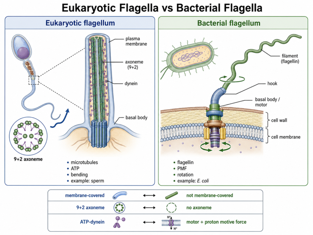
Flagella are long external appendages found in many motile bacteria. They are absent in a large number of species.
- The main function of bacterial flagella is movement of the cell through liquid or over particular environments.
- A bacterial flagellum works by rotation. The filament is connected to a rotary motor in the cell envelope, which is commonly driven by proton motive force and in some bacteria by sodium ion motive force.
- Flagellar movement can be controlled according to environmental chemical signals. Movement toward favourable chemicals or away from harmful chemicals is referred to as chemotaxis.
- Flagella are not present in every bacterial species. Non-motile bacteria may completely lack this structure.
- Motile Archaea possess a different structure called the archaellum. It performs a flagellum-like function but is structurally and evolutionarily different from bacterial flagella. Archaellar rotation is powered by ATP.
9. Fimbriae and Pili
Fimbriae and pili are thin surface structures found in different bacterial cells. Different forms have different functions.
- Fimbriae are generally associated with adhesion. They help bacterial cells to attach with host tissues, other cells or environmental surfaces.
- Surface attachment through fimbriae and pili can also take part in colonization and biofilm formation.
- Conjugative pili have role in bacterial conjugation. They help to establish contact between donor and recipient cells during transfer of genetic material.
- DNA transferred during conjugation is commonly plasmid DNA, although the exact conjugation systems and transferred DNA are different among bacteria.
- Some pili have other functions. Type IV pili can take part in adhesion, DNA uptake and surface movement known as twitching motility, where extension and retraction of pili moves the bacterial cell.
10. Inclusion Bodies and Storage Structures
Different inclusions are present within prokaryotic cells. Some are storage materials, whereas others are specialized cellular structures and do not function mainly for storage.
- Glycogen is accumulated in some bacteria as a carbon and energy reserve. It can be utilized again when the external carbon source becomes limited.
- Polyphosphate (polyP) can form intracellular granules. It is associated with phosphate storage and has several other roles in bacterial physiology.
- Sulfur granules are found in different sulfur-metabolizing bacteria. Elemental sulfur may accumulate within the cytoplasm, periplasm or other locations depending upon the organism.
- Gas vesicles are hollow protein structures found in some aquatic Bacteria and Archaea. They provide buoyancy, allowing cells to change their position within the water column. They are not storage granules.
- Magnetosomes are found in magnetotactic bacteria. These are membrane-bounded structures containing crystals of magnetic minerals, commonly magnetite or greigite, and they help the cells to align with magnetic fields.
- Carboxysomes are protein-bounded bacterial microcompartments associated with carbon fixation. They contain RuBisCO and components that concentrate carbon dioxide around this enzyme. These are specialized structures and not simple reserve granules.
11. Endospores
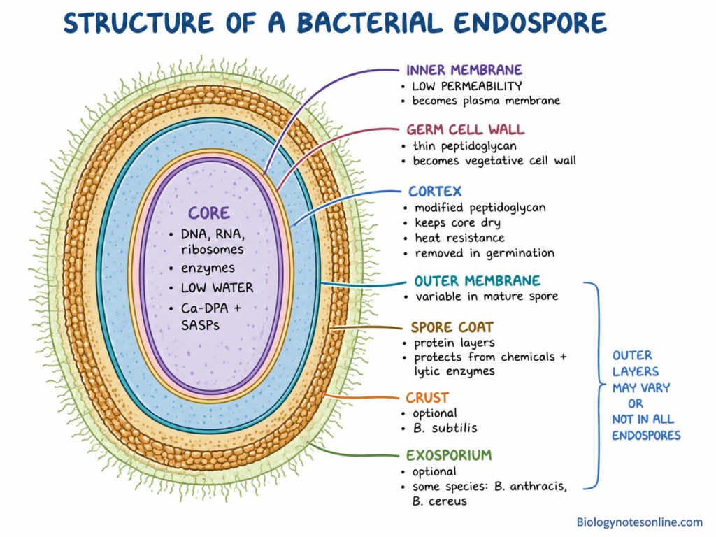
Endospores are resistant dormant structures produced by certain bacteria. This ability is restricted to particular bacterial groups.
- Endospore formation occurs in some bacteria, with Bacillus and Clostridium being the classical well-studied examples. It is formed inside the vegetative cell during the process of sporulation.
- The endospore functions mainly as a survival structure. It allows the bacterial genome and essential cellular materials to persist when the environmental condition becomes unfavourable.
- Mature endospores remain in a dormant state with extremely low cellular activity. They show much greater resistance to heat, desiccation and different chemical or physical stresses than ordinary vegetative cells.
- When favourable conditions return, germination takes place and the dormant spore changes toward an active vegetative cell. Outgrowth follows germination.
- An endospore is not a reproductive spore. Formation of one endospore from a vegetative bacterial cell does not increase the number of organisms. It is mainly formed for persistence and survival.
For detailed structure, formation and return to vegetative growth, see Endospore – Structure, Sporulation, Germination and Examples.
Prokaryotic Cell Diagram
DNA and Genetic Organization of a Prokaryotic Cell
The genetic material of prokaryotic cells is present inside the cell without formation of a membrane-bound nucleus. DNA organization is however not exactly same in Bacteria and Archaea.
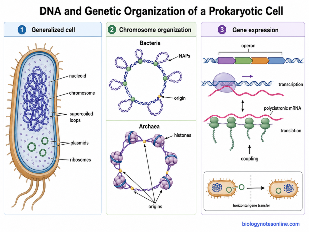
- Chromosomal DNA- The major genetic material is present as chromosomal DNA. In many bacteria, it is a single circular double-stranded DNA molecule. But this is not same for all members. Linear chromosomes and more than one chromosome are also present in some bacterial species.
- Nucleoid- The major chromosome occupies an irregular cellular region called the nucleoid. No nuclear envelope is formed around it. The nucleoid is therefore directly present within the cytoplasmic region.
- DNA compaction- Prokaryotic chromosome is much longer than the cell in which it is present and has to remain in a highly compacted form. DNA bending, looping and different levels of organization are involved. It is not simply an unorganized mass of DNA inside the cell.
- Supercoiling- DNA supercoiling is one of the important processes involved in bacterial chromosome compaction. DNA gyrase and other topoisomerases control the topological condition of DNA. Supercoiling also changes during processes such as transcription and replication.
- DNA-binding proteins- Different nucleoid-associated proteins (NAPs) bind with bacterial chromosome and help in its organization. Proteins such as HU can bend, bridge or compact DNA. These proteins also have roles in regulation of gene expression.
- Archaeal chromatin- Chromosome organization in Archaea has several differences from bacteria. Histone proteins are present in many archaeal groups and these can bind and wrap DNA. Some Archaea instead depend more strongly on other small DNA-binding proteins. Thus, one bacterial model of nucleoid organization cannot be applied to all prokaryotes.
- DNA replication- Replication of chromosome begins from specific regions called origins of replication. Well-studied bacteria such as Escherichia coli use a single chromosomal origin, whereas several archaeal chromosomes contain multiple replication origins. Considerable variation is present among Archaea.
- Plasmids- These are extrachromosomal genetic elements present in many prokaryotic cells. Plasmids can replicate independently of the main chromosome. They are often circular in bacteria and may carry different accessory genes, including genes for antibiotic resistance, virulence or other useful characteristics. Plasmids are not present in every cell.
- Gene arrangement- Prokaryotic genomes generally have a high proportion of coding DNA. Genes may occur singly or as groups, and their organization differs greatly among species.
- Operons- In bacteria, several functionally related genes can be arranged together in an operon and controlled as a transcriptional unit. One transcript may therefore contain coding regions for more than one protein, producing a polycistronic mRNA. Not all bacterial genes however are arranged into operons.
- Gene expression- Transcription and translation occur within the same cellular compartment because a nuclear membrane does not separate the DNA from cytoplasmic ribosomes. In many bacteria, translation can begin on an mRNA while its transcription is still taking place. This is referred to as transcription-translation coupling, although the degree of such coupling is not same in all bacterial groups.
- Archaeal expression- Archaea also lack a nuclear envelope, but their transcription machinery and chromosome-associated proteins have important differences from those of bacteria. Transcription and translation may occur in a coordinated manner in some archaeal species, however the process is not equally established for all archaeal groups.
- Genetic flexibility- Plasmids and other mobile genetic elements can contribute to movement of genes between prokaryotic cells. Such horizontal gene transfer has an important role in bacterial genetic variation, including spread of antimicrobial-resistance genes carried on transferable plasmids.
Types of Prokaryotic Cells
The two major types of prokaryotic cells are Bacteria and Archaea. Both have prokaryotic type of cellular organization, but they are different groups.
1. Bacteria
Bacteria are the members belonging to the Domain Bacteria. They are unicellular organisms and are widely distributed in nature.
- Members are found in soil, water, sediments and inside or over different living organisms. Some are also present in extreme environmental conditions.
- Most of the bacterial cells possess a cell wall containing peptidoglycan. The amount and arrangement of peptidoglycan is different in different bacterial groups. Mycoplasma however does not contain a cell wall.
- Bacteria occur in different shapes. Coccal, rod-shaped, curved and spiral forms are common.
- Escherichia coli and Bacillus subtilis are some of the common bacterial examples. Cyanobacteria are photosynthetic members and can carry out oxygenic photosynthesis.
- Different bacteria have different ecological functions. Many members are involved in decomposition and cycling of nutrients, while some remain in association with plants and animals.
- Some bacteria are pathogenic and cause diseases in humans, animals and plants. A large number of bacterial forms however are non-pathogenic, and many are also beneficial.
2. Archaea
Archaea are the prokaryotic organisms placed under the Domain Archaea. They were earlier grouped with bacteria because of having similar simple cellular organization. Molecular studies later separated them as a different major group.
- The membrane of Archaea is different from typical bacterial membrane. Archaeal membrane lipids mainly contain isoprenoid chains attached to glycerol through ether bonds, while bacterial membranes commonly have fatty acids with ester bonds.
- Typical bacterial peptidoglycan is absent in Archaea. Many members possess a protein or glycoprotein surface layer. In some methanogenic Archaea, pseudomurein may be present.
- Methanogens are the methane-producing members of Archaea. They commonly occur under anaerobic conditions such as sediments, wetlands and digestive tract of different animals.
- Some members are extreme halophiles. They grow in environments containing very high concentration of salts. Halobacterium is a common example.
- Thermophilic forms are also present. Some Archaea can grow at very high temperatures, and Sulfolobus is one of the well-known thermophilic examples.
- Archaea are not found only in extreme environments. Many members are also present in ordinary soils, marine water, freshwater and sediments. They are widely distributed in nature.
Differences Between Bacteria and Archaea
The major differences between Bacteria and Archaea are-
| Characteristics | Bacteria | Archaea |
|---|---|---|
| Domain | Belong to Domain Bacteria. | Belong to Domain Archaea. |
| Cell wall | Most members contain peptidoglycan in the cell wall. Wall is absent in some forms like Mycoplasma. | Typical bacterial peptidoglycan is absent. Protein S-layer, pseudomurein or other wall materials may be present. |
| Membrane lipids | Membrane commonly contains fatty acids joined to glycerol by ester bonds. | Membrane contains isoprenoid chains joined to glycerol mainly by ether bonds. |
| Membrane structure | Usually forms a phospholipid bilayer. | Bilayer is present in many members. Tetraether lipids can form a monolayer in some Archaea. |
| Ribosomes | 70S ribosomes are present. Their molecular features are typically bacterial. | Ribosomes are also 70S, but several proteins and molecular features are different and show similarity with eukaryotic systems. |
| RNA polymerase | Comparatively simpler bacterial RNA polymerase is present. | RNA polymerase is more complex and has several similarities with eukaryotic RNA polymerases. |
| DNA-associated proteins | DNA is associated with different nucleoid-associated proteins. Typical archaeal histones are absent in most bacteria. | Histones or histone-like DNA-binding proteins are present in many members. |
| Initiator amino acid | Protein synthesis generally starts with N-formylmethionine (fMet). | Protein synthesis starts with methionine (Met), similar to eukaryotic cytoplasmic translation. |
| Motility structure | Motile forms may contain bacterial flagella, which rotate with an ion-driven motor. | Motile forms possess archaella. These are structurally different from bacterial flagella and use ATP. |
| Methanogenesis | Methane production by true methanogenesis is not found. | Methanogenesis occurs only in particular archaeal groups. |
| Habitats | Found in soil, water, living organisms and many other environments. | Also widely distributed. Some are halophiles, thermophiles or methanogens, but many occur in ordinary environments. |
| Pathogenic forms | Many bacterial pathogens are known. | No archaeal species has been firmly established as a primary human pathogen. |
How Do Prokaryotic Cells Reproduce?
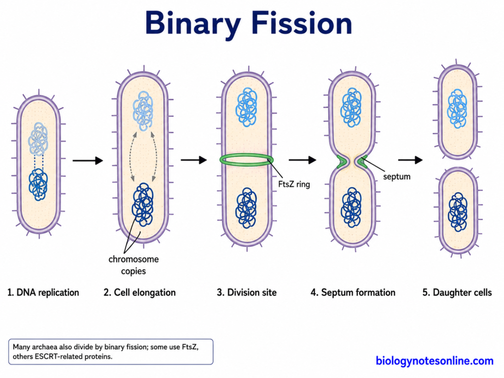
Prokaryotic cells reproduce mainly by asexual cell division. Binary fission is the common method in bacteria and is also found in many Archaea. The division machinery however is not same in every prokaryotic group.
A. Binary Fission
During binary fission, one parental cell divides to produce two daughter cells. The major steps are-
- The chromosomal DNA is first replicated. Replication produces two copies of the chromosome, which are required for the developing daughter cells.
- The cell increases in size and becomes elongated. New membrane and cell material are also formed during this period.
- The replicated chromosomes become separated toward different regions of the cell. Chromosome segregation often begins while DNA replication is still going on, rather than only after replication has completely finished.
- A division site is now established between the two chromosome regions. In many bacteria, FtsZ forms a ring-like structure at this site and different division proteins are assembled. New septal peptidoglycan is synthesized and the membrane begins to constrict.
- The septum becomes completed and the parental cell separates into two daughter cells. Each normally receives a copy of the chromosome. The two cells are genetically very similar, except for changes produced by mutation or other genetic processes.
Many Archaea also divide by binary fission. Some use an FtsZ-based system, whereas other archaeal groups use different machinery such as ESCRT-related proteins. Thus, the bacterial mechanism is not universal for all prokaryotic cells.
B. Horizontal Gene Transfer
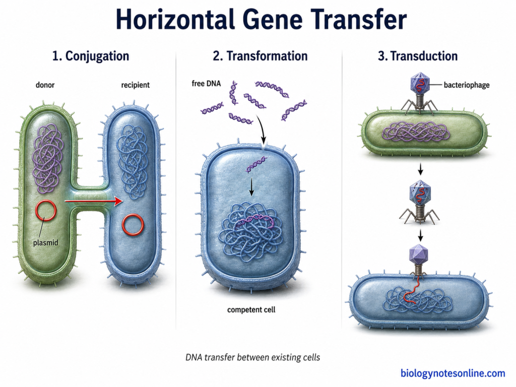
Genetic variation can also arise by movement of DNA from one cell to another. This process is called horizontal gene transfer (HGT). Conjugation, transformation and transduction are the three classical mechanisms in bacteria. These are not methods of reproduction, because transfer of DNA itself does not produce new daughter cells.
1. Conjugation
- During conjugation, DNA is transferred from a donor cell to a recipient by direct cell-to-cell contact.
- Plasmids are commonly transferred by this process. Conjugative systems establish contact between cells and transfer DNA from one cell to another.
- The recipient can therefore obtain new genetic characters without being produced as a new cell.
2. Transformation
- Transformation involves uptake of free or extracellular DNA from the surrounding environment.
- DNA released from other cells can be taken up by a bacterial cell when it is in a physiologically competent condition.
- The incoming DNA may become incorporated into the chromosome by recombination, or in some cases remain as an independently replicating genetic element. This provides another source of genetic variation.
3. Transduction
- Transduction is the transfer of bacterial DNA from one cell to another with the help of a bacteriophage.
- During infection, a bacteriophage can carry bacterial genetic material from a previous host and introduce it into another bacterial cell.
- The transferred DNA may then recombine with DNA of the recipient. Thus, bacteriophages can take part in horizontal movement of genes between bacteria.
Metabolism and Energy Production of Prokaryotic Cells
Prokaryotic cells have different types of metabolism for the production of energy. The source of energy is not same in all members. Some use organic compounds, some utilize light while others obtain energy by oxidation of inorganic substances. ATP (Adenosine Triphosphate) is formed during these processes.
Some of the important points about metabolism and energy production in prokaryotic cells are-
- Metabolic diversity- The mode of energy production is highly variable among prokaryotes. Bacteria and Archaea include organisms using organic compounds, inorganic compounds or light as their energy source. Thus, one type of metabolism is not present in all members.
- Aerobic respiration- Aerobic respiration takes place when oxygen (O₂) is used as the terminal electron acceptor. In this respiration, electrons pass through a membrane electron transport system. Energy is released during the process and is used for the formation of an ion gradient, which finally helps in ATP production.
- Anaerobic respiration- Oxygen is not used as the final electron acceptor. Instead, nitrate, sulfate, fumarate and several other substances can act as electron acceptors depending upon the organism. An electron transport chain is still involved in this respiration.
- Fermentation- In this process, an external terminal electron acceptor is not used. ATP is formed mainly by substrate-level phosphorylation. The end products are different in different fermentative prokaryotes and the energy yield is generally lower than respiration.
- Photosynthesis- Some prokaryotic groups can obtain energy from light. Cyanobacteria carry out oxygenic photosynthesis in which water acts as an electron donor and oxygen is released. Anoxygenic photosynthetic bacteria are also present. Oxygen is not produced in this type of photosynthesis.
- Chemolithotrophy- The major source of energy is the oxidation of reduced inorganic compounds. Hydrogen, ammonia, nitrite, reduced sulfur compounds and ferrous iron are some of the electron donors used by different members. Many chemolithotrophic prokaryotes also use CO₂ as their carbon source.
- ATP generation- ATP is formed by different mechanisms. During fermentation and some metabolic reactions, it is produced by substrate-level phosphorylation. In respiration, most ATP is formed with the help of an electrochemical ion gradient and ATP synthase. Phototrophic prokaryotes can also generate ATP during light-driven electron transport.
- Cytoplasmic membrane- Prokaryotic cells do not possess mitochondria. The respiratory electron transport components in bacteria are present in the cytoplasmic membrane, instead of an inner mitochondrial membrane. During this process, movement of electrons is coupled with transport of protons or other ions across the membrane, producing an electrochemical gradient.
- ATP without mitochondria- The protons move back across the cytoplasmic membrane through ATP synthase. Energy of this movement is used to synthesize ATP from ADP and inorganic phosphate. This is referred to as oxidative phosphorylation. Thus, prokaryotes can make ATP without mitochondria because the cytoplasmic membrane carries the membrane-associated processes required for respiratory energy production.
Do Prokaryotic Cells Have Organelles?
Prokaryotic cells do not contain the common membrane-bound cell organelles that are found in eukaryotic cells. A true nucleus is absent. Mitochondria, endoplasmic reticulum (ER), Golgi apparatus and chloroplasts are also not present. This is the general cellular organization of prokaryotic cells.
Ribosomes are present. These are non-membranous cellular structures made up of rRNA and proteins and are used for protein synthesis. In bacterial cells, 70S ribosomes occur throughout the cytoplasm. They are not attached with any rough endoplasmic reticulum like those present in eukaryotic cells.
Apart from these, some bacterial cells also possess specialized compartments inside the cell. These are now referred to as bacterial organelles in different modern cell biology studies. The structure is not same in all. Some are surrounded by lipid membrane while some others are covered by a protein shell.
Carboxysomes are protein-bounded microcompartments present in cyanobacteria and different autotrophic bacteria. They contain RuBisCO and carbonic anhydrase, which are involved in carbon fixation.
Magnetosomes are another type of specialized bacterial structure. These are present in magnetotactic bacteria and have magnetic mineral crystals enclosed by a lipid membrane. The crystals are commonly magnetite or greigite. Magnetosomes are used by these bacteria for orientation according to magnetic field.
Prokaryotic vs Eukaryotic Cells
The major differences between prokaryotic cells and eukaryotic cells are-
| Characteristics | Prokaryotic Cells | Eukaryotic Cells |
|---|---|---|
| Cell type | Simple type of cellular organization. | More complex cellular organization is present. |
| Occurrence | Found in Bacteria and Archaea. | Found in animals, plants, fungi and protists. |
| Cell size | Generally smaller. Most bacterial cells occur in micrometre range. | Usually larger than prokaryotic cells, although size varies greatly. |
| Nucleus | A true membrane-bound nucleus is absent. | A true membrane-bound nucleus is present. |
| Nuclear envelope | Not present around the genetic material. | Nuclear envelope surrounds the nuclear material. |
| Nucleoid | Major DNA remains in a region called the nucleoid. | Nucleoid is absent. DNA is mainly enclosed within nucleus. |
| Chromosomes | In many bacteria, the major chromosome is circular. Linear chromosomes and other arrangements also occur in some prokaryotes. | Nuclear chromosomes are generally linear and more than one chromosome is commonly present. |
| Histones | Typical bacterial chromosomes do not use eukaryotic-type histones. Many Archaea however possess histones or related DNA-binding proteins. | DNA is associated with histone proteins to form chromatin. |
| Plasmids | Plasmids are common in many prokaryotic organisms, but not present in every cell. | Plasmids are uncommon in most eukaryotic cells, although they occur naturally in some eukaryotes. |
| Membrane-bound organelles | Typical membrane-bound organelles are absent. Some prokaryotes however contain specialized intracellular compartments. | Different membrane-bound organelles are present. |
| Mitochondria | Absent. | Present in most eukaryotic cells, with some specialized exceptions. |
| Endoplasmic reticulum | Absent. | Endoplasmic reticulum (ER) is present. |
| Golgi apparatus | Absent. | Present. |
| Chloroplasts | Chloroplasts are absent. Photosynthetic prokaryotes perform photosynthesis without chloroplasts. | Present in plants and many photosynthetic eukaryotes. |
| Ribosomes | Cytoplasmic ribosomes are generally 70S. | Cytoplasmic ribosomes are mainly 80S. |
| Ribosomal subunits | 70S ribosome consists of 30S and 50S subunits. | 80S cytoplasmic ribosome consists of 40S and 60S subunits. |
| Cell wall | Present in most bacteria and many Archaea. Composition is different between the two domains. | Present in plants, fungi and some other eukaryotes, but absent in animal cells. |
| Peptidoglycan | Present in most bacterial cell walls. Typical peptidoglycan is absent in Archaea. | Peptidoglycan is absent. |
| Plasma membrane | Present around the cytoplasm. | Present around the cell. |
| Membrane lipids | Bacteria mainly have ester-linked fatty acid lipids. Archaea characteristically contain ether-linked isoprenoid lipids. | Membranes mainly contain ester-linked fatty acid phospholipids. |
| Cytoplasm | Present. Typical eukaryotic membrane-bound organelles are not present within it. | Cytoplasm contains different membrane-bound organelles and cytoskeletal structures. |
| Cytoskeleton | Cytoskeletal proteins are present, but the organization differs among different prokaryotic groups. | Well-developed cytoskeleton containing microtubules, microfilaments and intermediate filaments. |
| Cell division | Commonly takes place by binary fission. Other division mechanisms also occur in some groups. | Cell division generally takes place by mitosis. |
| Sexual reproduction | True eukaryotic-type sexual reproduction with meiosis and gamete fusion is absent. | Sexual reproduction involving meiosis and fertilization occurs in many eukaryotes. |
| DNA replication | Takes place in cytoplasmic nucleoid region. | Nuclear DNA replication takes place inside nucleus. |
| Transcription | Takes place in the cytoplasm or nucleoid region. | Nuclear transcription takes place inside nucleus. |
| Translation | Takes place in cytoplasm. In many bacteria, translation can begin while transcription is still taking place. | Cytoplasmic translation is spatially separated from nuclear transcription. |
| Operons | Operons are common in bacteria and also occur in Archaea. | Operon-like arrangements are uncommon in most eukaryotes, although they occur in some groups. |
| Introns | Less common in bacterial protein-coding genes. Introns and intron-related elements are present in different Archaea. | Introns are common in many eukaryotic genes. |
| Flagella | Bacterial flagella are rotary structures. Archaeal archaella are different in structure and evolution. | Eukaryotic flagella have a microtubule-based structure and move by bending. |
| Fimbriae and pili | Present in many bacterial cells. | Typical bacterial fimbriae and pili are absent. |
| Endospores | Highly resistant endospores are produced by certain bacteria. | Bacterial-type endospores are absent. |
| Respiration | Respiratory electron transport commonly occurs on the cytoplasmic membrane. | Aerobic respiratory electron transport mainly takes place in the inner mitochondrial membrane. |
| ATP production | ATP is formed by substrate-level phosphorylation and, in respiratory cells, membrane-associated oxidative phosphorylation. | ATP is produced by different processes, with oxidative phosphorylation mainly occurring in mitochondria. |
| Photosynthesis | Some bacteria perform photosynthesis using specialized internal or plasma membrane systems. | Photosynthesis takes place in chloroplasts of plants and many algae. |
| Level of organization | Mostly unicellular forms. Colonies and multicellular-like arrangements also occur in some groups. | Unicellular as well as highly organized multicellular organisms are present. |
| Examples | Escherichia coli, Bacillus subtilis, cyanobacteria and different Archaea. | Amoeba, yeast, plant cells and animal cells. |
Examples of Prokaryotic Cells
The following are some of the examples of prokaryotic cells–
Bacterial Examples
- Escherichia coli- Escherichia coli is a rod-shaped bacterium commonly found in intestinal tract of humans and other animals. Most strains remain harmless in the intestine. Some strains however can cause intestinal and other infections.
- Bacillus subtilis- It is a Gram-positive rod-shaped bacterium. Bacillus subtilis is commonly found in soil and is used widely for study of bacterial cells. Endospore is formed during unfavourable conditions.
- Staphylococcus aureus- The cells are Gram-positive and coccal in shape. They generally occur in irregular clusters. Staphylococcus aureus can remain on skin and nasal region of humans, and some strains cause different infections such as skin infection and abscess.
- Vibrio cholerae- Vibrio cholerae is a curved rod-shaped Gram-negative bacterium. The cells are motile. Certain strains produce cholera in humans.
- Cyanobacteria- These are photosynthetic members of Bacteria. Members contain chlorophyll and perform oxygenic photosynthesis. Oxygen is released during this process. They do not possess chloroplasts.
- Mycoplasma spp.- Members are very small bacterial forms and a cell wall is absent. Due to absence of rigid cell wall, the cells show variable shapes. Some Mycoplasma species are pathogenic in humans and animals.
Archaeal Examples
- Methanogens- These members produce methane during their metabolism. They are anaerobic forms in general and are found in wetlands, sediments and digestive tract of different animals. Methanobrevibacter smithii is an example found in human intestine.
- Halophilic Archaea- Members grow in environments having very high salt concentration. Halobacterium salinarum is one of the common examples. The organism is found in highly saline environments.
- Thermophilic Archaea- These Archaea grow at high temperatures. Some members can also grow under acidic conditions. Sulfolobus acidocaldarius is a thermoacidophilic archaeal example.
Functions and Biological Importance of Prokaryotes
Prokaryotes have several important functions in the environment and for other living organisms. Some are decomposers, some are involved in different nutrient cycles, while different members are also producers and symbiotic forms.
Ecological Importance
Some of the important ecological functions of prokaryotes are-
- Decomposition- Dead plants, animals and different organic substances are decomposed by many bacterial members into simpler materials. During this process, nutrients again become available in soil and water.
- Nutrient cycling- Prokaryotes are involved in cycling of carbon, nitrogen, phosphorus, sulfur and other elements. Different members carry out different reactions of these cycles and the same group is not involved in every process.
- Carbon cycle- Carbon dioxide is fixed by photosynthetic and chemoautotrophic prokaryotes into organic compounds. On the other hand, decomposition and respiration again release carbon dioxide. Methanogenic Archaea produce methane under anaerobic condition. Thus, different forms occur at different steps of carbon cycle.
- Nitrogen cycle- It is one of the important cycles where prokaryotes have a major role. Nitrogen fixation, nitrification, ammonia oxidation and denitrification are carried out by different bacterial and archaeal groups. Atmospheric nitrogen is also converted into biologically available form by nitrogen-fixing members.
- Primary production- Cyanobacteria are photosynthetic bacteria and produce organic matter using light energy. Oxygen is released. Chemoautotrophic Bacteria and Archaea can also produce organic substances by using energy obtained from oxidation of inorganic compounds.
- Oxygen production- Cyanobacteria carry out oxygenic photosynthesis. They are important producers in many aquatic environments and also contribute to oxygen production.
- Symbiotic relationships- Many bacterial and archaeal members remain associated with other organisms. Rhizobium forms nodules in roots of leguminous plants and fixes atmospheric nitrogen, which is then available for plant metabolism. Different prokaryotes are also found in digestive tract of animals and perform various functions there.
Importance to Humans
The following are some of the important roles of prokaryotes for humans-
- Human microbiome- Bacteria are present normally on skin, mouth, intestine and several other regions of human body. The intestinal members help in digestion of different food substances and production of some vitamins. They also interact with immune system. Methanogenic Archaea are also present in human intestine.
- Food production- Different bacterial forms are used in fermented foods. Lactic acid bacteria are used in production of yogurt, cheese and other fermented products, where acids and other compounds are formed during fermentation.
- Biotechnology- Bacteria are widely used for production of enzymes, recombinant proteins and several useful compounds. Escherichia coli is one of the common organisms used in recombinant DNA technology. Some archaeal enzymes are also used because they can remain active under high temperature and other extreme conditions.
- Agriculture- Soil prokaryotes decompose organic matter and make different nutrients available to plants. Some members also remain associated with roots. Their activities are important for soil fertility and plant growth.
- Nitrogen fixation- Atmospheric nitrogen (N₂) cannot be directly used by most plants. Nitrogen-fixing prokaryotes convert it into ammonia during this process. Rhizobium and related bacteria associated with legume roots are some of the important agricultural examples.
- Disease-causing forms- Some bacterial species are pathogenic. Mycobacterium tuberculosis, Vibrio cholerae and Staphylococcus aureus are some examples. Many bacterial members however do not cause disease and several are beneficial.
Frequently Asked Questions (FAQs)
1. What is a prokaryotic cell?
A prokaryotic cell is a cell which does not contain a true membrane-bound nucleus. The genetic material remains inside the cell without a nuclear envelope. Bacteria and Archaea have this type of cellular organization.
2. What are the main characteristics of prokaryotic cells?
Prokaryotic cells are generally small and mostly unicellular. A membrane-bound nucleus and typical membrane-bound cell organelles are absent. They contain 70S ribosomes, DNA within a nucleoid region and a plasma membrane.
3. What organisms have prokaryotic cells?
The organisms belonging to Domain Bacteria and Domain Archaea have prokaryotic cells. They are two different evolutionary groups.
4. What are the main parts of a prokaryotic cell?
The major parts include plasma membrane, cytoplasm, nucleoid, ribosomes and usually a cell wall. Plasmids, capsule, pili, flagella and different inclusions may also be present in some forms.
5. Do prokaryotic cells have a nucleus?
No true membrane-bound nucleus is present. The DNA remains within a cytoplasmic region called the nucleoid.
6. Where is DNA located in a prokaryotic cell?
The major chromosomal DNA is present in the nucleoid region. No nuclear membrane surrounds it. Plasmid DNA may also occur separately in many prokaryotic cells.
7. Do prokaryotic cells have mitochondria?
Prokaryotic cells do not possess mitochondria. In respiratory bacteria, many of the reactions associated with electron transport and ATP formation take place at the cytoplasmic membrane.
8. Do prokaryotic cells have membrane-bound organelles?
The typical membrane-bound organelles of eukaryotic cells such as mitochondria, Golgi apparatus and ER are absent. Some prokaryotes however possess specialized intracellular compartments, including magnetosomes and carboxysomes.
9. Do prokaryotic cells have ribosomes?
Yes. Ribosomes are present in the cytoplasm and are used for protein synthesis. They are non-membranous structures.
10. What type of ribosomes are found in prokaryotic cells?
Prokaryotic cytoplasmic ribosomes are generally 70S ribosomes. They consist of a 30S smaller subunit and a 50S larger subunit.
11. Do all prokaryotic cells have a cell wall?
No. Most bacteria contain a cell wall having peptidoglycan, but wall-less bacteria such as Mycoplasma are also present. Archaeal walls are different and do not contain typical bacterial peptidoglycan.
12. What is the difference between bacteria and archaea?
Both are prokaryotic but they are different groups. Bacteria generally have ester-linked membrane lipids and most contain peptidoglycan walls. Archaea mainly possess ether-linked isoprenoid membrane lipids and typical bacterial peptidoglycan is absent.
13. How do prokaryotic cells reproduce?
Binary fission is the common method of reproduction. Chromosomal DNA is replicated, the cell increases in size and finally separates into daughter cells. Conjugation, transformation and transduction transfer genetic material but are not methods of reproduction.
14. What are some examples of prokaryotic cells?
Escherichia coli, Bacillus subtilis, Staphylococcus aureus, cyanobacteria and Mycoplasma are bacterial examples. Methanogens, halophilic Archaea and thermophilic Archaea are other prokaryotic forms.
15. What is the difference between prokaryotic and eukaryotic cells?
Prokaryotic cells lack a membrane-bound nucleus and the common membrane-bound organelles. Eukaryotic cells possess a true nucleus and organelles such as mitochondria, ER and Golgi apparatus. Prokaryotic cells are also generally smaller.
16. How do prokaryotic cells produce ATP without mitochondria?
Mitochondria are not required for ATP production in prokaryotes. In respiratory bacteria, electron transport components are present in the cytoplasmic membrane. An ion gradient is produced across this membrane and ATP synthase uses the gradient for formation of ATP.
References
- Armache, J.-P., Anger, A. M., Márquez, V., Franckenberg, S., Fröhlich, T., Villa, E., Berninghausen, O., Thomm, M., Arnold, G. J., Beckmann, R., & Wilson, D. N. (2013). Promiscuous behaviour of archaeal ribosomal proteins: Implications for eukaryotic ribosome evolution. Nucleic Acids Research, 41(2), 1284–1293. https://doi.org/10.1093/nar/gks1259
- Baron, S. (Ed.). (1996). Medical microbiology (4th ed.). University of Texas Medical Branch at Galveston. https://www.ncbi.nlm.nih.gov/books/NBK7627/
- Beeby, M., & Daum, B. (2025). How does the archaellum work? Biomolecules, 15(4), 465. https://doi.org/10.3390/biom15040465
- Beh, J. Q., Wick, R. R., Howden, B. P., Connor, C. H., & Webb, J. R. (2025). Challenges and considerations for whole-genome-based antimicrobial resistance plasmid investigations. Antimicrobial Agents and Chemotherapy, 69(12), e01097-25. https://doi.org/10.1128/aac.01097-25
- Beskrovnaya, P., Sexton, D. L., Golmohammadzadeh, M., Hashimi, A., & Tocheva, E. I. (2021). Structural, metabolic and evolutionary comparison of bacterial endospore and exospore formation. Frontiers in Microbiology, 12, 630573. https://doi.org/10.3389/fmicb.2021.630573
- Buckel, W. (2021). Energy conservation in fermentations of anaerobic bacteria. Frontiers in Microbiology, 12, 703525. https://doi.org/10.3389/fmicb.2021.703525
- Burkhardt, D. H., Rouskin, S., Zhang, Y., Li, G.-W., Weissman, J. S., & Gross, C. A. (2017). Operon mRNAs are organized into ORF-centric structures that predict translation efficiency. eLife, 6, e22037. https://doi.org/10.7554/eLife.22037
- Cannon, G. C., Bradburne, C. E., Aldrich, H. C., Baker, S. H., Heinhorst, S., & Shively, J. M. (2001). Microcompartments in prokaryotes: Carboxysomes and related polyhedra. Applied and Environmental Microbiology, 67(12), 5351–5361. https://doi.org/10.1128/AEM.67.12.5351-5361.2001
- Clark, M. A., Douglas, M., & Choi, J. (2018). Biology 2e. OpenStax. https://openstax.org/books/biology-2e/pages/1-introduction
- Geerlings, N. M. J., Kienhuis, M. V. M., Hidalgo-Martinez, S., Hageman, R., Vasquez-Cardenas, D., Middelburg, J. J., Meysman, F. J. R., & Polerecky, L. (2022). Polyphosphate dynamics in cable bacteria. Frontiers in Microbiology, 13, 883807. https://doi.org/10.3389/fmicb.2022.883807
- Greening, C., & Lithgow, T. (2020). Formation and function of bacterial organelles. Nature Reviews Microbiology, 18(12), 677–689. https://doi.org/10.1038/s41579-020-0413-0
- Guo, H., Suzuki, T., & Rubinstein, J. L. (2019). Structure of a bacterial ATP synthase. eLife, 8, e43128. https://doi.org/10.7554/eLife.43128
- Higgins, N. P. (2016). Species-specific supercoil dynamics of the bacterial nucleoid. Biophysical Reviews, 8(Suppl. 1), 113–121. https://doi.org/10.1007/s12551-016-0207-9
- Hoshino, T., & Inagaki, F. (2019). Abundance and distribution of Archaea in the subseafloor sedimentary biosphere. The ISME Journal, 13(1), 227–231. https://doi.org/10.1038/s41396-018-0253-3
- Jogler, C., & Schüler, D. (2009). Genomics, genetics, and cell biology of magnetosome formation. Annual Review of Microbiology, 63, 501–521. https://doi.org/10.1146/annurev.micro.62.081307.162908
- Jurtshuk, P., Jr. (1996). Bacterial metabolism. In S. Baron (Ed.), Medical microbiology (4th ed.). University of Texas Medical Branch at Galveston. https://www.ncbi.nlm.nih.gov/books/NBK7919/
- Khadka, S., Kinney, E. L., Ryan, B. E., & Mike, L. A. (2025). Mechanisms governing bacterial capsular polysaccharide attachment and chain length. Annals of the New York Academy of Sciences, 1548(1), 80–98. https://doi.org/10.1111/nyas.15364
- Krawczyk, S. J., Leśniczak-Staszak, M., Gowin, E., & Szaflarski, W. (2024). Mechanistic insights into clinically relevant ribosome-targeting antibiotics. Biomolecules, 14(10), 1263. https://doi.org/10.3390/biom14101263
- Levin, P. A., & Angert, E. R. (2015). Small but mighty: Cell size and bacteria. Cold Spring Harbor Perspectives in Biology, 7(7), a019216. https://doi.org/10.1101/cshperspect.a019216
- Lucius, S., & Hagemann, M. (2024). The primary carbon metabolism in cyanobacteria and its regulation. Frontiers in Plant Science, 15, 1417680. https://doi.org/10.3389/fpls.2024.1417680
- Maguire, B. A. (2009). Inhibition of bacterial ribosome assembly: A suitable drug target? Microbiology and Molecular Biology Reviews, 73(1), 22–35. https://doi.org/10.1128/MMBR.00030-08
- Mahone, C. R., & Goley, E. D. (2020). Bacterial cell division at a glance. Journal of Cell Science, 133(7), jcs237057. https://doi.org/10.1242/jcs.237057
- Mencía, M. (2020). The archaeal-bacterial lipid divide, could a distinct lateral proton route hold the answer? Biology Direct, 15, 7. https://doi.org/10.1186/s13062-020-00262-7
- Mousavi, S. A., Willems, A., Nesme, X., de Lajudie, P., & Lindström, K. (2020). Revised phylogeny of Rhizobiaceae: Proposal of the delineation of Pararhizobium gen. nov., and 13 new species combinations. Systematic and Applied Microbiology, 43(2), 126046. https://doi.org/10.1016/j.syapm.2019.126046
- Parker, N., Schneegurt, M., Thi Tu, A.-H., Forster, B. M., & Lister, P. (2016). Microbiology. OpenStax. https://openstax.org/books/microbiology/pages/1-introduction
- Pearson, A. (2019). Resolving a piece of the archaeal lipid puzzle. Proceedings of the National Academy of Sciences of the United States of America, 116(45), 22423–22425. https://doi.org/10.1073/pnas.1916583116
- Pende, N., Sogues, A., Megrian, D., Sartori-Rupp, A., England, P., Palabikyan, H., Rittmann, S. K.-M. R., Graña, M., Wehenkel, A. M., Alzari, P. M., & Gribaldo, S. (2021). SepF is the FtsZ anchor in archaea, with features of an ancestral cell division system. Nature Communications, 12, 3214. https://doi.org/10.1038/s41467-021-23099-8
- Piepenbrink, K. H., & Sundberg, E. J. (2016). Motility and adhesion through type IV pili in Gram-positive bacteria. Biochemical Society Transactions, 44(6), 1659–1666. https://doi.org/10.1042/BST20160221
- Royzenblat, S. K., & Freddolino, P. L. (2024). Spatio-temporal organization of the E. coli chromosome from base to cellular length scales. EcoSal Plus, 12(1), eESP-0001-2022. https://doi.org/10.1128/ecosalplus.esp-0001-2022
- Singh, P. K., Little, J., & Donnenberg, M. S. (2022). Landmark discoveries and recent advances in type IV pilus research. Microbiology and Molecular Biology Reviews, 86(3), e00076-22. https://doi.org/10.1128/mmbr.00076-22
- Vasconcelos, D., Schuster, B., & Kyaw, C. M. (2018). Archaeal S-layers: Overview and current state of the art. Frontiers in Microbiology, 8, 2597. https://doi.org/10.3389/fmicb.2017.02597
- van Wolferen, M., Pulschen, A. A., Baum, B., Gribaldo, S., & Albers, S.-V. (2022). The cell biology of archaea. Nature Microbiology, 7(11), 1744–1755. https://doi.org/10.1038/s41564-022-01215-8
- Volland, J.-M., Gonzalez-Rizzo, S., Gros, O., Tyml, T., Ivanova, N., Schulz, F., Goudeau, D., Elisabeth, N. H., Nath, N., Udwary, D., Malmstrom, R. R., Guidi-Rontani, C., Bolte-Kluge, S., Davies, K. M., Jean, M. R., Mansot, J.-L., Mouncey, N. J., Angert, E. R., Woyke, T., & Date, S. V. (2022). A centimeter-long bacterium with DNA contained in metabolically active, membrane-bound organelles. Science, 376(6600), 1453–1458. https://doi.org/10.1126/science.abb3634
- Walsby, A. E. (1994). Gas vesicles. Microbiological Reviews, 58(1), 94–144. https://doi.org/10.1128/mr.58.1.94-144.1994
- Wang, S., Walker, R., Schicklberger, M., Nico, P. S., Fox, P. M., Karaoz, U., Chakraborty, R., & Brodie, E. L. (2021). Microbial phosphorus mobilization strategies across a natural nutrient limitation gradient and evidence for linkage with iron solubilization traits. Frontiers in Microbiology, 12, 572212. https://doi.org/10.3389/fmicb.2021.572212
- Weixlbaumer, A., Grünberger, F., Werner, F., & Grohmann, D. (2021). Coupling of transcription and translation in Archaea: Cues from the bacterial world. Frontiers in Microbiology, 12, 661827. https://doi.org/10.3389/fmicb.2021.661827
- Woodgate, J., & Zenkin, N. (2023). Transcription–translation coupling: Recent advances and future perspectives. Molecular Microbiology, 120(4), 539–546. https://doi.org/10.1111/mmi.15076
- Woese, C. R., Kandler, O., & Wheelis, M. L. (1990). Towards a natural system of organisms: Proposal for the domains Archaea, Bacteria, and Eucarya. Proceedings of the National Academy of Sciences of the United States of America, 87(12), 4576–4579. https://doi.org/10.1073/pnas.87.12.4576
- Wu, Z., Liu, J., Yang, H., & Xiang, H. (2014). DNA replication origins in archaea. Frontiers in Microbiology, 5, 179. https://doi.org/10.3389/fmicb.2014.00179