

An endospore is a dormant, highly resistant and non-reproductive structure formed inside the cell of certain bacteria. It is formed for survival of the bacterial cell during unfavourable conditions, and not for reproduction.
Endospores are mostly formed by some members of Bacillota, especially the genera Bacillus and Clostridium. Some other related bacterial genera also have this ability. But every Gram-positive bacterium does not form an endospore.
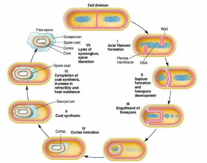
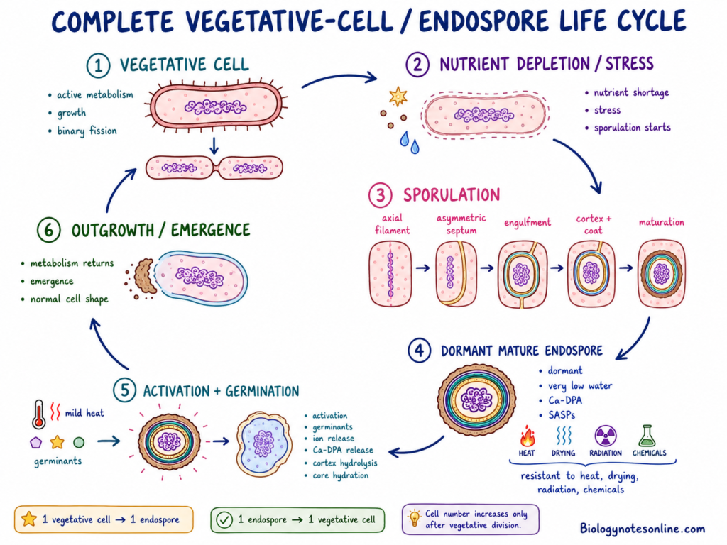
The process of endospore formation is referred to as sporulation. It generally begins during deficiency of nutrients or other unfavourable growth conditions. During this process, unequal division of the bacterial cell takes place and a smaller forespore is formed inside the mother cell. The forespore later develops different protective layers and forms the mature endospore.
The mature endospore remains in a dormant condition with very little metabolic activity. It is highly resistant to heat, drying, radiation and many chemical agents as compared to the vegetative bacterial cell. One bacterial cell generally forms one endospore. Thus, this process does not increase the number of bacterial cells.
When suitable nutrient, water and other growth conditions are again available, the dormant endospore changes into an active vegetative cell. This process is known as germination. After germination, outgrowth takes place and the vegetative bacterial cell again starts its normal growth and multiplication.
What is an Endospore?
An endospore is a dormant, non-reproductive and highly resistant structure formed inside certain bacteria. It protects the bacterial genome and essential cellular components during conditions unsuitable for active growth.
The term endospore has the word “endo”, which means within or inside. Thus, the endospore is formed internally within the bacterial cell and not outside the cell.
During favourable growth condition, the bacterium remains in its vegetative state. It is the active form of bacterial cell, which carries out metabolism, growth and cell division. When the environmental condition becomes unsuitable, certain bacterial cells can form an endospore for their survival.
One vegetative bacterial cell generally produces one endospore. After germination, this endospore again gives rise to one vegetative cell. Therefore, endospore formation does not increase the number of bacterial cells and it is not a reproductive process.
The bacterial endospore is different from the spores formed by fungi. Fungal spores are reproductive units and may be formed by sexual or asexual process. The bacterial endospore is mainly a survival structure, which protects the cell during heat, drying, lack of nutrients and other harmful conditions.

Characteristics of Bacterial Endospores
The following are the important characteristics of bacterial endospores–
- Formation– Endospore is formed inside a vegetative bacterial cell. The cell containing the developing spore is known as the mother cell or sporangium.
- Number– Usually, one bacterial cell produces one endospore. It does not form many spores from a single cell.
- Survival– The major function of an endospore is survival during unfavourable condition. It is not used for reproduction.
- Dormancy– Mature endospore remains in metabolically dormant condition. It has no detectable metabolic activity during this state.
- Core water– The core contains very low amount of water as compared to vegetative bacterial cell. This condition is important for its dormancy and heat resistance.
- Resistance– Endospores show greater resistance than the vegetative cells. They can resist heat, drying, radiation and many chemical agents.
- Division– A dormant endospore does not grow or divide. Therefore, its formation does not increase the number of bacterial cells.
- Core contents– The spore core contains DNA, ribosomes and different essential enzymes. These are used again during formation of vegetative cell.
- Refractility– Mature endospores are highly refractile under phase-contrast microscope. They generally appear bright or phase-bright.
- Staining– Endospores are not stained clearly by ordinary bacterial staining methods. Special endospore staining is required for their clear observation under light microscope.
- Germination– During favourable environmental condition, the dormant endospore undergoes germination. It is followed by outgrowth and development of an active vegetative bacterial cell.
Which Bacteria Form Endospores?
Endospore formation is found in some bacteria of the phylum Bacillota (Firmicutes). Bacillus and Clostridium are the major genera. The ability is also present in some other related bacterial forms. Every Gram-positive bacteria does not have this property.
The following are some important endospore-forming bacteria-
- Bacillus– It is the best-known aerobic or facultatively anaerobic spore-forming genus. Bacillus subtilis, Bacillus cereus and Bacillus anthracis are included in this group.
- Clostridium– Most of the members grow under anaerobic condition and produce resistant endospores. Some important forms include Clostridium tetani, Clostridium botulinum and Clostridium perfringens.
- Clostridioides– Clostridioides difficile is an anaerobic spore-forming bacterium. Earlier, this organism was placed under the genus Clostridium.
- Paenibacillus– Several soil and plant-associated bacteria are present in this genus. Endospore formation is found in species such as Paenibacillus polymyxa and Paenibacillus larvae.
- Geobacillus– Thermophilic bacteria are included under this genus, which can grow at higher temperature. Geobacillus stearothermophilus is an important spore-forming species.
- Alicyclobacillus– These bacteria can tolerate acidic condition. Their spores may survive heating and some species are related with spoilage of fruit juices.
- Brevibacillus– This genus was previously included within Bacillus. Several members have the ability of endospore formation.
- Lysinibacillus– Endospore-forming bacteria are also present in this genus. Lysinibacillus sphaericus is one commonly known species.
- Sporosarcina– Some species of Sporosarcina produce endospores and are commonly present in soil. The cells may occur in coccal or rod-like form depending on the species.
- Other genera– Some endospore-forming members are also present in Anoxybacillus, Desulfotomaculum, Moorella, Sporolactobacillus, Thermoanaerobacter and Thermoactinomyces.
Structure of a Bacterial Endospore
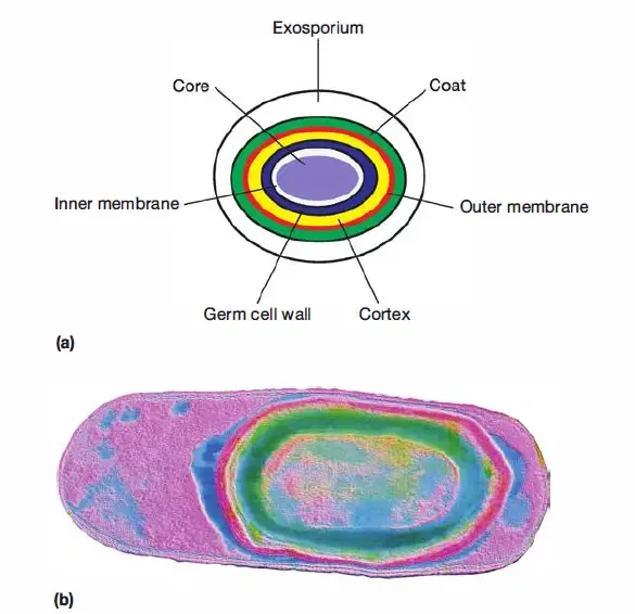
A bacterial endospore has a complex multilayered structure. The arrangement is not fully same in every species. From the inner region to outside, the important parts are as follows-
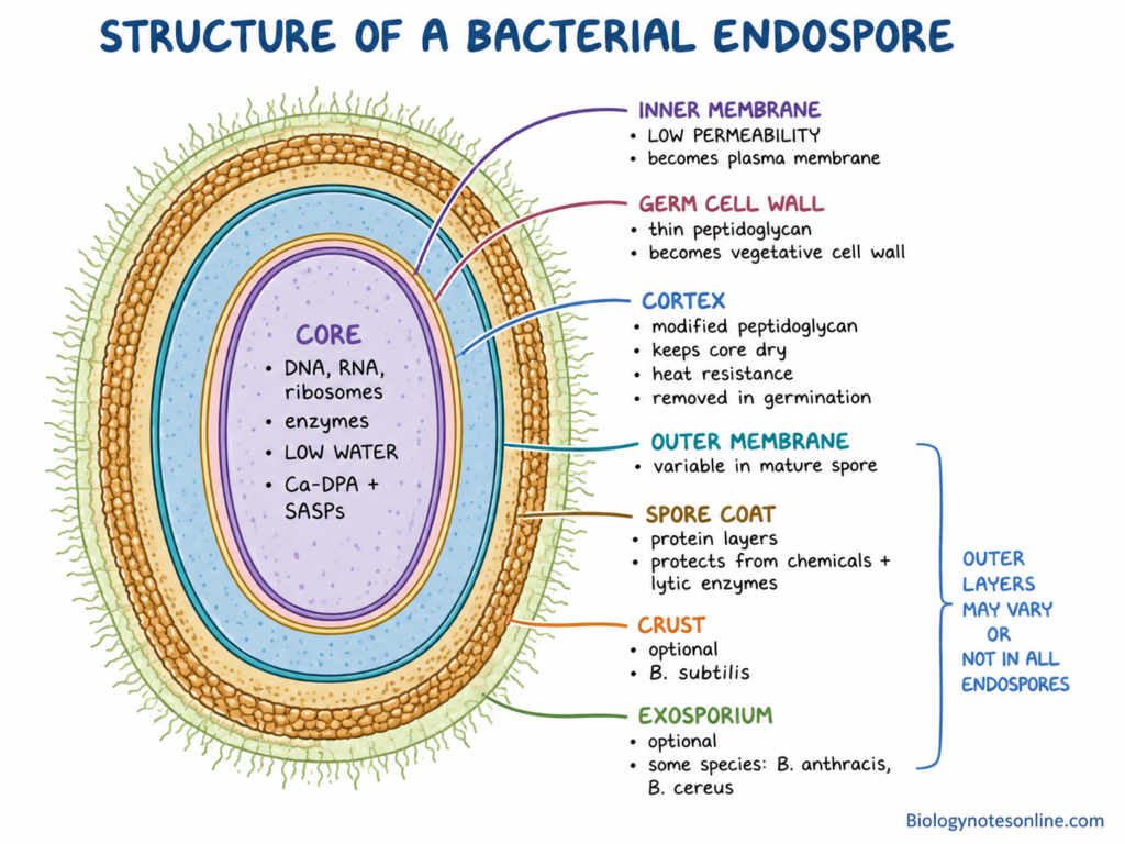
- Core– The central portion of endospore is referred to as the core. It contains bacterial DNA, RNA, ribosomes and different essential enzymes. Water content is very low in this region. Calcium-dipicolinate (Ca-DPA) and small acid-soluble spore proteins (SASPs) are also present. SASPs remain attached with the spore DNA and protect it from different damages.
- Inner membrane– It surrounds the spore core and is also known as the core membrane. This membrane has very low permeability to many chemicals. During germination, it develops into the plasma membrane of vegetative bacterial cell.
- Germ cell wall– A thin peptidoglycan layer is present just outside the inner membrane. It is similar to the cell wall of vegetative bacteria. After germination, the same layer becomes the cell wall of newly formed vegetative cell.
- Cortex– This is a thick layer of modified peptidoglycan. The cortex is present outside the germ cell wall. It helps in maintaining the low water content of core, which is important for dormancy and heat resistance. During germination, this layer is hydrolysed and removed.
- Outer membrane– The developing forespore becomes enclosed by an inner and an outer membrane during engulfment. Outer membrane lies between the cortex and spore coat. Its condition in fully mature endospores may differ and it is not always clearly retained.
- Spore coat– It is a thick protein covering present around the outer portions of endospore. Several protein layers can be present in the coat. These layers protect the spore from lytic enzymes and many toxic chemical agents. Coat organization shows variations between different spore-forming bacteria.
- Crust– In Bacillus subtilis, an outermost coat layer is known as the crust. It is present above the outer coat. The crust is not considered as a common structure of every bacterial endospore.
- Exosporium– Some bacterial endospores possess an additional loose outer covering called the exosporium. It is present in species such as Bacillus anthracis and Bacillus cereus. The exosporium has a basal layer and an outer hair-like region. Many endospores do not contain this layer.
Chemical Composition and Protective Components
Different chemical substances are present in the core, cortex, membranes and coat of bacterial endospore. Their amount is not same in all bacterial spores. The following are some important chemical and protective components-
- Core water– The amount of water is very low in the spore core. Due to this dehydrated condition, movement of the core molecules becomes reduced. It has an important role in dormancy and wet heat resistance.
- Calcium-dipicolinate– A large amount of dipicolinic acid (DPA) is present in combination with calcium ion. This complex is referred to as calcium-dipicolinate (Ca-DPA). It is accumulated inside the core and helps in core stability, dehydration and heat resistance.
- SASPs– Small acid-soluble spore proteins (SASPs) are found closely bound with the spore DNA. These proteins protect DNA from dry heat, ultraviolet radiation and different harmful chemicals. During germination, SASPs are degraded.
- Mineral ions– Calcium is the major mineral ion present with DPA. Some other divalent ions are also found in the core. The mineral content may change with bacterial species and condition of sporulation.
- Core materials– The core contains bacterial DNA, ribosomes and different essential enzymes. These remain in inactive condition within the dormant spore. They are again used when germination and outgrowth take place.
- Energy reserve– A large depot of 3-phosphoglyceric acid (3-PGA) may be present inside the core. It remains unused during dormancy. After germination, it is broken down for early formation of ATP.
- Cortex peptidoglycan– The cortex is made up of a special modified peptidoglycan. Muramic-δ-lactam is one of its characteristic component and peptide cross-linking is less. This layer maintains the dehydrated state of core. During germination, the cortex is hydrolysed.
- Coat proteins– Spore coat is formed mainly of proteins arranged in different layers. It prevents the entry of large molecules and gives protection from lytic enzymes and many chemical agents. Some coat proteins can also inactivate damaging chemicals.
- Inner membrane– It contains phospholipids and different membrane proteins. The lipids have very little mobility in dormant endospore. Its low permeability acts as an important barrier against entry of toxic chemicals into the core.
- Repair enzymes– Complete prevention of DNA damage is not possible under every condition. Spore photoproduct lyase and other DNA repair systems remove the damage during germination and early outgrowth.
Types of Endospores Based on Position and Morphology
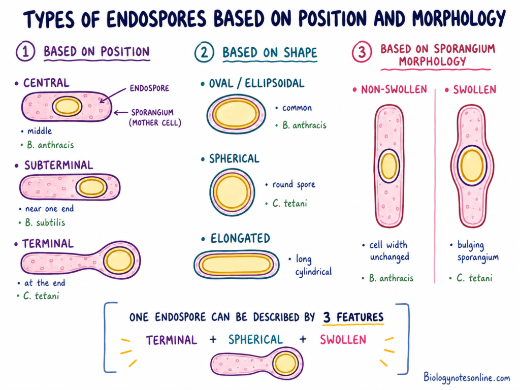
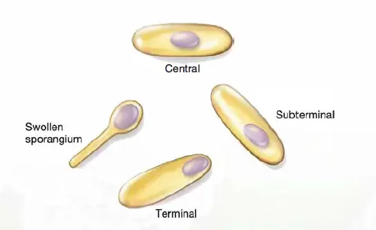
The position and shape of endospore are different among bacterial species. Swelling of the mother cell is also considered during the identification. The types are as follows-
Based on Position
- Central– The endospore remains around the middle region of bacterial cell. It may be oval or ellipsoidal. In Bacillus anthracis, the spore is generally central, but a subterminal position may also occur.
- Subterminal– It is present between the centre and terminal end. More close to one end, but not exactly at the pole. Bacillus subtilis commonly produces subterminal endospores.
- Terminal– Here, the endospore is located at the end of vegetative cell. A round terminal spore is present in Clostridium tetani. The end portion becomes swollen, producing a drumstick or tennis-racket appearance.
Based on Shape
- Oval– Oval or ellipsoidal endospores are found in many spore-forming bacteria. Bacillus anthracis produces an ellipsoidal spore. It does not generally cause swelling of the sporangium.
- Spherical– The spore is round or nearly round in shape. In Clostridium tetani, the spherical spore occurs terminally. A distinct bulge is formed at one end of the cell.
- Elongated– Some bacterial spores are longer and more cylindrical in appearance. This form is less common than oval or spherical endospores. Spore shape can differ between species and sometimes between strains.
Based on Sporangium Morphology
- Swollen– The diameter of endospore becomes greater than the width of mother cell. Due to this, swelling or bulging of the sporangium takes place. Terminal spherical spore of Clostridium tetani is one important example.
- Non-swollen– In this type, the endospore does not enlarge the mother cell. The original width of sporangium remains almost unchanged. Bacillus anthracis contains central or subterminal ellipsoidal spores without swelling of the cell.
A single endospore may be described by position, shape and sporangium appearance together. For example, terminal, spherical and swollen.
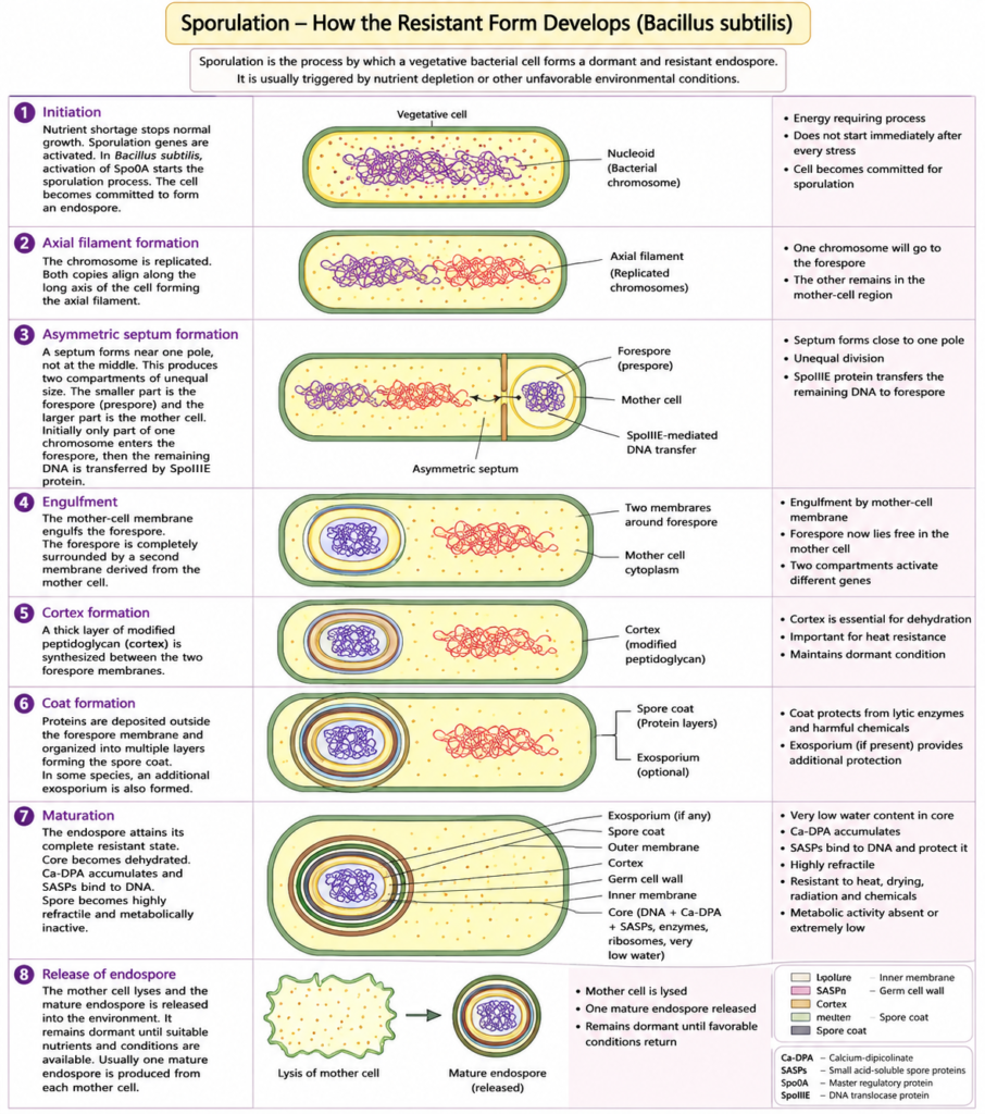
Sporulation – How the Resistant Form Develops
Sporulation is the developmental process where a vegetative bacterial cell is changed into a resistant endospore. The process is mainly studied in Bacillus subtilis. Exact signals and some developmental events can be different in other spore-forming bacteria.
a. Conditions That Trigger Sporulation
The following conditions are involved in initiation of sporulation-
- Nutrient depletion– Sporulation generally starts when nutrients required for active growth become limited. It commonly takes place near the end of exponential growth.
- Nutrient type– Limitation of carbon, nitrogen or phosphate can induce sporulation. The effective nutrient is not same under every growth condition.
- Stationary phase– The bacterial population enters into stationary phase after depletion of an essential nutrient. Cell division becomes reduced. Sporulation genes are now activated in some cells.
- Other signals– Population density, secreted signalling peptides and the physiological condition of cell also affect the process. Different environmental informations are combined before sporulation starts.
- Regulated decision– Sporulation does not begin immediately after every stress. It needs high amount of energy and many genes are involved. Some stressed cells may enter other survival states rather than forming spores.
- Species difference– The inducing condition varies among spore-forming bacteria. Nutrient limitation is common, but the sensing proteins and regulatory pathways are not fully same in every species.
b. Initiation and Chromosome Organization
- DNA replication– Before formation of the spore septum, bacterial DNA is replicated. Two copies of the chromosome are produced.
- Axial chromosome– Both chromosome copies become arranged along the long axis of bacterial cell. This arrangement is called the axial filament or axial chromosome arrangement.
- Developmental control– Sporulation-specific regulatory genes become active. The normal vegetative division programme is changed into an unequal division process.
- Commitment– After sufficient activation of the developmental programme, the cell becomes committed to sporulation. It now continues towards forespore formation instead of normal binary fission.
c. Asymmetric Septum and Forespore Formation
- Polar septum– A septum is formed close to one pole of bacterial cell. It is not produced at the central region like normal cell division.
- Two compartments– The unequal septum forms one small compartment and another larger part. The small compartment is called the forespore or prespore. Larger portion becomes the mother cell.
- Genetic material– Both compartments finally receive a copy of the same bacterial chromosome. At first, only a portion of chromosome may enter the forespore. Remaining part is transferred through the septum by the SpoIIIE protein.
- Different programmes– The two cells have similar genetic information, but they do not perform the same function. Separate groups of genes become active in forespore and mother cell.
d. Engulfment of the Forespore
- Membrane movement– The membrane of mother cell begins to move around the forespore. It slowly covers the forespore from both sides.
- Engulfment– Both ends of the moving membrane finally meet and fuse. The forespore is now released within the cytoplasm of mother cell.
- Cell within cell– A cell-within-a-cell arrangement is produced in this stage. The forespore is surrounded by an inner and an outer membrane.
- Communication– Mother cell and forespore remain connected through different signalling proteins and membrane protein complexes. This communication controls further development of both compartments.
e. Cortex and Coat Formation
- Germ cell wall– A thin peptidoglycan layer is formed around the inner forespore membrane. After germination, it develops into the cell wall of new vegetative cell.
- Cortex– Specialized peptidoglycan is deposited between the two forespore membranes. This thick layer is called the cortex. It is different from normal vegetative peptidoglycan.
- Coat assembly– Proteins formed mainly in the mother cell are deposited outside the forespore. They become arranged into different layers of the spore coat.
- Exosporium– Some bacterial species form one additional outer covering called the exosporium. It is present in spores of Bacillus anthracis and some other species, but absent from many bacterial endospores.
f. Core Dehydration and Maturation
- Calcium dipicolinate– Large amount of calcium-dipicolinate (Ca-DPA) is accumulated inside the developing spore core. It takes part in core dehydration and development of resistance.
- SASP formation– Small acid-soluble spore proteins (SASPs) are produced and become attached with spore DNA. DNA is protected from heat, radiation and different damaging chemicals by these proteins.
- Core water– Water is gradually removed from the core. The core now contains much lower water than the vegetative bacterial cytoplasm.
- Resistance– Resistance against wet heat, drying, radiation and many chemical agents develops during maturation. The coat, inner membrane, cortex and core components have different role in it.
- Dormancy– Metabolic activity becomes absent or extremely low. The endospore is now in its mature dormant condition.
g. Mother-Cell Lysis and Spore Release
- Final maturation– Cortex formation, coat assembly and maturation of core are completed inside the mother cell. The spore becomes highly refractile.
- Mother-cell breakdown– The wall and membrane of mother cell are hydrolysed. Mother-cell lysis takes place after completion of spore development.
- Spore release– A free mature endospore is released into the surrounding region. One mother cell generally produces one endospore.
- Persistence– The released spore remains dormant for a long period. It can germinate when suitable nutrients and other germination conditions become available.
Role of Spo0A and Sporulation-Specific Sigma Factors
- Spo0A– Spo0A is the major initiation regulator of sporulation in Bacillus subtilis. Its active phosphorylated form (Spo0A~P) controls the expression of early sporulation genes. A sufficient level is required before the cell enters into spore development.
- Sigma F– σF becomes active in the early forespore. It controls the first forespore-specific group of genes.
- Sigma E– In the mother cell, early developmental genes are controlled by σE. Its activity starts after formation of the asymmetric septum.
- Sigma G– σG acts later in the forespore. It controls genes involved in maturation, SASP formation and different core properties.
- Sigma K– The late mother-cell programme is controlled by σK. Many genes required for spore coat formation and final maturation are expressed in this stage.
- Sequential activity– These sigma factors do not work together at the same time. σF and σG act in the forespore, whereas σE and σK act in the mother cell. Their sequential activity keeps development of both compartments properly connected.
Germination and Return to Vegetative Growth
Germination is the process by which a dormant endospore loses its dormant and highly resistant condition. It begins to change into an active vegetative bacterial cell. Germination is followed by outgrowth, where normal metabolism and cell growth are again started.
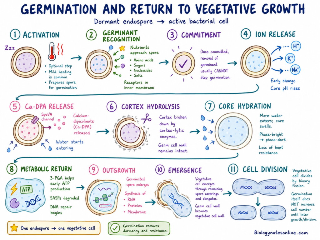
The following are the major events during this process-
- Activation– Some endospores require an activation treatment before germination. Mild heating is commonly used for this purpose. Activation makes the spore more ready to respond to germinants, but it does not complete the germination process. It is also not required by every spore.
- Germinant recognition– Suitable nutrients are detected by the dormant spore. Amino acids, sugars, nucleosides and some mineral salts may act as germinants. In many Bacillus spores, germinant receptors are present within the inner membrane. The type of germinant and its sensing system are different among bacterial species.
- Commitment– After receiving sufficient germination signal, the endospore becomes committed to germinate. Removal of the germinant after this stage generally cannot stop the process. The spore now moves out from its dormant condition.
- Ion release– Hydrogen, potassium and sodium ions are released from the spore core during early germination. Due to these changes, the internal pH of core starts to increase. Release of ions is one of the first measurable changes in the dormant spore.
- Ca-DPA release– The stored calcium-dipicolinate (Ca-DPA) is released from the core through proteins of the SpoVA channel. Water begins to enter in its place. Core dehydration is gradually lost during this step.
- Cortex hydrolysis– The special peptidoglycan of spore cortex is broken down by cortex-lytic enzymes. The germ cell wall remains protected, as it later forms the cell wall of vegetative cell. Removal of cortex allows expansion of the spore core.
- Core hydration– More water now enters into the core and swelling takes place. Mobility of core proteins and inner-membrane lipids is restored. During this process, the endospore loses much of its heat resistance and changes from phase-bright to phase-dark appearance.
- Metabolic return– Different enzymes inside the spore again become functional after rehydration. Stored 3-phosphoglycerate (3-PGA) can be used for early ATP formation. The small acid-soluble spore proteins (SASPs) are degraded and their amino acids become available. Damaged DNA and cellular components are also repaired.
- Outgrowth– Germination is now followed by outgrowth. RNA, proteins, membrane materials and other cellular components are formed. The germinated spore becomes larger and begins to develop the normal structure of vegetative bacterial cell.
- Emergence– The developing vegetative cell comes out through the remaining spore coverings. It elongates and obtains its normal bacterial shape. The germ cell wall now acts as the vegetative cell wall.
- Cell division– After completion of outgrowth, the vegetative cell carries out normal metabolism and growth. Binary fission is again started under suitable condition. One germinated endospore gives rise to one vegetative cell, so germination itself does not increase the number of cells.
Why Bacterial Spores Are Highly Resistant
The high resistance of bacterial endospores is not produced by one component only. Core substances, membranes and outer layers act in different ways. One protective component may be important against heat, while another acts against chemicals or radiation.
The following are the major reasons for resistance of bacterial spores-
- Low core water– The spore core contains very low amount of water. Movement of proteins and other core molecules is reduced in this condition. It is one of the major factor for resistance against moist heat.
- Calcium dipicolinate– A large amount of dipicolinic acid (DPA) occurs with calcium as calcium-dipicolinate (Ca-DPA). It is accumulated inside the core. Ca-DPA helps in core dehydration and stability, and has an important role in heat resistance. Its protective effect is not same against every damaging agent.
- SASPs– Small acid-soluble spore proteins (SASPs) become closely attached with the spore DNA. DNA structure and its chemical reaction are changed by this binding. It gives protection from dry heat, ultraviolet radiation, drying and many DNA-damaging chemicals.
- Spore coat– Several protein layers are present around the cortex. These layers form the spore coat. It prevents entry of large molecules and protects the spore from lytic enzymes, oxidizing substances and many harmful chemicals. Coat is not the main cause of moist-heat resistance.
- Inner membrane– The inner membrane of dormant spore is highly compressed and has very low permeability. Many toxic chemicals cannot easily reach the core due to this membrane. Damage of the inner membrane is also one important cause of spore killing by some oxidizing agents.
- Cortex– A thick layer of specialized peptidoglycan is present outside the germ cell wall. This is referred to as the cortex. It helps in keeping the core in dehydrated condition, which is required for development of resistance against moist heat.
- DNA protection– The bacterial chromosome remains protected by SASPs and the dehydrated condition of core. Due to this, DNA damage caused by heat, radiation and chemicals becomes reduced. The protection is not complete under every treatment.
- Repair system– Some damage may remain within a treated spore. During germination and early outgrowth, DNA-repair enzymes become active and repair many of these damages. Spore photoproduct formed by ultraviolet radiation is repaired by spore photoproduct lyase.
- Dormant core– Metabolic activity is absent or extremely low in a mature endospore. Its enzymes, ribosomes and other important components remain in an inactive state. Dormancy alone does not produce complete resistance, but it keeps the internal components in a stable condition.
- Multiple barriers– Heat, radiation and chemical agents do not damage endospores by the same mechanism. Low core water is more important for moist heat, SASPs mainly protect DNA, while the coat and inner membrane give greater protection from many chemicals. Thus, different protective parts act together.
Resistance Mechanism of Bacterial Spores
Bacterial spores resist different harmful conditions by several protective mechanisms. The same mechanism does not work against every treatment. Wet heat, dry heat, chemicals and radiation affect different parts of the spore.
Resistance to Wet and Dry Heat
- Core dehydration– Water content of the spore core is very low. Due to this, movement of proteins becomes greatly reduced. Core proteins are not easily denatured by wet heat. It is the major factor for wet-heat resistance.
- Calcium-DPA– Dipicolinic acid (DPA) is mainly present as calcium-dipicolinate (Ca-DPA) inside the core. It helps to maintain low core-water content. The role in wet-heat resistance is mainly through this dehydrated condition.
- SASPs– Spore DNA remains covered by small acid-soluble spore proteins (SASPs). These proteins prevent loss of DNA bases and other heat damages. Wet heat generally kills spores by damage of important core proteins, not by direct DNA damage.
- Dry heat– The killing action of dry heat is different. Here, DNA damage is the major event. SASPs give strong protection to DNA, while Ca-DPA also takes part in dry-heat resistance.
- DNA repair– Some DNA damage is still produced by dry heat. It is repaired during germination and early outgrowth. RecA and other repair proteins are involved in it.
Resistance to Desiccation
- Dehydrated state– The core is already present in a highly dehydrated condition. Thus, removal of external water causes less change in its internal components.
- DNA binding– SASPs remain attached with the bacterial DNA during dormancy. They protect DNA during drying and repeated rehydration. Spores without these proteins show greater DNA damage after desiccation.
- DPA content– DPA is another important component for desiccation resistance. Loss of both DPA and major SASPs causes very low survival during drying.
- Repeated drying– Mature spores can survive several cycles of desiccation and rehydration. Vegetative bacterial cells are generally damaged after much less drying.
Resistance to Chemicals and Enzymes
- Spore coat– The coat is made up of several protein layers. It protects the internal spore from many aldehydes, halogens, oxidizing agents and lytic enzymes. Lysozyme cannot easily reach the cortex of an intact spore.
- Chemical reaction– Large amount of coat protein can react with some harmful chemicals before they reach the inner regions. In some cases, movement of the chemical through the coat is also slow.
- Inner membrane– This membrane has extremely low permeability. Small toxic molecules do not easily pass through it to the core. Damage of the inner membrane is one important way by which several oxidizing agents kill spores.
- Protective enzymes– Catalase, superoxide dismutase and some other enzymes are present in the outer layers of spores of certain species. They can destroy hydrogen peroxide or reactive oxygen compounds. This mechanism is not same in every bacterial spore.
- DNA protection– SASPs protect spore DNA from hydrogen peroxide, nitrous acid, formaldehyde and some other genotoxic chemicals. But all alkylating chemicals are not blocked by these proteins.
- Repair after damage– Formaldehyde, nitrous acid and some alkylating agents may produce DNA damage. Repair of this damage takes place during spore outgrowth.
Resistance to UV and Ionizing Radiation
- Altered DNA structure– SASPs bind with the spore DNA and change its normal structure. Due to this, ultraviolet radiation does not mainly produce the usual cyclobutane pyrimidine dimers found in vegetative cells.
- Spore photoproduct– UV radiation mainly forms a special DNA lesion called spore photoproduct (SP). It is less difficult to repair than many other UV-induced lesions.
- Photoproduct repair– During germination, SP is repaired by spore photoproduct lyase (Spl). Nucleotide-excision repair and recombination repair also take part in removal of radiation damage.
- Outer pigments– Pigments present in the coat or other outer layers of some spores absorb UV radiation. This protection is species-specific and is not found equally in every spore.
- Ionizing radiation– Gamma rays and other ionizing radiations produce strand breaks and different DNA damages. SASPs provide some protection. Repair of DNA during outgrowth, including repair of double-strand breaks, has a major role in survival.
Are Endospores Indestructible?
No. Bacterial endospores are not indestructible. They are highly resistant structures, but a proper sterilization treatment can kill or inactivate them.
Resistance does not indicate complete protection. Endospores can survive drying, ordinary heating and exposure to many chemicals which easily destroy vegetative bacterial cells. Some spores may also survive normal cooking temperature. The level of resistance is not same in every bacterial species and strain.
Steam sterilization is commonly used for destruction of bacterial spores. Saturated steam is applied at the required temperature and pressure for a fixed exposure period. The cycle must be selected according to the material, packaging and sterilizer. Incomplete heating or short exposure may allow some spores to remain viable.
Endospores can also be inactivated by sufficient dry heat, incineration, ionizing radiation and some chemical sporicides. Hypochlorite, peracetic acid and certain hydrogen peroxide preparations have sporicidal activity. Proper concentration and contact time are required. Many ordinary surface disinfectants are not able to destroy bacterial spores.
Spore resistance depends on core dehydration, spore proteins, mineral content and the condition under which sporulation occurred. Age of the spore, treatment condition and number of spores also affect their survival. Thus, a treatment effective against one spore preparation may not show the same killing effect against another.
Functions and Biological Importance of Endospores
Endospore formation has one major function, survival of the bacterial cell during conditions not suitable for active growth. It is not a reproductive process. One vegetative cell generally forms one endospore, which again gives one vegetative cell after germination.
Some of the important functions and biological importance of endospores are-
- Survival– Endospores allow certain bacteria to survive during heat, drying, nutrient deficiency and exposure to many harmful chemicals. Vegetative cells are easily destroyed under many of these conditions.
- Genome protection– The bacterial DNA and other essential cell materials remain protected within the spore core. Different spore layers and protective proteins take part in it. Thus, genetic information is carried through the unfavourable period.
- Dormancy– A mature endospore remains metabolically dormant. Growth and cell division do not take place. Very little energy is required during this condition, which allows the bacterium to remain without available nutrients for a long period.
- Persistence– Spores may remain present in soil, dust, water, food materials and different surfaces. Their resistant nature helps the bacterial species to persist where the vegetative form cannot remain alive.
- Germination– When suitable nutrients and environmental condition return, the endospore undergoes germination. Metabolism is now started and an active vegetative bacterial cell is formed. This allows normal growth of the bacterium to begin again.
- Dispersal– Dormant spores can be carried through dust, water, animals and contaminated materials. After reaching a suitable region, they may germinate. In this way, endospores also support movement of bacterial species between different regions.
- Transmission– In some pathogenic bacteria, the spore acts as an important transmissive form. Spores of Clostridioides difficile remain on hospital surfaces and help in spread of infection. Bacillus anthracis spores can enter the host through skin, inhalation or ingestion.
- Disease recurrence– Dormant spores are not affected by many antibiotics which act against actively growing bacterial cells. In Clostridioides difficile infection, surviving spores may later germinate and are involved in persistence and recurrence of the disease.
- Food spoilage– Spores of different Bacillus and Clostridium species may survive some food-processing treatments. After germination, the vegetative cells can multiply and produce food spoilage. Under suitable condition, some species also produce toxins and cause foodborne disease.
- Sterilization testing– Because of their high resistance, bacterial spores are used as biological indicators of sterilization. Spores of Geobacillus stearothermophilus are commonly used to check steam-sterilization or autoclave process. Their destruction indicates that the required sterilizing condition has been reached.
Endospore Staining and Microscopic Identification
Endospore staining is a differential staining method used to detect endospores within bacterial cells and free spores outside the cells. It also shows their shape, position and whether swelling of the sporangium has taken place.
Why a Special Stain Is Required
Endospore has several resistant coverings around its core. Ordinary aqueous stains do not easily pass through these layers. Due to this, the spore may remain colourless or appear as a bright refractile body inside the stained vegetative cell.
Simple staining and Gram staining may sometimes show an unstained spore region. But the result is not always clear. Gram staining alone is therefore not the preferred method for confirmation of endospore. Heat, steam or some specialized treatment is used for entry of the primary stain.
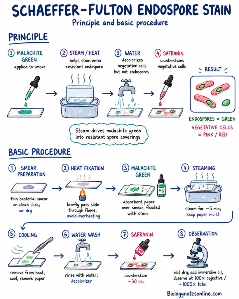
Principle of the Schaeffer–Fulton Method
The Schaeffer–Fulton method is commonly used for staining bacterial endospores. Malachite green is used as the primary stain. Steam is applied, which helps the stain to enter through the resistant spore coverings.
After cooling, water is used as the decolorizer. Malachite green is removed from vegetative cells, but remains within the endospores. Safranin is then applied as counterstain. Endospores and free spores appear green, while vegetative bacterial cells become pink to red.
Basic Procedure
The following are the basic steps of the Schaeffer–Fulton endospore staining method–
- Smear preparation– A thin bacterial smear is prepared on a clean grease-free glass slide. It is allowed to dry completely.
- Heat fixation– The dried smear is heat-fixed carefully. Excess heating should be avoided because cell shape may become damaged.
- Malachite green– A small piece of absorbent paper is placed over the smear. The paper is saturated with malachite green stain.
- Steaming– The slide is steamed for about 5 minutes. During this period, the paper must remain moist. More stain is added when drying starts.
- Cooling– The slide is removed from heat and allowed to cool. Absorbent paper is now removed carefully.
- Water washing– Tap water is used for washing the slide. In this method, water acts as the decolorizing agent.
- Counterstaining– Safranin is applied for about 30 seconds. Vegetative cells take up this counterstain.
- Final washing– The slide is washed with water. It is then blotted dry without rubbing the stained smear.
- Observation– A drop of immersion oil is placed over the stained region. The slide is examined with the oil-immersion objective (100× objective, about 1000× total magnification).
Safety– Gloves, laboratory coat and eye protection should be used. The slide, steam bath and staining rack become hot. Absorbent paper should not be allowed to dry or burn during heating. Approved non-pathogenic teaching cultures must be used, and stain waste is discarded according to laboratory instruction.
Interpretation of Results
The stained slide is examined for colour of the spore and vegetative cell. Position, shape and swelling are also recorded.
- Intracellular spore– A green structure is observed within a pink or red vegetative bacterial cell. It represents an endospore still present inside the mother cell.
- Free spore– A separate green body is seen without the surrounding red cell. It is referred to as a free mature spore.
- Vegetative cell– The non-spore portion of bacterial cell appears pink, reddish-pink or brownish red after safranin counterstaining.
- Spore position– Endospore may be central, subterminal or terminal in position. The location is useful during preliminary bacterial identification.
- Spore shape– The structure may be oval, ellipsoidal or spherical. Shape should be recorded together with its position.
- Cell swelling– Some endospores distend the mother cell and form a clear swelling. In other bacteria, the width of sporangium remains almost unchanged.
- Positive result– Green intracellular spores or free green spores are found on the slide. Pink or red vegetative cells may occur with them.
- Negative result– Only pink to red bacterial cells are present and no clear green spore is seen. This result should be interpreted carefully.
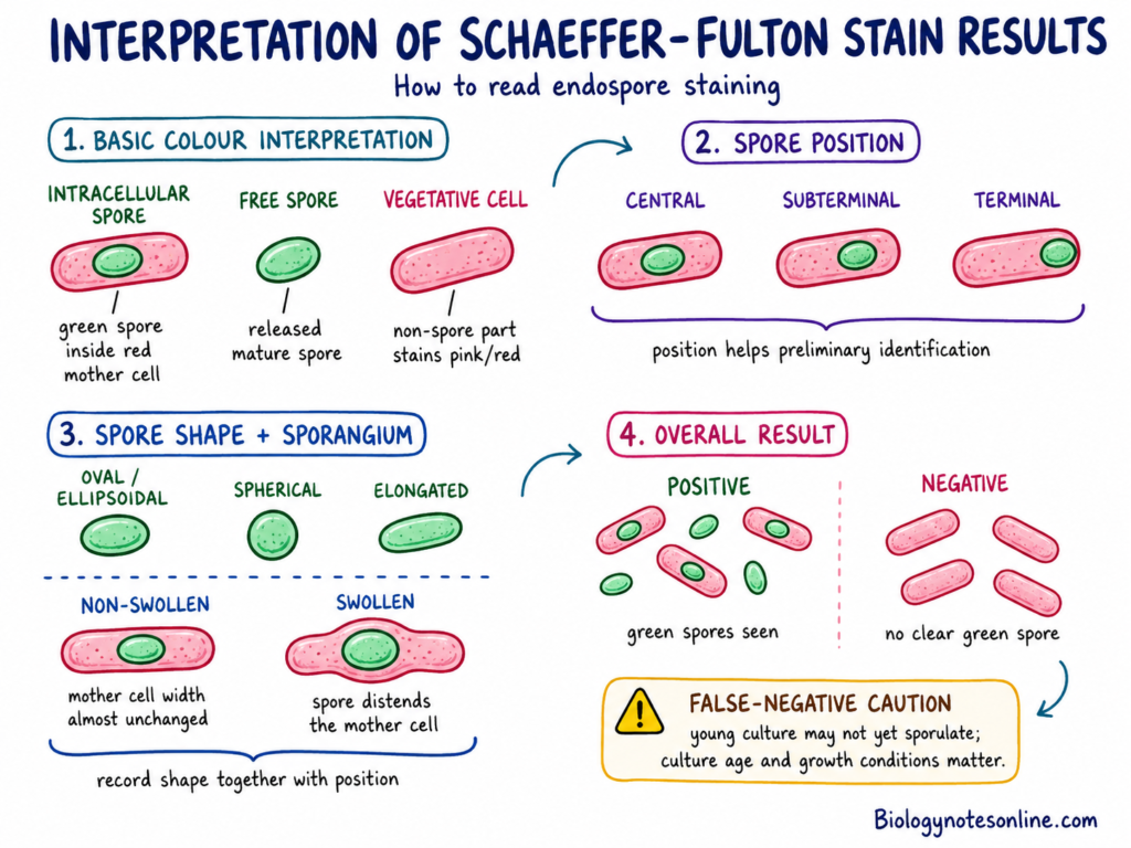
A young culture of a spore-forming bacterium may give a false-negative observation. Sporulation may not have started, or only a small number of cells are forming spores. Culture age and growth condition are therefore important.
Other Methods of Detection
- Dorner stain– It is another differential method for bacterial spores. Carbol fuchsin is driven into the spores by heating, followed by acid-alcohol treatment and a nigrosin background. Endospores appear red, vegetative cells remain colourless and the background becomes dark.
- Moeller stain– This is an older carbol-fuchsin method. Strong pretreatment and heating are used to increase spore permeability. The vegetative cells are generally counterstained with methylene blue.
- Phase-contrast microscopy– Unstained mature spores can be observed directly. They appear highly refractile or phase-bright. This method also shows intracellular and free spores without carrying out a staining procedure.
- Fluorescence methods– Fluorescent dyes, labelled proteins or specific probes can be used for detection and study of spore structures. These techniques are useful for spore counting, viability studies and observation of coat development. They are mainly used in research and specialized laboratories.
- Electron microscopy– Transmission electron microscopy (TEM) shows the internal cortex, coat and other spore layers. Scanning electron microscopy (SEM) is used for surface morphology. It provides greater structural detail, but is not a routine staining test.
- Molecular detection– PCR and genome-based methods can detect genes related to sporulation. But detection of one sporulation gene does not itself prove that mature spores are present. Some non-sporulating bacteria may retain several sporulation-related genes.
- Culture method– Samples may be given a controlled heat treatment before culture. Many vegetative cells are destroyed, while surviving spores are later germinated and grown on a suitable medium. The heating condition must be selected carefully because severe treatment can also reduce spore recovery.
- Resistance test– Resistance to heat, ethanol, lysozyme or a sporicidal treatment may be used along with microscopy and culture. This gives evidence of resistant spores, but should not be used alone for complete bacterial identification.
Destruction, Sterilization and Control of Endospore
Bacterial endospores are highly resistant, but they can be destroyed by a properly selected sterilization process. Ordinary cleaning or disinfection is not always sufficient. The method must reach the spore and remain effective for the required exposure period.
Sterilization vs Disinfection
Sterilization and disinfection are not the same process. Sterilization destroys all forms of microbial life, including bacterial spores. Disinfection removes or destroys many pathogenic microorganisms on an inanimate object, but spores may remain alive.
The following are the major differences-
- Sterilization– It is the complete destruction or removal of microorganisms, including highly resistant bacterial spores. Steam, dry heat, ethylene oxide and some low-temperature systems are used for this purpose.
- Disinfection– This process is used mainly on surfaces and non-living objects. Vegetative bacteria, fungi and many viruses are destroyed. But normal disinfection does not necessarily destroy bacterial endospores.
- Sporicidal– A chemical or physical treatment capable of killing bacterial spores is referred to as sporicidal. Some chemical disinfectants become sporicidal only after a longer contact time or under a specified concentration.
- Ordinary disinfectant– Alcohols, quaternary ammonium compounds and several routine surface disinfectants do not show reliable activity against mature spores. Thus, a surface may appear clean but viable spores can still be present.
- Prior cleaning– Dirt, blood, proteins and other organic substances may cover the spores or reduce activity of a chemical agent. Cleaning is therefore carried out before disinfection or sterilization. It also removes a large amount of contamination by physical action.
Steam Sterilization and Autoclaving
Autoclaving is one of the common methods used for destruction of bacterial spores. It uses saturated steam at a high temperature under pressure. Moist heat damages essential proteins and other important components of the spore.
- Moist heat– Steam transfers heat efficiently to the contaminated material. Condensation of steam over a cooler surface also releases heat.
- Pressure– Pressure alone does not kill the bacterial spore. It allows steam to remain at a temperature above the normal boiling point of water. The killing action is mainly produced by moist heat.
- Cycle parameters– A validated combination of temperature, pressure and exposure time is required. One fixed cycle cannot be used for every material or load.
- Load type– Liquids, wrapped instruments, porous materials and laboratory waste do not heat at the same rate. Large or tightly packed loads require proper arrangement so that steam can enter all regions.
- Air removal– Trapped air reduces direct contact between steam and the load. It may form a cooler region and incomplete sterilization can take place.
- Exposure time– The exposure period is counted after the required sterilizing condition has been reached. Heating and cooling time are not always part of the actual exposure period.
- Protocol– Autoclave cycles must be selected according to the equipment, load type and manufacturer instructions. Institutional biosafety procedures are also followed. Opening, loading and unloading of an autoclave should be performed by trained persons.
Chemical Sporicides and Low-Temperature Sterilization
Heat cannot be used for every instrument or material. Heat-sensitive items may be processed by a validated chemical or low-temperature sterilization system. The action depends on concentration, temperature, humidity and contact time.
- Hydrogen peroxide– Hydrogen peroxide vapour and gas-plasma systems are used for some heat-sensitive medical devices. They produce reactive substances which damage proteins, membranes and nucleic acids. Material and lumen limitations are present in these systems.
- Peracetic acid– It is a strong oxidizing agent with sporicidal activity. Liquid peracetic-acid systems can sterilize some immersible medical instruments at a low temperature. The item must be compatible with the process.
- Ethylene oxide– Ethylene oxide (EtO) is a gas sterilant for moisture-sensitive and heat-sensitive materials. It can move through many packaging materials. But it is toxic and requires controlled processing followed by aeration of the sterilized item.
- Chlorine agents– Some chlorine-based formulations can destroy bacterial spores when used under a validated concentration and contact period. Organic matter may reduce their activity. Corrosion, bleaching and material damage can also occur.
- Contact time– A sporicidal chemical must remain wet and in contact with the contaminated surface for the labelled period. Early drying or wiping can reduce the killing effect.
- Compatibility– Oxidizing chemicals may damage metals, rubber, adhesives or electronic components. EtO and other gaseous agents also need special equipment and occupational controls.
- Safety– These chemicals should be used according to the product label and approved laboratory or healthcare procedure. Laboratory sporicidal concentrations should not be converted into home-use mixtures. Mixing chlorine products with acids, ammonia or other chemicals can produce dangerous gases.
Why Boiling and Alcohol May Be Insufficient
Vegetative bacterial cells and mature endospores do not show the same resistance. A treatment which kills an active bacterial cell may not destroy its dormant spore.
- Boiling– Boiling can destroy many vegetative bacteria and other microorganisms. Some bacterial spores can survive at 100°C for a considerable period. Thus, boiling is not equal to a validated sterilization cycle.
- Uneven treatment– Material below the water surface, trapped air, large objects and organic matter may receive an incomplete treatment. Boiling also does not provide controlled steam penetration like a sterilizer.
- Alcohol– Ethanol and isopropanol have very poor or no reliable sporicidal activity. They are useful against many vegetative microorganisms, but dormant bacterial spores may remain viable.
- Physical removal– Washing with water and detergent may remove spores from a surface even when they are not killed. This can reduce the number of spores. The contaminated wash material must still be managed properly.
- Survival variation– Spore resistance is affected by bacterial species, strain, sporulation condition, temperature and surrounding material. A spore protected by dried organic matter may survive longer than a clean laboratory spore.
Biological Indicators
A biological indicator (BI) contains a known number of standardized bacterial spores with high resistance to a particular sterilization process. It is placed at a difficult-to-sterilize location within the load. After the cycle, survival or growth of the test spores is checked.
- Process challenge– Biological indicators provide a direct microbial challenge to the sterilization cycle. The spores are generally more resistant and present in greater numbers than normal contamination.
- Steam indicator– Spores of Geobacillus stearothermophilus are commonly used for monitoring moist-heat or steam sterilization. They are also used with some selected hydrogen peroxide and liquid peracetic-acid systems when specified.
- Other indicator– Bacillus atrophaeus spores are used for specified ethylene oxide and dry-heat sterilization systems. Some low-temperature systems may also use this organism according to their validated procedure.
- Correct selection– The biological indicator must be suitable for the sterilizer type and cycle. A BI selected for steam should not be automatically used for every chemical sterilization system.
- Cycle evaluation– A temperature record only shows that a measured temperature was reached at one location. Biological indicators test whether the complete process was capable of inactivating resistant spores under the load condition.
- Positive result– Growth from the processed indicator suggests failure of the cycle, loading problem, equipment defect or improper handling. The sterilization record and affected load are then evaluated according to the institutional protocol.
Differences Between Endospore and Vegetative Cell
The major differences between endospore and vegetative bacterial cell are as follows-
| Characteristic | Endospore | Vegetative Cell |
|---|---|---|
| Nature | It is a dormant and highly resistant structure. | Vegetative cell is the normal active form of bacterium. |
| Formation | Formed inside the mother cell during sporulation. Usually, one cell forms one endospore. | It is formed from another vegetative cell by binary fission or from a germinated endospore. |
| Major role | It is mainly used for survival during unfavourable condition. | Growth, metabolism and multiplication are carried out by this form. |
| Reproduction | Endospore formation is not a reproductive process. It does not increase the number of cells. | Vegetative cells reproduce by binary fission under suitable condition. |
| Metabolism | Metabolic activity remains absent or extremely low. | Active metabolism, respiration and enzyme activities are present. |
| Growth and division | It does not grow or divide during dormant condition. | Cell growth takes place and division is also continued when nutrients are available. |
| Water content | The spore core has very low amount of water. | Cytoplasm contains much higher amount of water. |
| Structure | Core is surrounded by inner membrane, germ cell wall, cortex and protein coat. An exosporium may also be present in some species. | It has the usual plasma membrane, cytoplasm and bacterial cell wall. Spore cortex and coat are absent. |
| Cell components | DNA, ribosomes and essential enzymes are present inside the core. They remain mainly inactive. | DNA, ribosomes and enzymes remain active and perform normal cell functions. |
| Calcium-DPA | A large amount of calcium-dipicolinate (Ca-DPA) is present in many endospores. | Calcium-DPA is not a characteristic component of vegetative cell. |
| Resistance | It has greater resistance to heat, drying, radiation and many chemicals. | Vegetative cell is much less resistant and is destroyed more easily. |
| Microscopic nature | Mature spores are highly refractile and generally appear phase-bright under phase-contrast microscope. | Vegetative cells are not highly refractile like mature spores. |
| Staining | Ordinary bacterial stains do not enter clearly. Special endospore staining is generally required. | It can be stained by simple staining and Gram-staining methods. |
| Return to activity | Under suitable condition, it undergoes germination and outgrowth. | It remains active and continues normal growth when favourable condition is present. |
| Environmental survival | It may persist for a long period without growth or available nutrients. | Long survival is difficult when water, nutrients or suitable condition are absent. |
Endospore vs Other Spore-Like Structures
Different bacteria and fungi form dormant or spore-like structures. Their formation and biological function are not same. Endospore, fungal spore, exospore, cyst, akinete and myxospore are formed by separate developmental process.
Endospore vs Fungal Spore
Bacterial endospores mainly act as survival structures. Fungal spores are generally formed for reproduction and dispersal, although some thick-walled fungal spores also help in survival.
| Characteristic | Endospore | Fungal Spore |
|---|---|---|
| Origin | It is formed by certain bacterial species. | Formed by fungi, which are eukaryotic organisms. |
| Major role | Survival during condition not suitable for active bacterial growth. | Mainly reproduction and dispersal. Some types also remain resistant. |
| Reproduction | Endospore formation is not a reproductive process. | Sexual or asexual spores take part in fungal reproduction. |
| Cell number | One bacterial cell generally forms one endospore. It later gives one vegetative cell. | A fungal structure can form many spores. Thus, the number of reproductive units is increased. |
| Formation | Forespore is formed internally within a mother cell. It is then engulfed by mother-cell membrane. | Spores develop in sporangia, on specialized hyphae or over reproductive structures. |
| Structure | Core, inner membrane, germ cell wall, cortex and protein coat are present. | Fungal spore contains a eukaryotic cell with a fungal wall, mainly containing chitin and glucans. Spore structure differs among fungal groups. |
| Calcium-DPA | Calcium-dipicolinate (Ca-DPA) is an important core component of many bacterial endospores. | This endospore component is not a normal feature of fungal spores. |
| Release | Mature endospore is commonly released after lysis of mother cell. | Spores are released from sporangia or separated from spore-producing hyphae and fruiting structures. |
| Dispersal | It may be carried through soil, dust, water or contaminated materials. But it is not formed for multiplication. | Many fungal spores are specially formed for movement through air, water or animals. |
| Germination | One endospore germinates and returns into one vegetative bacterial cell. | Fungal spore germinates and forms a germ tube, yeast cell or new fungal thallus. |
Endospore vs Exospore
An exospore is formed externally by some bacteria, mainly filamentous members of Actinobacteria such as Streptomyces. Exospore is not the same as exosporium, which is only an outer covering present around some bacterial endospores.
| Characteristic | Endospore | Exospore |
| Position of formation | Develops inside the mother cell. | Formed externally or by subdivision of aerial reproductive hyphae. |
| Major bacteria | Common in some members of Bacillota, such as Bacillus and Clostridium. | Found in selected Actinobacteria, particularly Streptomyces. |
| Initial development | Polar septum forms a small forespore and larger mother cell. | Specialized aerial hypha is produced. It later becomes divided into many spore compartments. |
| Engulfment | Mother-cell membrane moves around and completely engulfs the forespore. | Engulfment does not take place. Repeated septa are formed along the aerial hypha. |
| Number | Generally one endospore from one mother cell. | Many exospores may be formed in a chain from one aerial hypha. |
| Arrangement | A cell-within-a-cell stage is present during development. | Spores remain arranged in chains before their separation. |
| Envelope | It has a dehydrated core, specialized cortex and several coat layers. | Spore wall and outer protein layers are formed. A hydrophobic rodlet layer may also occur. |
| Resistance | Very high resistance to wet heat, drying, radiation and many chemicals. | More resistant than vegetative hyphae, but generally does not show the broad resistance of bacterial endospore. |
| Function | Mainly long-term survival. | Survival and dispersal of the bacterial colony. |
| Germination | Germination produces a vegetative bacterial cell. | Exospore swells and forms one or more germ tubes, which develop into vegetative hyphae. |
Endospore vs Bacterial Cyst
A bacterial cyst is another resistant resting cell. The best-known examples are produced by Azotobacter species. It develops by conversion of the complete vegetative cell and not by internal forespore formation.
| Characteristic | Endospore | Bacterial Cyst |
| Example | Bacillus subtilis, Clostridium species and other endospore formers. | Mainly studied in Azotobacter vinelandii and related species. |
| Development | Unequal cell division produces a forespore and mother-cell compartment. | Vegetative cell becomes rounded and is changed into the cyst. No separate forespore is formed. |
| Internal formation | Formed internally and remains inside the mother cell during maturation. | Complete vegetative cell develops into one cyst. |
| Structure | Core, inner membrane, germ cell wall, cortex and protein coat. | A central body is surrounded by an intine, exine and an outer capsule. |
| Cortex | Specialized endospore cortex is present. | Typical endospore cortex is absent. |
| Calcium-DPA and SASPs | Ca-DPA and small acid-soluble spore proteins (SASPs) are major protective core substances. | The same endospore-type chemical arrangement is not present. Other protective materials are used. |
| Resistance | High resistance against heat, desiccation, radiation and many chemicals. | Strong resistance to desiccation and some environmental stresses. Heat resistance remains lower than bacterial endospores. |
| Dormancy | Metabolically dormant. | Metabolism becomes greatly reduced during the resting state. |
| Major role | Survival during severe unfavourable condition. | Survival mainly during drying, nutrient limitation and other soil stresses. |
| Return to growth | Germination and outgrowth form a vegetative bacterial cell. | Cyst germinates by breakdown of its outer layers, and the central body develops into a vegetative cell. |
Endospore vs Akinete and Myxospore
Akinetes are formed by certain filamentous Cyanobacteria, mainly members of Nostocales and Stigonematales. Myxospores are formed by myxobacteria such as Myxococcus xanthus. All three remain dormant, but their structure and development are different.
| Characteristic | Endospore | Akinete | Myxospore |
| Organism | Certain endospore-forming bacteria of Bacillota. | Certain filamentous Cyanobacteria. | Myxobacteria, especially Myxococcus species. |
| Cell of origin | A vegetative cell divides unequally and forms a forespore. | One vegetative cyanobacterial cell enlarges and directly changes into an akinete. | A rod-shaped vegetative cell differentiates into a rounded myxospore. |
| Condition | Commonly formed during nutrient limitation and entry into stationary phase. | Light, temperature and nutrient conditions can induce formation. The signal differs with species. | Starvation and high population density are the major developmental signals. |
| Formation pattern | Polar septation, forespore engulfment and maturation inside the mother cell. | Cell enlargement, storage-material accumulation and thickening of the envelope. No engulfment. | Cells aggregate and produce fruiting bodies. Some cells inside them become myxospores. |
| Shape | Usually oval or spherical. Position may be central, subterminal or terminal. | Enlarged, non-motile, oval or spherical cell. | Vegetative rod changes into a shortened or spherical resting cell. |
| Covering | Cortex and multilayered protein coat are present. | Thick multilayered envelope develops around the complete cell. | A resistant wall and extracellular coverings are formed. |
| Storage material | 3-phosphoglycerate, Ca-DPA and different core components are present. | Glycogen and cyanophycin reserves are commonly accumulated. | Storage materials and protective cell-wall components increase during differentiation. |
| Resistance | Shows the greatest and most broad resistance among these forms. | Resistant to desiccation, cold, darkness and different environmental stresses. | More resistant to heat, drying, UV and sonication than vegetative myxobacterial cells. |
| Dormancy | Metabolic activity is absent or extremely low. | It remains as a dormant resting cell. | Dormant and non-growing condition occurs. |
| Location | Mature spore is released after lysis of mother cell. | Generally remains within the cyanobacterial filament before separation or germination. | Mostly present within a multicellular fruiting body. |
| Return to growth | One endospore forms one vegetative bacterial cell. | Akinete germinates and produces new vegetative cyanobacterial cells or filaments. | Myxospore germinates and returns into a rod-shaped vegetative myxobacterial cell. |
Frequently Asked Questions
What is an endospore?
An endospore is a dormant, non-reproductive and highly resistant structure formed inside certain bacterial cells. It protects the bacterial DNA and other essential components during unfavourable condition.
What is the primary function of an endospore?
The major function is survival of bacterial cell. It is not formed for increasing cell number.
Why do bacteria form endospores?
Some bacteria form endospores during nutrient shortage or other unsuitable growth condition. The active vegetative form cannot continue its normal growth during this period.
Are endospores reproductive?
No. One vegetative bacterial cell generally forms one endospore. After germination, the same spore produces one vegetative cell.
Which bacteria produce endospores?
Endospore formation is mainly found in some members of Bacillota. The common genera include Bacillus, Clostridium, Clostridioides, Paenibacillus and Geobacillus.
Do all Gram-positive bacteria form endospores?
No. This property is present only in certain bacterial groups. Most Gram-positive bacteria do not form an endospore.
Can Gram-negative bacteria form true endospores?
True endospore formation is very uncommon among Gram-negative-staining bacteria. Acetonema longum is one unusual example, but it belongs to the endospore-forming phylum Bacillota.
What are the major layers of a bacterial endospore?
The major parts are the core, inner membrane, germ cell wall, cortex and spore coat. Some species also contain an additional exosporium.
What is the role of calcium dipicolinate?
Calcium-dipicolinate (Ca-DPA) is accumulated inside the spore core. It is involved in core dehydration, stability and heat resistance.
What are SASPs?
Small acid-soluble spore proteins (SASPs) are protective proteins of the spore core. They bind with DNA and protect it from heat, drying, radiation and different chemicals.
What are central, terminal and subterminal endospores?
These terms indicate the position of endospore inside the mother cell. A central spore occurs near the middle, terminal at one end and subterminal between the middle and end.
What triggers sporulation?
Limitation of carbon, nitrogen, phosphate or other nutrients can start sporulation. Entry into stationary phase and different environmental signals are also involved.
What is a forespore?
A forespore is the smaller compartment produced after unequal division of the sporulating cell. It later develops into a mature endospore.
What is the difference between sporulation and germination?
Sporulation is the formation of an endospore from a vegetative cell. Germination is the return of the dormant spore towards an active vegetative form.
What occurs during endospore germination?
Calcium-DPA is released and water enters into the core. The cortex is hydrolysed, metabolism returns and outgrowth of vegetative cell begins.
Why are endospores resistant to heat?
The spore core contains very low amount of water. Calcium-DPA, SASPs, cortex and other protective layers also take part in heat resistance.
Can boiling destroy bacterial endospores?
Boiling can destroy many vegetative bacterial cells. But all bacterial endospores are not reliably destroyed by boiling, so it is not considered as a proper sterilization method.
Does alcohol kill endospores?
Alcohol does not show reliable sporicidal activity. Mature endospores may remain viable after exposure.
How does an autoclave destroy bacterial spores?
An autoclave uses saturated steam at a validated temperature and exposure time. Moist heat damages important spore proteins and other essential components. Pressure itself is not the main killing agent.
Why is malachite green used in endospore staining?
Malachite green is used as the primary stain. Steam helps this stain to enter through the resistant spore coverings.
What color are endospores after Schaeffer–Fulton staining?
Endospores and free spores appear green. Vegetative bacterial cells become pink to red after safranin counterstaining.
Can an endospore be observed in a Gram-stained preparation?
It may appear as an unstained or refractile body inside the bacterial cell. But Gram staining is not the preferred method for clear confirmation of endospores.
What is the difference between an endospore and a fungal spore?
A bacterial endospore is mainly a non-reproductive survival structure. Fungal spores are generally formed for reproduction and dispersal.
How long can an endospore remain dormant?
An endospore can remain dormant for many years under suitable condition. The exact period depends on bacterial species and surrounding environment.
Why are bacterial spores important in food safety?
Some spores can survive food-processing treatments and later germinate during storage. The formed vegetative cells may cause spoilage, toxin production or foodborne disease.
Why are endospores used as biological indicators?
Standardized resistant spores are used to check sterilization cycles. Their destruction gives evidence that the complete sterilization process was effective.
References
- Baron, S. (Ed.). (1996). Medical microbiology (4th ed.). University of Texas Medical Branch at Galveston. https://www.ncbi.nlm.nih.gov/books/NBK7627/
- Basta, M., & Annamaraju, P. (2022). Bacterial spores. In StatPearls. StatPearls Publishing. https://www.ncbi.nlm.nih.gov/books/NBK556071/
- Driks, A., & Eichenberger, P. (Eds.). (2016). The bacterial spore: From molecules to systems. ASM Press. https://doi.org/10.1128/9781555819323
- McGinnis, M. R., & Tyring, S. K. (1996). Introduction to mycology. In S. Baron (Ed.), Medical microbiology (4th ed.). University of Texas Medical Branch at Galveston. https://www.ncbi.nlm.nih.gov/books/NBK8125/
- Salton, M. R. J., & Kim, K.-S. (1996). Structure. In S. Baron (Ed.), Medical microbiology (4th ed.). University of Texas Medical Branch at Galveston. https://www.ncbi.nlm.nih.gov/books/NBK8477/
- Turnbull, P. C. B. (1996). Bacillus. In S. Baron (Ed.), Medical microbiology (4th ed., Chapter 15). University of Texas Medical Branch at Galveston. https://www.ncbi.nlm.nih.gov/books/NBK7699/
- Wells, C. L., & Wilkins, T. D. (1996). Clostridia: Sporeforming anaerobic bacilli. In S. Baron (Ed.), Medical microbiology (4th ed., Chapter 18). University of Texas Medical Branch at Galveston. https://www.ncbi.nlm.nih.gov/books/NBK8219/
- Bauda, E., Gallet, B., Moravcova, J., Effantin, G., Chan, H., Novacek, J., Jouneau, P.-H., Rodrigues, C. D. A., Schoehn, G., Moriscot, C., & Morlot, C. (2024). Ultrastructure of macromolecular assemblies contributing to bacterial spore resistance revealed by in situ cryo-electron tomography. Nature Communications, 15, 1376. https://doi.org/10.1038/s41467-024-45770-6
- Checinska, A., Paszczynski, A., & Burbank, M. (2015). Bacillus and other spore-forming genera: Variations in responses and mechanisms for survival. Annual Review of Food Science and Technology, 6, 351–369. https://doi.org/10.1146/annurev-food-030713-092332
- Chen, Y., Barat, B., Ray, W. K., Helm, R. F., Melville, S. B., & Popham, D. L. (2019). Membrane proteomes and ion transporters in Bacillus anthracis and Bacillus subtilis dormant and germinating spores. Journal of Bacteriology, 201(6), e00662-18. https://doi.org/10.1128/JB.00662-18
- Colas de la Noue, A., Natali, F., Fekraoui, F., Gervais, P., Martinez, N., Perrier-Cornet, J.-M., & Peters, J. (2020). The molecular dynamics of bacterial spore and the role of calcium dipicolinate in core properties at the sub-nanosecond time-scale. Scientific Reports, 10, 8265. https://doi.org/10.1038/s41598-020-65093-y
- Custer, G. F., Bresciani, L., & Dini-Andreote, F. (2022). Ecological and evolutionary implications of microbial dispersal. Frontiers in Microbiology, 13, 855859. https://doi.org/10.3389/fmicb.2022.855859
- Dalla Vecchia, E., Visser, M., Stams, A. J. M., & Bernier-Latmani, R. (2014). Investigation of sporulation in the Desulfotomaculum genus: A genomic comparison with the genera Bacillus and Clostridium. Environmental Microbiology Reports, 6(6), 756–766. https://doi.org/10.1111/1758-2229.12200
- Driks, A., & Eichenberger, P. (2016). The spore coat. Microbiology Spectrum, 4(2), TBS-0023-2016. https://doi.org/10.1128/microbiolspec.TBS-0023-2016
- Fritze, D. (2004). Taxonomy of the genus Bacillus and related genera: The aerobic endospore-forming bacteria. Phytopathology, 94(11), 1245–1248. https://doi.org/10.1094/PHYTO.2004.94.11.1245
- Galperin, M. Y. (2013). Genome diversity of spore-forming Firmicutes. Microbiology Spectrum, 1(2), TBS-0015-2012. https://doi.org/10.1128/microbiolspectrum.TBS-0015-2012
- Galperin, M. Y., Mekhedov, S. L., Puigbò, P., Smirnov, S., Wolf, Y. I., & Rigden, D. J. (2012). Genomic determinants of sporulation in Bacilli and Clostridia: Towards the minimal set of sporulation-specific genes. Environmental Microbiology, 14(11), 2870–2890. https://doi.org/10.1111/j.1462-2920.2012.02841.x
- Garg, R., Luckner, M., Berger, J., & Maldener, I. (2022). Changes in envelope structure and cell–cell communication during akinete differentiation and germination in filamentous cyanobacterium Trichormus variabilis ATCC 29413. Life, 12(3), 429. https://doi.org/10.3390/life12030429
- Garti-Levi, S., Eswara, A., Smith, Y., Fujita, M., & Ben-Yehuda, S. (2013). Novel modulators controlling entry into sporulation in Bacillus subtilis. Journal of Bacteriology, 195(7), 1475–1483. https://doi.org/10.1128/JB.02160-12
- Gilmore, M. E., Bandyopadhyay, D., Dean, A. M., Linnstaedt, S. D., & Popham, D. L. (2004). Production of muramic δ-lactam in Bacillus subtilis spore peptidoglycan. Journal of Bacteriology, 186(1), 80–89. https://doi.org/10.1128/JB.186.1.80-89.2004
- Henriques, A. O., & Moran, C. P., Jr. (2007). Structure, assembly, and function of the spore surface layers. Annual Review of Microbiology, 61, 555–588. https://doi.org/10.1146/annurev.micro.61.080706.093224
- Higgins, D., & Dworkin, J. (2012). Recent progress in Bacillus subtilis sporulation. FEMS Microbiology Reviews, 36(1), 131–148. https://doi.org/10.1111/j.1574-6976.2011.00310.x
- Hutchison, E. A., Miller, D. A., & Angert, E. R. (2014). Sporulation in bacteria: Beyond the standard model. Microbiology Spectrum, 2(5), TBS-0013-2012. https://doi.org/10.1128/microbiolspec.TBS-0013-2012
- Kaieda, S., Setlow, B., Setlow, P., & Halle, B. (2013). Mobility of core water in Bacillus subtilis spores by ²H NMR. Biophysical Journal, 105(9), 2016–2023. https://doi.org/10.1016/j.bpj.2013.09.022
- Khanna, K., Lopez-Garrido, J., & Pogliano, K. (2020). Shaping an endospore: Architectural transformations during Bacillus subtilis sporulation. Annual Review of Microbiology, 74, 361–386. https://doi.org/10.1146/annurev-micro-022520-074650
- Laue, M., Han, H.-M., Dittmann, C., & Setlow, P. (2018). Intracellular membranes of bacterial endospores are reservoirs for spore core membrane expansion during spore germination. Scientific Reports, 8, 11388. https://doi.org/10.1038/s41598-018-29879-5
- Li, W., Mednick, S., Setlow, P., & Li, Y.-Q. (2025). Modeling heterogeneity, commitment, and memory of bacterial spore germination. mBio, 16(5), e00596-25. https://doi.org/10.1128/mbio.00596-25
- Maughan, H., & Van der Auwera, G. (2011). Bacillus taxonomy in the genomic era finds phenotypes to be essential though often misleading. Infection, Genetics and Evolution, 11(5), 789–797. https://doi.org/10.1016/j.meegid.2011.02.001
- McKenney, P. T., Driks, A., & Eichenberger, P. (2013). The Bacillus subtilis endospore: Assembly and functions of the multilayered coat. Nature Reviews Microbiology, 11(1), 33–44. https://doi.org/10.1038/nrmicro2921
- McSpadden Gardener, B. B. (2004). Ecology of Bacillus and Paenibacillus spp. in agricultural systems. Phytopathology, 94(11), 1252–1258. https://doi.org/10.1094/PHYTO.2004.94.11.1252
- Najar, I. N., & Thakur, N. (2020). A systematic review of the genera Geobacillus and Parageobacillus: Their evolution, current taxonomic status and major applications. Microbiology, 166(9), 800–816. https://doi.org/10.1099/mic.0.000945
- Nicholson, W. L. (2002). Roles of Bacillus endospores in the environment. Cellular and Molecular Life Sciences, 59, 410–416. https://doi.org/10.1007/s00018-002-8433-7
- Nicholson, W. L., Munakata, N., Horneck, G., Melosh, H. J., & Setlow, P. (2000). Resistance of Bacillus endospores to extreme terrestrial and extraterrestrial environments. Microbiology and Molecular Biology Reviews, 64(3), 548–572. https://doi.org/10.1128/MMBR.64.3.548-572.2000
- Onyenwoke, R. U., Brill, J. A., Farahi, K., & Wiegel, J. (2004). Sporulation genes in members of the low G+C Gram-type-positive phylogenetic branch (Firmicutes). Archives of Microbiology, 182, 182–192. https://doi.org/10.1007/s00203-004-0696-y
- Riley, E. P., Lopez-Garrido, J., Sugie, J., Liu, R. B., & Pogliano, K. (2021). Metabolic differentiation and intercellular nurturing underpin bacterial endospore formation. Science Advances, 7(4), eabd6385. https://doi.org/10.1126/sciadv.abd6385
- Rodríguez-Salazar, J., Moreno, S., & Espín, G. (2017). LEA proteins are involved in cyst desiccation resistance and other abiotic stresses in Azotobacter vinelandii. Cell Stress and Chaperones, 22(3), 397–408. https://doi.org/10.1007/s12192-017-0781-1
- Romero-Rodríguez, A., Ruiz-Villafán, B., Martínez-de la Peña, C. F., & Sánchez, S. (2023). Targeting the impossible: A review of new strategies against endospores. Antibiotics, 12(2), 248. https://doi.org/10.3390/antibiotics12020248
- Sarker, M. R., & Setlow, P. (2007). Effect of a small, acid-soluble spore protein from Clostridium perfringens on the resistance properties of Bacillus subtilis spores. Journal of Bacteriology, 189(19), 7047–7052. https://doi.org/10.1128/JB.01179-07
- Setlow, B., Atluri, S., Kitchel, R., Koziol-Dube, K., & Setlow, P. (2006). Role of dipicolinic acid in resistance and stability of spores of Bacillus subtilis with or without DNA-protective α/β-type small acid-soluble proteins. Journal of Bacteriology, 188(11), 3740–3747. https://doi.org/10.1128/JB.00212-06
- Setlow, P. (2014a). Germination of spores of Bacillus species: What we know and do not know. Journal of Bacteriology, 196(7), 1297–1305. https://doi.org/10.1128/JB.01455-13
- Setlow, P. (2014b). Spore resistance properties. Microbiology Spectrum, 2(5), TBS-0003-2012. https://doi.org/10.1128/microbiolspec.TBS-0003-2012
- Setlow, P., & Christie, G. (2023). New thoughts on an old topic: Secrets of bacterial spore resistance slowly being revealed. Microbiology and Molecular Biology Reviews, 87(2), e00080-22. https://doi.org/10.1128/mmbr.00080-22
- Setlow, P., Wang, S., & Li, Y.-Q. (2017). Germination of spores of the orders Bacillales and Clostridiales. Annual Review of Microbiology, 71, 459–477. https://doi.org/10.1146/annurev-micro-090816-093558
- Shen, A. (2020). Clostridioides difficile spores: Bile acid sensors and Trojan horses of transmission. Clinics in Colon and Rectal Surgery, 33(2), 58–66. https://doi.org/10.1055/s-0040-1701230
- Stewart, G. C. (2015). The exosporium layer of bacterial spores: A connection to the environment and the infected host. Microbiology and Molecular Biology Reviews, 79(4), 437–457. https://doi.org/10.1128/MMBR.00050-15
- Tocheva, E. I., Matson, E. G., Morris, D. M., Moussavi, F., Leadbetter, J. R., & Jensen, G. J. (2011). Peptidoglycan remodeling and conversion of an inner membrane into an outer membrane during sporulation. Cell, 146(5), 799–812. https://doi.org/10.1016/j.cell.2011.07.029
- Wells-Bennik, M. H. J., Eijlander, R. T., den Besten, H. M. W., Berendsen, E. M., Warda, A. K., Krawczyk, A. O., Nierop Groot, M. N., Xiao, Y., Zwietering, M. H., Kuipers, O. P., & Abee, T. (2016). Bacterial spores in food: Survival, emergence, and outgrowth. Annual Review of Food Science and Technology, 7, 457–482. https://doi.org/10.1146/annurev-food-041715-033144
- Zhang, Y., Palma, C. S. D., Chen, Z., Zarazúa-Osorio, B., Fujita, M., & Igoshin, O. A. (2025). Biophysical modeling reveals the transcriptional regulatory mechanism of Spo0A, the master regulator in starving Bacillus subtilis. mSystems, 10(5), e00072-25. https://doi.org/10.1128/msystems.00072-25
- Zhu, D., Sorg, J. A., & Sun, X. (2018). Clostridioides difficile biology: Sporulation, germination, and corresponding therapies for C. difficile infection. Frontiers in Cellular and Infection Microbiology, 8, 29. https://doi.org/10.3389/fcimb.2018.00029