

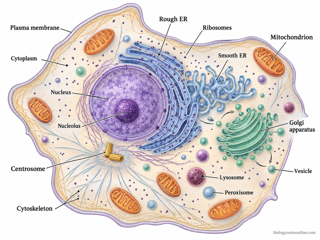
Animal cell is a type of eukaryotic cell, which is found in animals and it has a membrane-bound nucleus. It is enclosed by the plasma membrane or cell membrane, and inside it cytoplasm is present with nucleus and different specialised cellular structures called organelles.
Animal cell has no cell wall. This is one of the important feature that separates it from plant cell, because the outer boundary of animal cell is mainly the flexible plasma membrane.
The cytoplasm is the region present between the cell membrane and nuclear envelope. In this region, the cell organelles are suspended and many cellular activities take place. The nucleus contains the main genetic material of the cell and controls many cellular processes. Organelles are small cell structures, which are used to perform specific functions inside the cell. They work in a coordinated way for maintaining the normal activity of the animal cell.
A general animal cell shown in books is only a basic model. All animal cells do not look same. Some specialised animal cells may have more of some organelles, or may modify and even lack some structures that are shown in the generalized animal cell.
The shape and internal organization of animal cells also varies according to the function they perform. In the search for “animal cell parts”, “animal cell structure”, or “12 parts of an animal cell”, different textbooks may give different numbers like 12, 13 or 14 parts because they count or group the structures in different ways.
Labeled Animal Cell Diagram
Animal Cell Size and Shape
Animal cell size and shape are not fixed in all animal cells. They change according to cell type, function, cytoskeleton, and surrounding condition.
- Animal cells are generally microscopic. Many eukaryotic animal cells fall between 10-100 µm in diameter, but all animal cells do not remain in this same size.
- A typical animal cell is about 10-20 µm in diameter. This is only a general value. Some animal cells are smaller and some are much larger according to their special function.
- Human red blood cell is very small and about 7-8 µm in diameter. It is biconcave in shape and this shape helps in gas transport through narrow blood vessels.
- Some animal cells become very long without becoming wide. Neuron is an example. Its cell body may be small, but its axon can extend far for carrying nerve impulse.
- Animal cells have no rigid cell wall. So, their shape is more flexible than plant cells. The plasma membrane forms the outer boundary.
- The shape of animal cell may be round, oval, flat, elongated, branched, spindle-shaped, or irregular. It depends on the work of that cell.
- Cell shape is largely maintained by cytoskeleton. Microfilaments, intermediate filaments, and microtubules give internal support and also help in changing the cell shape.
- The cytoskeleton also arranges the cytoplasm. It keeps organelles in position and helps in movement of materials inside the cell.
- Surface area and volume affect the size of cell. As the cell becomes larger, the surface area does not increase as fast as the volume. So, very large cells may face difficulty in exchange of materials.
- Small cells exchange materials more easily. Nutrients, oxygen, and waste products move across the cell membrane in shorter distance.
- Cells that perform different functions show different shapes. Muscle cells are elongated for contraction. Nerve cells are branched for transmission of signals. Epithelial cells may be flat or cuboidal for covering and lining.
- The surrounding cells also affect cell shape. In tissues, animal cells may become pressed, attached, or arranged in layers. So, shape is not decided by the cell alone.
- Some cells change their shape during movement. White blood cells can change shape while passing through tissues and during engulfing of foreign particles.
- Animal cell size and shape are also related to internal organelles. A cell with high energy need may contain many mitochondria. A secretory cell may have more rough endoplasmic reticulum and Golgi apparatus.
- The generalized animal cell diagram is only a model. Real animal cells vary in size, shape, and internal organization according to their function.
Types and Examples of Animal Cells
There are different specialized cells found in animal body. These cells are modified in shape, size, and internal structure according to their work.
- Epithelial cells- These cells form covering and lining surfaces of the body. They are found in skin surface, inner lining of organs, ducts, and body cavities. Some epithelial cells are flat, some are cuboidal, and some are column-like in shape.
- Nerve cells- Nerve cells or neurons are specialized for receiving and transmitting signals. They have cell body, dendrites, and axon. The dendrites receive messages and the axon carries impulse away from the cell body.
- Muscle cells- Muscle cells are elongated cells used for contraction. These cells form skeletal muscle, smooth muscle, and cardiac muscle. Skeletal muscle helps in body movement, smooth muscle is found in walls of internal organs, and cardiac muscle forms the heart wall.
- Red blood cells- Red blood cells or erythrocytes are blood cells used for transport of oxygen and carbon dioxide. In mammals, mature red blood cells lack nucleus. Their biconcave shape helps in movement through blood vessels.
- White blood cells- White blood cells or leukocytes are defence cells of the body. They are present in blood and take part in immune response. Some white blood cells engulf pathogens and cellular debris.
- Bone cells- Bone cells are present in bone tissue. Osteocytes are mature bone cells and remain inside small spaces of bone matrix. Osteoblasts form new bone matrix, while osteoclasts help in bone breakdown and remodelling.
- Cartilage cells- Cartilage cells are called chondrocytes. They are present inside lacunae in cartilage matrix. These cells help in maintaining cartilage, which gives flexible support in different body parts.
- Fat cells- Fat cells or adipocytes are cells of adipose tissue. They store fat droplets inside the cytoplasm. The nucleus is pushed to one side because of stored fat in mature adipocyte.
- Fibroblasts- Fibroblasts are connective tissue cells. They produce collagen fibres, elastic fibres, and reticular fibres. These fibres give strength and support to connective tissue.
- Sperm cells- Sperm cells are male gametes. They are small and motile. A flagellum is present, which helps sperm to move towards egg cell during fertilization.
- Egg cells- Egg cell or ovum is the female gamete. It is larger than sperm and non-motile in most animals. After fertilization, egg and sperm form zygote, from which animal development begins.
Animal Cell Structure
Animal cell structure is organized from the outer plasma membrane toward the inner cytoplasm and nucleus. A typical animal cell has a cell membrane, cytoplasm, nucleus, mitochondria, ribosomes, endoplasmic reticulum, Golgi apparatus, lysosomes, peroxisomes, cytoskeleton, centrosome, vesicles, and some surface specializations in particular cells. These are not present as same number in every animal cell. The general animal cell is a model. Actual animal cells differ according to their work.
| Animal cell part | Basic structure | Main function |
|---|---|---|
| Cell membrane | Thin phospholipid boundary with proteins | Controls entry and exit of materials and receives signals |
| Cytoplasm | Cytosol with organelles inside it | Site for many reactions and internal cell organization |
| Nucleus | Double membrane-bound structure with chromatin and nucleolus | Stores deoxyribonucleic acid (DNA) and regulates cell activities |
| Mitochondria | Double-membrane organelles with folded inner membrane | Produces adenosine triphosphate (ATP) by cellular respiration |
| Ribosomes | Small non-membranous particles | Protein synthesis |
| Endoplasmic reticulum (ER) | Membrane network near nucleus | Protein processing and lipid-related functions |
| Golgi apparatus | Stacked flattened membrane sacs | Modifies, sorts, and packs molecules |
| Lysosomes | Enzyme-containing vesicles | Digestion and recycling inside cell |
| Peroxisomes | Oxidative enzyme-containing organelles | Breakdown of fatty acids and harmful reactive compounds |
| Cytoskeleton | Protein fibres in cytoplasm | Gives shape, support, movement, and transport |
| Centrosome and centrioles | Microtubule-organizing region with centrioles in many animal cells | Helps in microtubule arrangement and cell division |
| Vesicles and small vacuoles | Small membrane-bound sacs | Transport and temporary storage |
| Surface structures | Cilia, flagella, or microvilli in selected cells | Movement or surface absorption |
Cell Membrane
Cell Membrane Structure
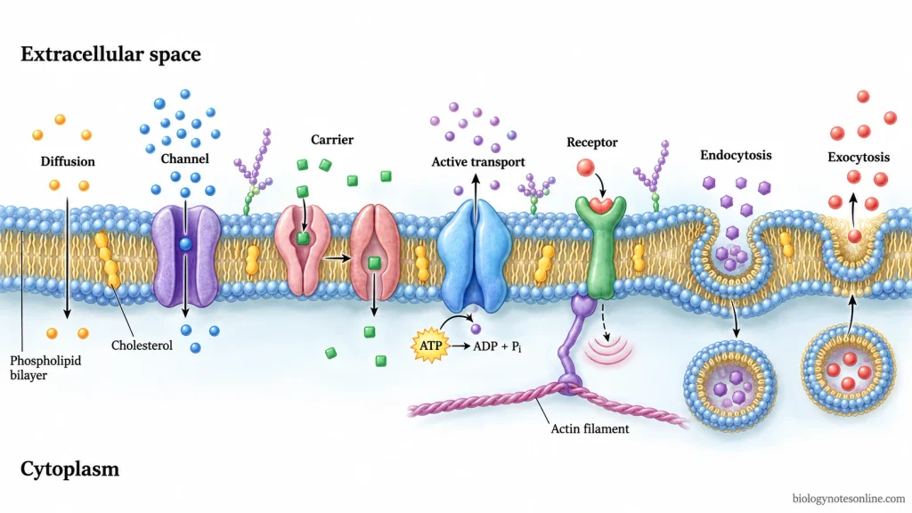
- Cell membrane or plasma membrane is the thin living boundary of an animal cell. It covers the cytoplasm from outside. It is flexible and no cell wall is present outside it.
- The cell membrane is mainly made up of phospholipid bilayer. It means two layers of phospholipid molecules are present. The phosphate heads face the watery side and fatty acid tails are turned inside.
- The inner part of the bilayer is hydrophobic. So, many charged particles and large water-soluble molecules cannot pass through it directly. Small non-polar molecules pass more easily.
- The structure of cell membrane is based on fluid mosaic model. The phospholipid layer is fluid, and proteins are present in it like scattered pieces. These molecules are not fixed at one place.
- Cholesterol is found between phospholipids in animal cell membrane. It gives stability to the membrane. It also controls fluid nature of membrane in different condition.
- Different membrane proteins are present in the bilayer. Some proteins pass through the whole membrane. Some are present only on the outer or inner surface.
- These proteins act as channels, carriers, pumps, receptors, enzymes, and attachment sites. Some of them hold cytoskeleton from inner side.
- Carbohydrates are present on the outer surface of cell membrane. They remain attached with proteins and lipids. These form glycoproteins and glycolipids.
- The carbohydrate layer helps in recognition of cells. It works like surface mark of the cell.
- In some animal cells, the membrane forms surface modifications like microvilli, cilia, or flagella. These are not found in all animal cells.
Cell Membrane Functions
- Cell membrane protects the inner content of cell.
- It separates cytoplasm from the outer medium.
- It controls entry and exit of substances.
- It is selectively permeable. So, all substances cannot pass in same way.
- Oxygen and carbon dioxide diffuse through the membrane.
- Water moves by osmosis.
- Ions, glucose, amino acids, and many polar molecules need transport proteins.
- In passive transport, substances move without using direct energy.
- In active transport, energy from adenosine triphosphate (ATP) is used. The substance may move against concentration gradient.
- Channel proteins form passage for ions and water.
- Carrier proteins bind the particular molecule and change their shape.
- Pumps move ions or molecules by using ATP.
- Cell membrane receives chemical signals by receptor proteins.
- It helps in cell communication. Hormones or other signalling molecules bind to membrane receptors.
- Endocytosis takes materials inside the cell by folding of membrane.
- Exocytosis releases materials outside when vesicles fuse with membrane.
- The membrane gives attachment to cytoskeleton. This helps in cell shape.
- In tissues, membrane proteins connect one cell with another cell.
- Carbohydrate marks help in cell recognition and self or non-self identification.
Cytoplasm Structure and Functions
Cytoplasm Structure
- Cytoplasm is the region present between the plasma membrane and the nuclear envelope. In an animal cell, it surrounds the nucleus and fills the inner part of the cell. It is not only a watery liquid. It contains cytosol, organelles, cytoskeleton, and different dissolved substances.
- Cytosol is the gel-like fluid part of the cytoplasm. The organelles are suspended in this cytosol. So, cytoplasm is the wider term, whereas cytosol is only the fluid portion outside the organelles.
- Cytoplasm contains many cell organelles. Mitochondria, ribosomes, endoplasmic reticulum, Golgi apparatus, lysosomes, peroxisomes, vesicles, and small vacuoles are present in it. These organelles do not float without arrangement. They are supported by cytoskeleton.
- The cytoskeleton is present through the cytoplasm. It is made up of protein fibres. These fibres help in maintaining cell shape, holding some organelles in place, moving vesicles, and movement of cytoplasm inside the cell.
- Cytoplasm contains water, ions, enzymes, nutrients, waste products, and many small molecules. These substances make the cytoplasm suitable for cellular reactions.
- In eukaryotic animal cells, cytoplasm is compartmentalized by organelles. Each organelle has its own membrane and internal condition. This makes different reactions to occur in different places.
- The cytoplasm is continuous with many cell activities. Vesicles move through it. Ribosomes work in it. Mitochondria remain embedded in it. It is the internal medium of the cell.
Cytoplasm Functions
- Cytoplasm holds the organelles inside the cell.
- It gives space for cellular reactions.
- Many metabolic reactions take place in the cytosol.
- It helps in movement of materials inside the cell.
- Cytoplasm supports transport of vesicles and organelles with the help of cytoskeleton.
- It stores ions, nutrients, enzymes, and other useful molecules.
- It helps in maintaining the shape of the cell.
- Cytoplasm keeps the organelles in proper cellular position.
- Protein synthesis starts on ribosomes present in the cytoplasm.
- It helps in communication between nucleus and other parts of the cell.
- The cytoplasm provides medium for breakdown and formation of many molecules.
- It also helps in distributing substances after they are made in one part of the cell.
- During cell division, cytoplasm is divided into two daughter cells.
- Cytoplasmic organelles carry out special functions like energy production, protein processing, digestion, and detoxification.
- The cytoplasm keeps the internal environment of the cell suitable for normal cell activity.
Nucleus Structure and Functions
Nucleus is a membrane-bound organelle of eukaryotic animal cell that contains the main genetic material of the cell. It acts as the control region for many cell activities because deoxyribonucleic acid (DNA) is kept inside it.
Nucleus Structure
- Nucleus is generally round or oval in shape. It is commonly present near the central region of the cell, but its position may change according to cell type.
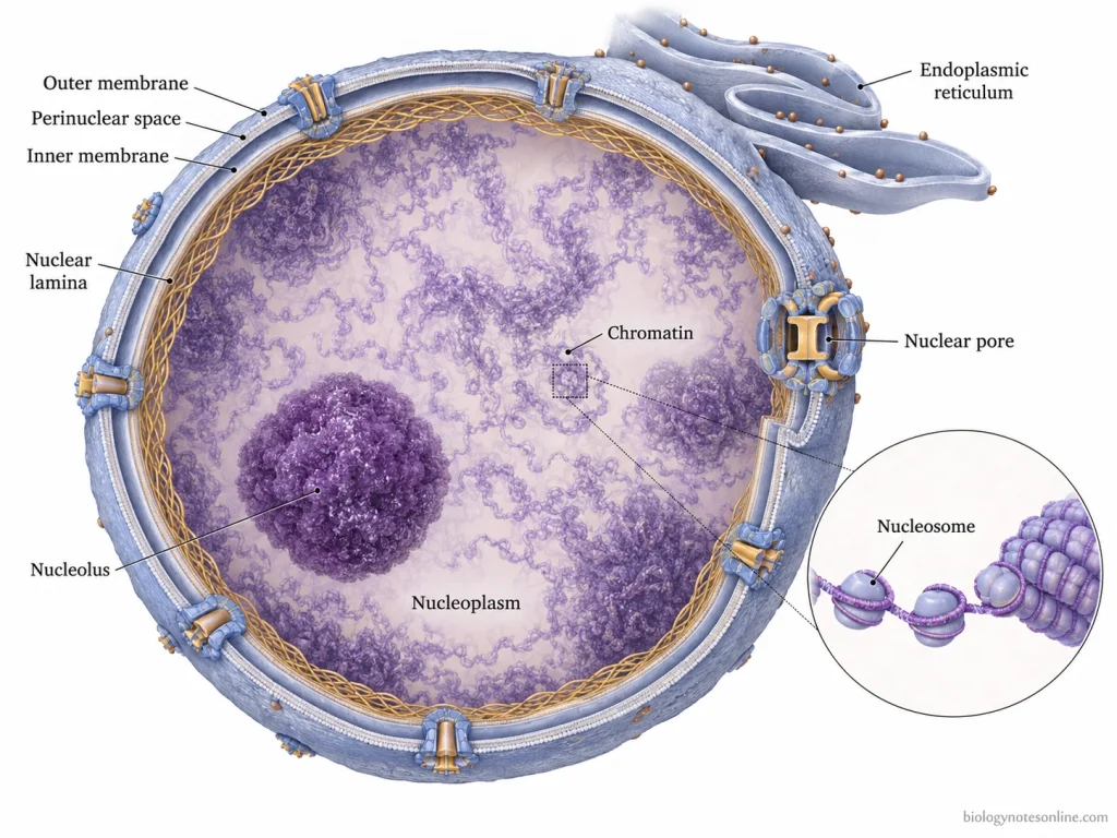
- The nucleus is surrounded by nuclear envelope. It is a double membrane covering. This envelope separates the nuclear content from cytoplasm and keeps nucleus as a distinct compartment.
- The nuclear envelope has two membranes. The outer nuclear membrane is continuous with endoplasmic reticulum in many cells. The inner membrane is present towards the nucleoplasm.
- Nuclear pores are present in the nuclear envelope. These pores act as passage for selected movement of proteins, ribonucleic acid (RNA), and other molecules between nucleus and cytoplasm.
- Inside the nucleus, a fluid-like substance is present. It is called nucleoplasm. Chromatin, nucleolus, enzymes, and many nuclear proteins remain suspended in it.
- Chromatin is made up of DNA and proteins. It remains spread inside the nucleus in non-dividing cell. During cell division, chromatin becomes condensed and forms chromosomes.
- The DNA present in chromatin carries hereditary information. It has genes, which give instructions for formation of proteins and other cell products.
- Nucleolus is a dense body present inside the nucleus. It is not surrounded by membrane. It is the region where ribosomal RNA (rRNA) is produced and ribosomal subunits are assembled.
- One nucleus may contain one or more nucleoli. This may vary with the activity of the cell. Cells making more protein may have more active nucleolus.
- All animal cells do not keep nucleus at maturity. Mammalian red blood cells lose their nucleus during maturation. So, they are mature cells without nucleus.
Nucleus Functions
- Nucleus stores DNA.
- It controls most of the cell activities.
- It regulates gene expression.
- DNA replication takes place inside the nucleus before cell division.
- Transcription takes place in the nucleus. In this process, DNA information is copied into RNA.
- Messenger RNA (mRNA) is formed in the nucleus and then moves to cytoplasm for protein synthesis.
- Nucleus controls protein synthesis indirectly by sending mRNA instructions.
- Nucleolus forms rRNA and helps in formation of ribosomal subunits.
- Nuclear pores control movement between nucleus and cytoplasm.
- The nuclear envelope protects genetic material from direct contact with cytoplasmic enzymes and other materials.
- Chromatin organization helps in packing DNA inside the small nuclear space.
- Nucleus helps in transfer of hereditary characters from one cell generation to next.
- During cell division, nuclear material is duplicated and distributed to daughter cells.
Mitochondria Structure and Functions
Mitochondria are double-membrane organelles of animal cell that produce adenosine triphosphate (ATP) during cellular respiration. They are commonly called the energy producing organelles of the cell.
Mitochondria Structure
- Mitochondria are mostly oval or rod-shaped organelles. Their shape and number may vary in different animal cells.
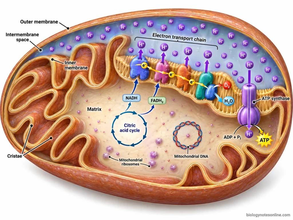
- Mitochondria has two membranes. One is outer membrane and another is inner membrane. Between these two membranes, a narrow space is present called intermembrane space.
- The outer membrane forms the outer covering of mitochondria. It separates mitochondria from the cytoplasm.
- The inner membrane is folded inside. These folds are called cristae. Cristae increases the surface area of inner membrane.
- The inner membrane contains many proteins needed for electron transport and ATP formation. It is the main site for oxidative phosphorylation.
- The inner space enclosed by inner membrane is called matrix. It contains enzymes, mitochondrial ribosomes, and mitochondrial deoxyribonucleic acid (DNA).
- Matrix is the site where many reactions of cellular respiration takes place. The citric acid cycle occurs in this region.
- Mitochondria have their own DNA and ribosomes. So, some mitochondrial proteins can be formed inside mitochondria itself. But most mitochondrial proteins are still made from nuclear genes and enter from cytoplasm.
- The cristae and matrix are not same in function. Cristae mainly increase area for ATP producing reactions, while matrix contains many enzymes of metabolic reactions.
- The number of mitochondria is not same in all cells. Cells with more energy requirement generally have more mitochondria.
Mitochondria Functions
- Mitochondria produce ATP.
- They carry out aerobic respiration.
- They convert energy from food molecules into usable cellular energy.
- Citric acid cycle takes place in mitochondrial matrix.
- Electron transport chain occurs on the inner mitochondrial membrane.
- ATP synthase present in inner membrane helps in ATP formation.
- Mitochondria use oxygen in the final steps of aerobic respiration.
- They help in oxidation of fatty acids in many animal cells.
- Mitochondria also store and regulate some calcium ions.
- They take part in controlled cell death, called apoptosis.
- They produce heat in some specialized cells.
- Mitochondria help in maintaining energy balance of the cell.
- They support active transport, cell division, movement, and biosynthesis by supplying ATP.
- The activity of mitochondria changes according to the energy need of cell.
Ribosomes Structure and Functions
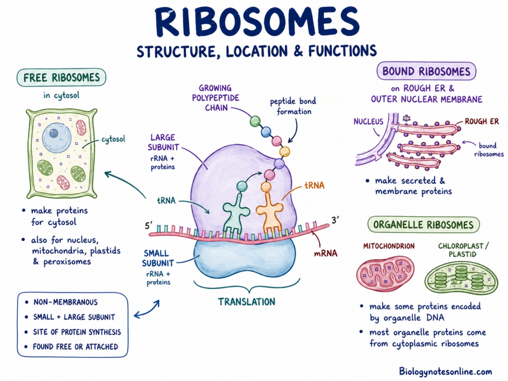
Ribosomes are small non-membranous cell structures, which are used for protein synthesis. They are present freely in cytoplasm or attached with rough endoplasmic reticulum (ER) in animal cells.
Ribosomes Structure
- Ribosomes are not covered by any membrane. They are small granular particles found in the cytoplasm.
- Ribosomes are made up of ribosomal ribonucleic acid (rRNA) and proteins. The rRNA forms the main functional part of ribosome. Proteins help in proper folding and stability of ribosome.
- In animal cells, cytoplasmic ribosome is 80S type. It is formed of one large 60S subunit and one small 40S subunit. These two subunits join during protein synthesis.
- The small subunit binds with messenger ribonucleic acid (mRNA). The large subunit helps in joining amino acids and forming the polypeptide chain.
- Ribosomal subunits are assembled in the nucleolus. After their formation, they come out to cytoplasm through nuclear pores.
- Ribosomes may remain free in cytosol. Some ribosomes are attached on the outer surface of rough ER.
- Free ribosomes and bound ribosomes have same basic structure. Their position is different, and the final destination of protein is also different.
- Many ribosomes may attach to one mRNA molecule at the same time. This structure is called polyribosome or polysome.
- Mitochondria also contain their own ribosomes. These ribosomes are used for synthesis of some mitochondrial proteins.
Ribosomes Functions
- Ribosomes synthesize proteins.
- They are the site of translation.
- Ribosomes read the codons present on mRNA.
- Transfer ribonucleic acid (tRNA) brings amino acids to ribosome.
- Amino acids are joined together to form polypeptide chain.
- rRNA helps in formation of peptide bond between amino acids.
- Free ribosomes generally make proteins used in cytosol.
- Bound ribosomes make proteins for secretion, membrane, lysosomes, and endomembrane system.
- Ribosomes help in expression of genetic information present in deoxyribonucleic acid (DNA).
- The protein formed by ribosome may work as enzyme, receptor, structural protein, hormone, or antibody.
- Polyribosomes help in making many copies of same protein from one mRNA.
- Ribosomes support cell growth, repair, division, and normal metabolic activity by producing required proteins.
Endoplasmic Reticulum
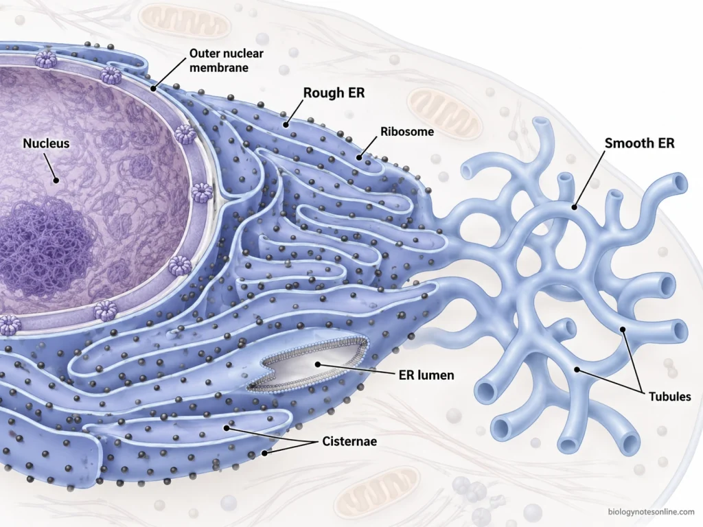
Endoplasmic reticulum (ER) is a membrane-bound network of tubules and flattened sacs present in the cytoplasm of eukaryotic animal cells. It is mainly used for protein processing, lipid formation, calcium storage, and transport of cell materials.
ER Structure
- ER is made up of membranous tubules and flattened sacs called cisternae. It forms a continuous internal network inside the cytoplasm.
- The ER membrane is connected with the outer membrane of the nuclear envelope. So, it lies close to the nucleus and also extends into different parts of the cytoplasm.
- ER is of two main types. These are rough endoplasmic reticulum (RER) and smooth endoplasmic reticulum (SER).
- RER has ribosomes attached on its outer cytoplasmic surface. Due to these ribosomes, the surface looks rough under microscope.
- RER is generally formed of flattened sacs. It is mostly present near the nucleus, but its amount varies in different cells.
- SER does not have ribosomes on its surface. It is more tubular in form. Its surface looks smooth because ribosomes are absent.
- The inner space of ER is called ER lumen. Proteins and other molecules can enter this space during their processing.
- The ER is a part of endomembrane system. Materials can move from ER to Golgi apparatus through small transport vesicles.
- Cells that produce more secretory proteins usually have more RER. Cells active in lipid synthesis or detoxification may contain more SER.
ER Functions
- ER helps in protein processing.
- RER takes part in synthesis of proteins.
- Ribosomes on RER make proteins that enter the ER lumen or ER membrane.
- Many secretory proteins and membrane proteins are first made on RER.
- Proteins formed in RER undergo folding and early modification.
- Some proteins are packed into vesicles and sent to Golgi apparatus.
- SER helps in lipid synthesis.
- Phospholipids and cholesterol are formed with help of ER membrane enzymes.
- SER also helps in steroid hormone synthesis in some specialized cells.
- SER takes part in detoxification of drugs and poisons, mainly in liver cells. OpenStax
- SER stores calcium ions in some animal cells.
- In muscle cells, a special form of SER helps in calcium release and storage for contraction.
- ER helps in transport of proteins and lipids inside the cell.
- It gives membrane material for many vesicles and organelles.
- ER also helps in maintaining internal compartmentalization of the animal cell.
Golgi Apparatus
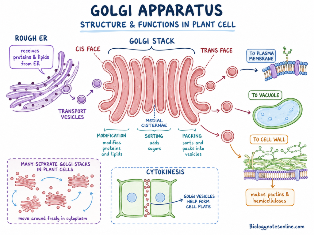
Golgi apparatus is a membrane-bound organelle of animal cell that modifies, sorts, packs, and sends proteins and lipids to their proper places. It is also called Golgi body or Golgi complex.
Golgi Apparatus Structure
- Golgi apparatus is made up of flattened membranous sacs. These sacs are called cisternae.
- The cisternae are arranged one above another like a stack. It is not a single sac. Several sacs are present together.
- Golgi apparatus has two main sides. One is the receiving side and another is the releasing side.
- The receiving side is called cis face. Vesicles coming from endoplasmic reticulum (ER) fuse with this side.
- The releasing side is called trans face. From this side, modified materials are packed into vesicles and sent to their destination.
- Between cis and trans face, middle cisternae are present. Proteins and lipids move through these regions and get modified step by step.
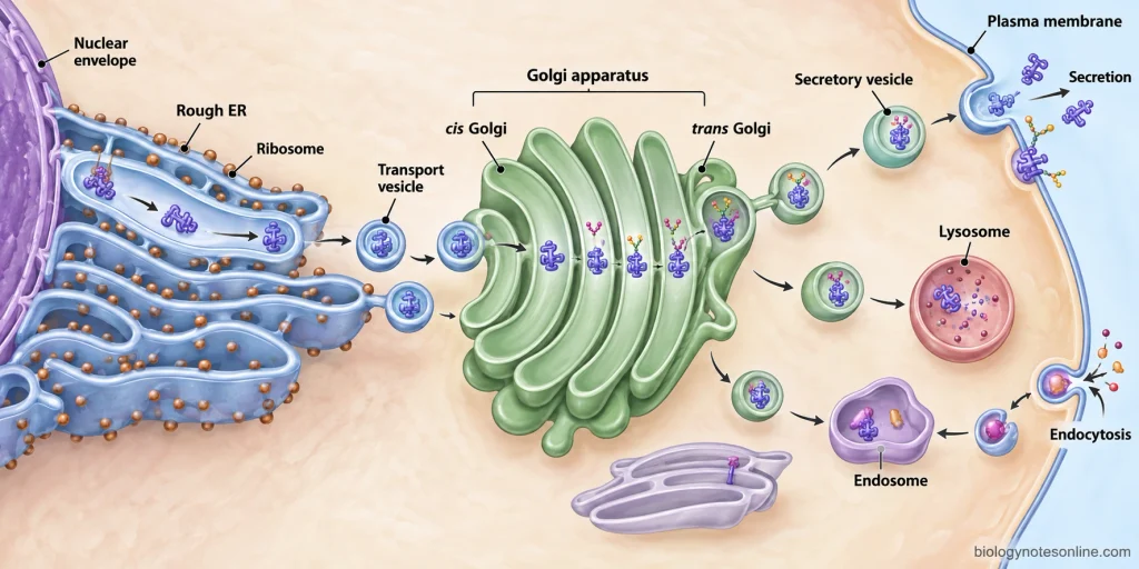
- Small vesicles are present around the Golgi apparatus. These vesicles carry materials from ER to Golgi apparatus, and from Golgi apparatus to plasma membrane, lysosomes, or outside of the cell.
- Golgi apparatus is usually present near the nucleus and ER in many animal cells. Its number and size may change according to cell type and secretory activity.
- The Golgi membrane contains enzymes. These enzymes help in modification of proteins and lipids.
Golgi Apparatus Functions
- Golgi apparatus modifies proteins and lipids.
- It sorts cell materials.
- It packs materials into vesicles.
- It receives proteins and lipids from ER.
- It sends processed materials to plasma membrane, lysosomes, secretory vesicles, and other cell regions.
- It adds or changes carbohydrate groups on some proteins.
- Glycoproteins and glycolipids are processed in Golgi apparatus.
- It helps in formation of lysosomes.
- It prepares secretory products for release from the cell.
- Secretory vesicles from Golgi apparatus fuse with plasma membrane during exocytosis.
- It helps in renewal of plasma membrane by supplying membrane proteins and lipids.
- It acts as the sorting and distribution centre of the endomembrane system.
Lysosomes
Lysosomes are single membrane-bound organelles of animal cell that contain digestive enzymes. They break down large molecules, worn out cell parts, and foreign materials inside the cell.
Lysosomes Structure
- Lysosomes are small vesicle-like organelles. They are surrounded by a single membrane.
- The lysosomal membrane separates digestive enzymes from the cytoplasm. This is important because these enzymes can break down many biological molecules.
- The inside of lysosome is acidic. This acidic condition helps the lysosomal enzymes to work properly.
- Lysosomes contain acid hydrolases. These enzymes can digest proteins, lipids, polysaccharides, deoxyribonucleic acid (DNA), and ribonucleic acid (RNA).
- The lysosomal membrane has transport proteins. These proteins move the digested products out of lysosome and back into cytoplasm.
- Hydrogen ion (H⁺) pumps are present in lysosomal membrane. These pumps use energy from adenosine triphosphate (ATP) to keep the lysosome acidic.
- Lysosomes are related with the endomembrane system. Enzymes of lysosome are made in rough endoplasmic reticulum (ER), modified in Golgi apparatus, and sent toward lysosomes in vesicles.
- Lysosomes may differ in size and shape. Their number is also not same in every animal cell.
- In many cells, lysosomes fuse with endosomes, phagosomes, or autophagosomes. After fusion, the enclosed material is exposed to lysosomal enzymes.
Lysosomes Functions
- Lysosomes digest intracellular materials.
- They break down proteins, lipids, carbohydrates, and nucleic acids.
- They digest food particles taken in by endocytosis.
- They destroy some pathogens after phagocytosis.
- They remove worn out organelles by autophagy.
- They recycle useful small molecules back to cytoplasm.
- Lysosomes help in cellular waste removal.
- They take part in turnover of cell components.
- They help in maintaining normal cell condition by clearing damaged material.
- In some cells, lysosomes also take part in cell death related processes.
- Lysosomal enzymes work best in acidic medium. In neutral cytoplasm, their activity becomes less.
- If lysosomal enzymes or transport steps are defective, undigested substances may accumulate inside cells. This condition is related with lysosomal storage diseases.
Peroxisomes
Peroxisomes are small single membrane-bound organelles that contain oxidative enzymes and catalase. They take part in fatty acid breakdown and detoxification of harmful reactive compounds inside animal cells.
Peroxisomes Structure
- Peroxisomes are small rounded organelles present in cytoplasm. They are surrounded by a single membrane.
- The peroxisomal membrane separates its enzymes from the cytoplasm. This keeps many oxidative reactions inside the organelle only.
- Peroxisomes contain oxidase enzymes. These enzymes use molecular oxygen and remove hydrogen atoms from different organic molecules.
- During many reactions, hydrogen peroxide (H₂O₂) is produced. It is a reactive compound and can damage cell parts if it spreads freely.
- Catalase is present inside peroxisomes. It breaks down H₂O₂ into water and oxygen, or uses H₂O₂ for oxidation of some organic compounds
- Peroxisomes do not have their own deoxyribonucleic acid (DNA) or ribosomes. Most peroxisomal proteins are made in cytosol and then transported into peroxisomes.
- The number and size of peroxisomes are not same in all cells. They change according to cell type and metabolic condition.
- Peroxisomes may divide from already existing peroxisomes. New membrane and proteins are added, then the organelle grows and separates.
Peroxisomes Functions
- Peroxisomes carry out oxidative metabolism.
- They break down selected fatty acids.
- They are involved in oxidation of very-long-chain fatty acids (VLCFAs).
- They help in breakdown of some amino acids and uric acid.
- They produce H₂O₂ during oxidation reactions.
- Catalase breaks down H₂O₂ and reduces its harmful effect.
- Peroxisomes help in detoxification of some harmful molecules.
- They protect cytoplasm from uncontrolled oxidative damage.
- They also take part in lipid metabolism.
- In animal cells, fatty acid oxidation takes place in both mitochondria and peroxisomes.
- Peroxisomes help in synthesis of some important lipids, including plasmalogens in animal cells.
- Peroxisomal activity varies from one tissue to another tissue. Cells active in lipid metabolism and detoxification may contain more active peroxisomes.
Cytoskeleton
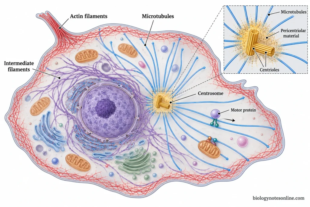
Cytoskeleton is a network of protein fibres present throughout the cytoplasm. It gives shape, support, internal arrangement, and movement to the animal cell.
Cytoskeleton Structure
- Cytoskeleton is not a membrane-bound organelle. It is made up of fine protein filaments spread inside the cytoplasm.
- The cytoskeleton has three main types of fibres. These are microfilaments, intermediate filaments, and microtubules.
- Microfilaments are the thinnest fibres of the cytoskeleton. They are mainly made up of actin protein.
- Actin filaments are present near the cell surface in many animal cells. They help in changing cell shape and in movement of cell surface.
- Microfilaments also take part in contraction with myosin. In muscle cells, actin and myosin are used for contraction.
- Intermediate filaments are thicker than microfilaments but thinner than microtubules. They are strong and rope-like fibres.
- Intermediate filaments give mechanical strength to the cell. They also help to hold the nucleus and some other structures in position.
- Microtubules are the thickest fibres of the cytoskeleton. They are hollow tube-like structures made up of tubulin protein.
- Microtubules are formed from alpha-tubulin and beta-tubulin subunits. They are dynamic and can grow or shrink according to the need of cell.
- In animal cells, many microtubules are organized from the centrosome near the nucleus. The arrangement may change during cell division.
- Motor proteins work along cytoskeletal fibres. Kinesin and dynein move on microtubules, while myosin moves on actin filaments. These proteins carry vesicles, organelles, or produce movement.
Cytoskeleton Functions
- Cytoskeleton gives shape to the cell.
- It supports the plasma membrane.
- It keeps some organelles in proper position.
- It helps in internal organization of cytoplasm.
- It provides tracks for movement of vesicles and organelles.
- Microtubules guide intracellular transport.
- Microfilaments help in cell movement and contraction.
- Intermediate filaments give mechanical support.
- Cytoskeleton helps the cell to resist mechanical stress.
- During cell division, microtubules form spindle fibres.
- Spindle fibres help in separation of chromosomes.
- Cytoskeleton takes part in cytokinesis with actin filaments.
- Cilia and flagella are supported by microtubules in animal cells.
- It helps in movement of materials over the cell surface in cells having cilia.
- Cytoskeleton also helps in changing cell shape during migration.
Centrosome and Centrioles
Centrosome is the main microtubule-organizing region of animal cells, and centrioles are cylindrical microtubule structures usually present inside it. They help in arrangement of microtubules and take part during cell division.
Centrosome and Centrioles Structure
- Centrosome is usually present near the nucleus. It is not covered by a membrane.
- In most animal cells, the centrosome contains two centrioles and surrounding pericentriolar material. The pericentriolar material is the protein-rich region around centrioles.
- The two centrioles are arranged nearly at right angle to each other. This pair is embedded in pericentriolar material.
- Each centriole is cylindrical in shape. It is made up of microtubules arranged in a definite pattern.
- A typical centriole has nine groups of microtubule triplets. This is called 9 triplet arrangement.
- The centrosome is the site from which many microtubules grow out into the cytoplasm. In animal cells, it works as the major microtubule-organizing center.
- Microtubules do not mainly start from the centriole wall itself. They are nucleated from the pericentriolar material around the centrioles.
- Before cell division, the centrosome duplicates. After duplication, two centrosomes help in forming the spindle apparatus.
- Centrioles are related to basal bodies of cilia and flagella. Basal bodies also have a centriole-like structure and help in organizing these surface projections.
- Centriole number and arrangement may differ in specialized cells. It should not be taken that every animal cell has the exact same centriole condition.
Centrosome and Centrioles Functions
- Centrosome organizes microtubules.
- It acts as a microtubule-organizing center.
- It helps in maintaining internal arrangement of cytoplasm.
- Microtubules grow from centrosome toward the cell periphery.
- Centrosome helps in positioning of some organelles.
- It takes part in cell division.
- Before mitosis, centrosome duplicates.
- Two centrosomes move apart and help in spindle formation.
- Spindle fibres attach with chromosomes and help in their separation.
- Centrioles have role in centrosome duplication and spindle organization.
- Centrioles also help in formation of basal bodies.
- Basal bodies organize cilia and flagella in cells where these structures are present.
- Centrosome activity changes during interphase and mitosis.
- In dividing animal cells, centrosomes work as principal microtubule-organizing centres.
Vesicles and Small Vacuoles
Vesicles and small vacuoles are membrane-bound sacs present in animal cells, which are used for transport, storage, secretion, and movement of materials between different cell parts. Vesicles are usually smaller and more active in membrane fusion, while vacuoles are comparatively larger storage compartments.
Vesicles and Small Vacuoles Structure
- Vesicles are small membrane-bound sacs. They are formed from cell membrane or from membranes of organelles.
- The vesicle membrane is made up of lipid bilayer. It can fuse with other membranes inside the cell.
- Vesicles may bud from endoplasmic reticulum (ER), Golgi apparatus, plasma membrane, endosomes, or other membrane compartments.
- Transport vesicles carry proteins and lipids from one organelle to another organelle. They are common in the endomembrane system.
- Secretory vesicles contain materials that are to be released outside the cell. They move towards plasma membrane.
- Endocytic vesicles are formed when the plasma membrane folds inward. In this way, materials from outside are taken into the cell.
- Some vesicles may contain enzymes or stored molecules. Some are used only for short distance movement of substances.
- Small vacuoles are also membrane-bound sacs. In animal cells, they are usually smaller and less prominent than the large central vacuole of plant cell.
- Vacuoles are somewhat larger than vesicles. Their membrane do not fuse with other cell membranes as commonly as vesicle membrane does.
- Animal cells may have many small vacuoles instead of one large central vacuole. Their number and size changes with cell type and activity.
Vesicles and Small Vacuoles Functions
- Vesicles transport materials inside the cell.
- They carry proteins from ER to Golgi apparatus.
- They carry modified proteins and lipids from Golgi apparatus to other sites.
- Vesicles help in secretion.
- Secretory vesicles fuse with plasma membrane and release materials outside the cell.
- Vesicles help in endocytosis.
- They bring substances from outside into the cytoplasm.
- Vesicles also take part in exocytosis.
- They help in renewal of plasma membrane by adding membrane material.
- Some vesicles deliver materials to lysosomes for digestion.
- Small vacuoles store water, ions, nutrients, and waste materials for short time.
- Small vacuoles help in temporary storage and transport.
- Vesicle movement is supported by cytoskeleton and motor proteins.
- They help in communication between organelles.
- Vesicles and small vacuoles keep different materials separated from cytosol.
Specialized Surface Structures
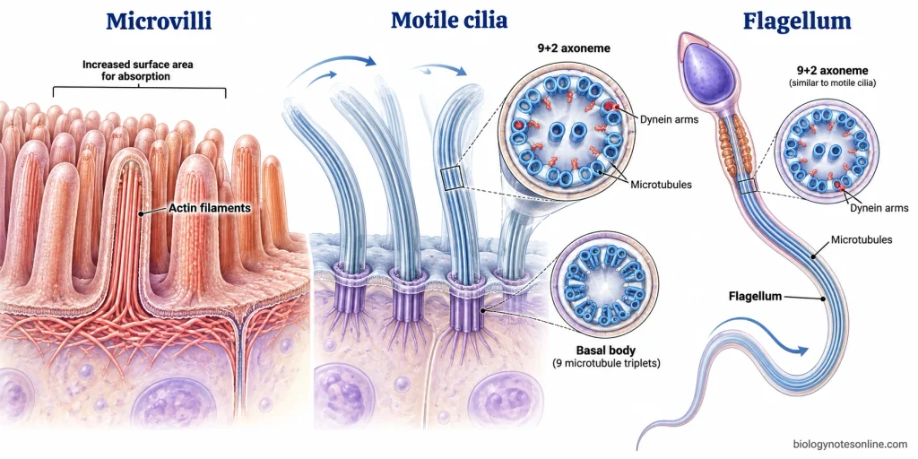
Specialized surface structures are special projections of plasma membrane found only in particular animal cells. Cilia, flagella, and microvilli are included in it. These are not universal parts of every animal cell.
Cilia
Cilia are short hair-like projections present on the surface of some animal cells. They are covered by plasma membrane and internally supported by microtubules. The basal part of cilia is attached with basal body. These are not present in every animal cell.
Motile cilia show beating movement. In respiratory tract cells, they move mucus and trapped particles over the cell surface. In some cells, cilia helps in movement of fluid along the tissue surface. Some cilia are non-motile and mainly sensory in function. They receive signals from the surrounding environment.
Flagella
Flagella are longer whip-like projections present only in some specialized animal cells. In animal cell, the common example is sperm cell. The sperm flagellum helps in movement of the whole cell.
Cilia and flagella are structurally related. Both are extensions of plasma membrane and contain microtubule-based inner core called axoneme. In many motile cilia and flagella, the axoneme has 9+2 arrangement of microtubules. Dynein protein helps in bending movement by acting on these microtubules.
Microvilli
Microvilli are small finger-like folds of the plasma membrane. They are shorter than cilia and are not used for beating movement. They are actin-based surface folds.
Microvilli are supported by actin filaments. These actin bundles keep the projection stable and maintain the folded surface. Microvilli are mostly found in cells used for absorption, such as intestinal epithelial cells. They increase the surface area of the cell and helps in faster uptake of materials.
Functions of Animal Cells
Animal cell functions are the basic activities by which animal cell remains alive, uses energy, forms materials, and keeps its internal condition controlled. OpenStax
- Cell boundary- Animal cell maintains a separate inner environment by the plasma membrane. It controls entry and exit of substances, so cytoplasm is kept different from the outside medium.
- Material exchange- Oxygen, water, ions, glucose, amino acids, and other useful substances enter the cell as required. Waste materials are removed out from the cell by controlled transport.
- Energy production- Mitochondria break down food molecules during cellular respiration and form adenosine triphosphate (ATP). This ATP is used for active transport, synthesis, movement, and many metabolic works.
- Protein synthesis- Ribosomes form proteins by joining amino acids in proper order. The information is carried by messenger ribonucleic acid (mRNA) and the ribosome reads it during translation.
- Genetic control- The nucleus stores deoxyribonucleic acid (DNA). DNA controls cell activities by giving instructions for protein formation and other cell processes.
- Cellular synthesis- Animal cells make new proteins, lipids, nucleic acids, and membrane materials. These materials are formed according to the requirement of cell.
- Internal transport- Materials move from one part of cell to another part. Endoplasmic reticulum, Golgi apparatus, vesicles, and cytoskeleton help in this movement and distribution.
- Processing and packaging- Proteins and lipids are modified, sorted, and packed inside the cell. Golgi apparatus receives materials from endoplasmic reticulum and sends them to proper place.
- Digestion and recycling- Lysosomes break down damaged organelles, large molecules, and some materials taken from outside. The useful small products may again be used by the cytoplasm.
- Oxidative metabolism- Peroxisomes break down selected fatty acids and harmful reactive compounds. Hydrogen peroxide (H₂O₂) is kept inside peroxisomes and is broken down there.
- Cell shape and support- Cytoskeleton maintains cell shape and gives internal support. It also helps in positioning of organelles and movement of vesicles inside cytoplasm.
- Cell signalling- Animal cells receive signals from outside. Receptors bind signalling molecules and the cell gives response according to the signal.
- Secretion- Some materials are released outside the cell. Secretory vesicles fuse with plasma membrane and release the material by exocytosis.
- Growth- Animal cells grow by increasing their cell materials. New proteins, membranes, and other molecules are added during this process.
- Cell division– Some animal cells divide when required. DNA is copied first, then the cell divides and forms daughter cells.
- Cell balance- Animal cells maintain balance of water, ions, nutrients, pH, and waste products. This is done by membrane transport and activity of different organelles.
Animal Cells – At a glance
| Point | Quick summary |
|---|---|
| Definition | Animal cell is a eukaryotic cell found in animals. It has a true nucleus and membrane-bound organelles. |
| Outer covering | It is covered by plasma membrane. Cell wall is absent. |
| Main internal region | Cytoplasm is present inside the plasma membrane. It contains cytosol, organelles, cytoskeleton, and dissolved molecules. |
| Control centre | Nucleus stores deoxyribonucleic acid (DNA) and controls many cell activities. |
| Energy production | Mitochondria form adenosine triphosphate (ATP) during cellular respiration. |
| Protein formation | Ribosomes synthesize proteins by joining amino acids. |
| Processing system | Endoplasmic reticulum (ER) helps in protein processing, lipid synthesis, and transport. |
| Packaging centre | Golgi apparatus modifies, sorts, and packs proteins and lipids into vesicles. |
| Digestion | Lysosomes break down large molecules, damaged organelles, and unwanted materials. |
| Detoxification | Peroxisomes break down selected fatty acids and harmful reactive compounds. |
| Cell support | Cytoskeleton maintains cell shape, internal arrangement, and movement of vesicles. |
| Cell division support | Centrosome and centrioles help in microtubule organization and spindle formation during division. |
| Transport sacs | Vesicles and small vacuoles help in transport, storage, secretion, and movement of materials. |
| Surface structures | Cilia, flagella, and microvilli are present only in some animal cells. They are not universal parts. |
| Size | Animal cells are mostly microscopic. Many are about 10-100 µm, but size varies with cell type. |
| Shape | Shape may be round, flat, elongated, branched, spindle-shaped, or irregular. Function affects shape. |
| Main functions | Animal cells carry out energy production, protein synthesis, transport, digestion, signalling, growth, and division. |
| Examples | Epithelial cells, nerve cells, muscle cells, red blood cells, white blood cells, bone cells, fat cells, sperm cells, and egg cells. |
References
- Alberts, B., Johnson, A., Lewis, J., Raff, M., Roberts, K., & Walter, P. (2002). Molecular biology of the cell (4th ed.). Garland Science. https://www.ncbi.nlm.nih.gov/books/NBK21054/
- Betts, J. G., Young, K. A., Wise, J. A., Johnson, E., Poe, B., Kruse, D. H., Korol, O., Johnson, J. E., Womble, M., & DeSaix, P. (2013). Anatomy and physiology. OpenStax. https://openstax.org/books/anatomy-and-physiology/pages/1-introduction
- Betts, J. G., Young, K. A., Wise, J. A., Johnson, E., Poe, B., Kruse, D. H., Korol, O., Johnson, J. E., Womble, M., & DeSaix, P. (2022). Anatomy and physiology 2e. OpenStax. https://openstax.org/books/anatomy-and-physiology-2e/pages/1-introduction
- Clark, M. A., Choi, J., & Douglas, M. (2016). Biology. OpenStax. https://openstax.org/books/biology/pages/1-introduction
- Clark, M. A., Douglas, M., & Choi, J. (2018). Biology 2e. OpenStax. https://openstax.org/books/biology-2e/pages/1-introduction
- Cooper, G. M. (2000). The cell: A molecular approach (2nd ed.). Sinauer Associates. https://www.ncbi.nlm.nih.gov/books/NBK9839/
- Fowler, S., Roush, R., & Wise, J. (2013). Concepts of biology. OpenStax. https://openstax.org/books/concepts-biology/pages/1-introduction
- Parker, N., Schneegurt, M., Tu, A.-H. T., Lister, P., & Forster, B. M. (2016). Microbiology. OpenStax. https://openstax.org/books/microbiology/pages/1-introduction
- Zedalis, J., & Eggebrecht, J. (2018). Biology for AP® courses. OpenStax. https://openstax.org/books/biology-ap-courses/pages/1-introduction
Excellent Article
Thank You