Staining technique notes covering principles, dyes, reagents, procedures, results, and applications of simple, differential, structural, and special staining methods used in microbiology and microscopy.
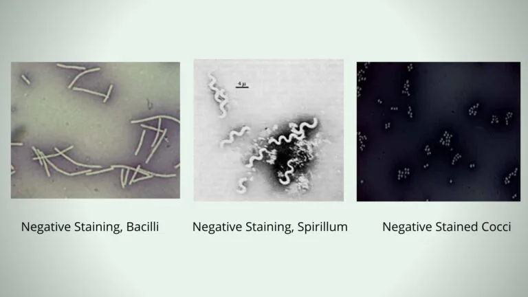
In negative staining method, an acidic dye is used known as India Ink or Nigrosin. When the bacterial cells are exposed to this stain, due to the presence of acidic nature it readily gives up a hydrogen ion (proton) and the chromophore. As a result, the dye becomes negatively charged, now the bacterial cell surface deflects the stain.
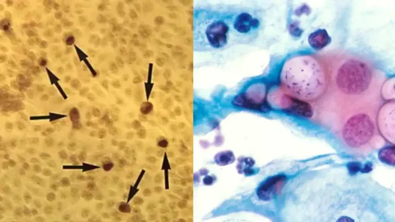
Chlamydia is caused by Chlamydia trachomatis which is a Gram-negative bacterium, and it is the organism that behave as an obligate intracellular parasite because it cannot produce its own metabolic energy. It is the process where the bacterium must survive and replicate inside the eukaryotic host cell, and this organism shows a biphasic developmental cycle
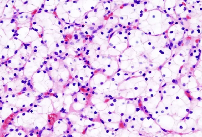
Hematoxylin and Eosin (H&E) stain is the routine staining method used in histology and histopathology for observing the basic structure of tissues. It is the process in which two dyes are applied on colorless tissue sections so that the cellular parts become clearly visible under the microscope. Hematoxylin behaves as a basic dye and it
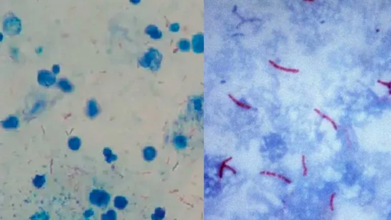
Acid fast stain is a differential staining method that is used to identify the acid fast bacteria which retain the primary dye even after treatment with acid alcohol. It is the process where the organisms having a high amount of mycolic acids and waxes in their cell wall show resistance to decolourisation. These are hydrophobic
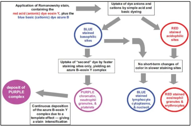
What are the Romanowsky Stains? Romanowsky stains are the polychromatic stains that are used in hematological and cytological studies to differentiate blood cells and bone marrow cells under the microscope. It is the process where a mixture of acidic dye (Eosin Y) and basic dyes (oxidized methylene blue or Azure B) is applied, and this
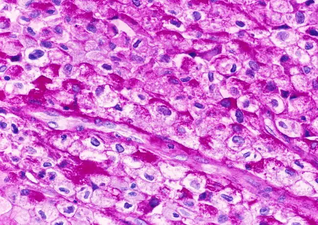
Periodic Acid-Schiff (PAS) staining is a histochemical method used to demonstrate carbohydrate and carbohydrate-rich compounds in tissue sections. It is the process where periodic acid is used to oxidize the 1,2-glycol groups of polysaccharides, mucin, glycogen and fungal cell wall components into aldehyde groups. These aldehydes then react with Schiff’s reagent to produce a characteristic
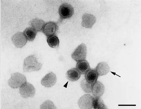
Negative staining of viruses is the process in which the viral particles is visualized by embedding them in an electron-dense stain rather than staining the virus itself. It is the method where the background gets heavily stained while the virus remains unstained, so the particles appear as light structures against a dark surrounding. It is
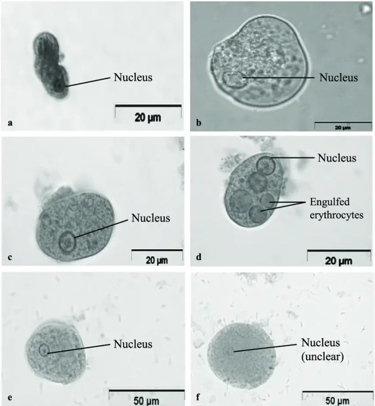
Wheatley Trichrome Staining is the permanent staining method used in parasitology for detecting intestinal protozoans from stool samples, and it is mainly applied when cysts and trophozoites is to be identified in a fixed smear. It is the process in which three dyes are used so that the parasite structures get a clear differential colour