Amoeba staining is the process used in laboratories for observing the structural details of free-living and intestinal amoebae. It is needed because most amoebae are transparent in fresh samples, and their internal parts cannot be seen clearly without color contrast. It is the process that helps in identifying pathogenic forms like Entamoeba histolytica from non-pathogenic
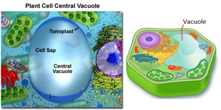
A light microscope may be used to see and examine the vacuole’s structure. While the vacuole doesn’t stain as the other organelles of the cell (because it does not contain many constituents that stain) tests have demonstrated that staining is possible for this organelle because the vacuole’s sap absorbs and stores dyes that are colored.
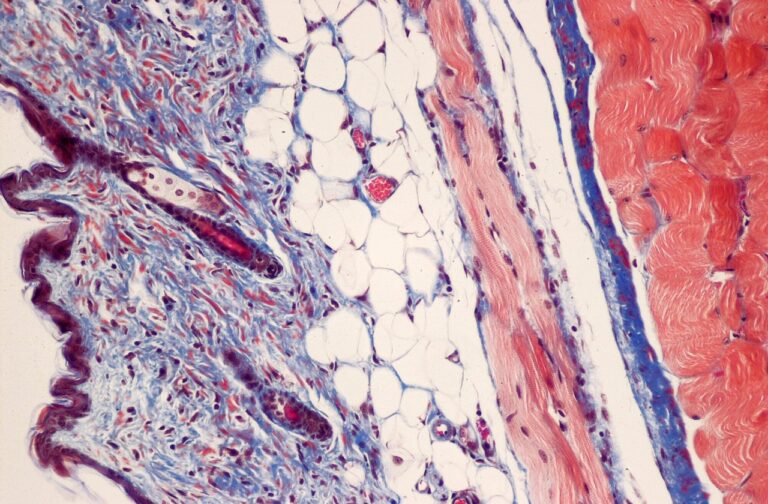
Masson’s Trichrome Staining (MTS) is a special histological staining method used to differentiate the connective tissue elements of a section. Principle of the Masson’s Trichrome Staining The principle of Masson’s Trichrome Staining is based on the differential penetration of dyes into tissue components of different density, and it is the process where electrostatic attraction and
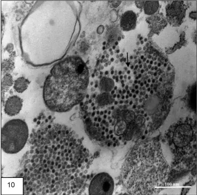
What is positive staining of Virus? Positive staining of viruses is the process in which the viral particle is made dark on a light background. It is the opposite of negative staining where the virus image is light and the background becomes dark. It is used widely to study the diverse morphology of viruses especially
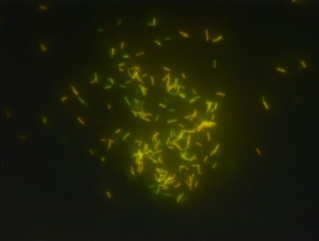
Auramine–Rhodamine staining is the process used to demonstrate acid-fast bacilli from clinical specimens and it is considered a modified form of the Acid-Fast staining technique. It is the process where fluorochrome dyes bind with the mycolic acid of the bacterial cell wall and it is visualized under a fluorescent microscope. It is the preferred screening
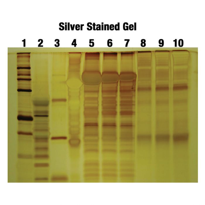
Silver staining is the process used for detecting and identifying proteins and nucleic acids in different gels, and it is highly sensitive in nature. It is the method where silver ions (Ag⁺) binds to different chemical groups present in proteins like carboxyl and sulfhydryl groups, and these ions is then reduced into metallic silver (Ag⁰).
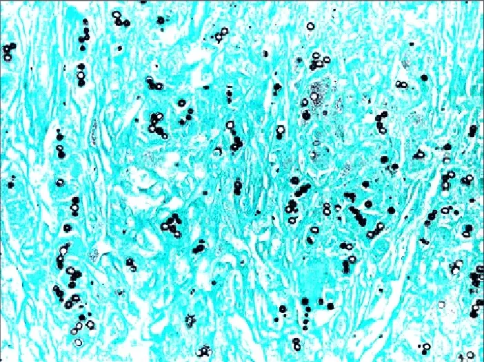
Grocott-Gomori’s Methenamine Silver (GMS) stain is the histological staining method that is used mainly for the detection of fungal microorganisms in tissue sections and smears. It is the process that was first developed by Gomori and later modified by Grocott, and it is used because of its high sensitivity to demonstrate carbohydrates present in the
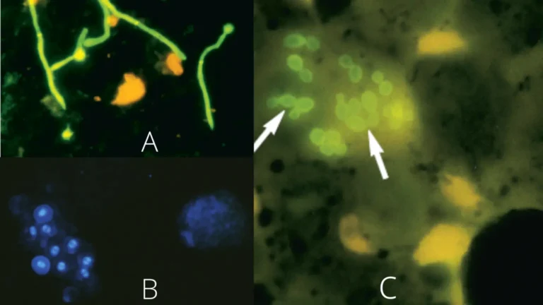
Calcofluor White (CFW) is a fluorescent dye that is used for the quick detection of fungi, yeasts, and some parasites. It is a stilbene derivative and it is the compound that absorbs ultraviolet light and then emits a visible blue light. It binds with β-linked polysaccharides like chitin in fungal cell walls and cellulose in
Different stains have been developed over time to distinguish bacteria species, separating them morphologically and the specific characteristics they possess. The most popular stain is Gram staining, acid-fast staining, and endospore staining. Each stain aims at identifying and defining bacteria according to their forms and morphologies.