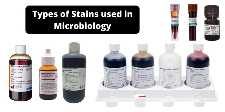
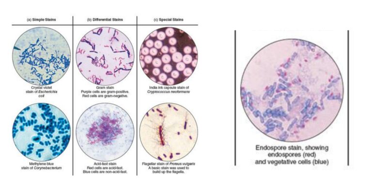
Different stains react or concentrate on different areas of a tissue or cell These properties can be utilized to highlight certain areas or regions. A few of the most well-known biological staining methods can be found below. If not otherwise indicated All of these dyes can be used on tissues and cells that are fixed as well as essential dyes (suitable for use in live organisms) are indicated.
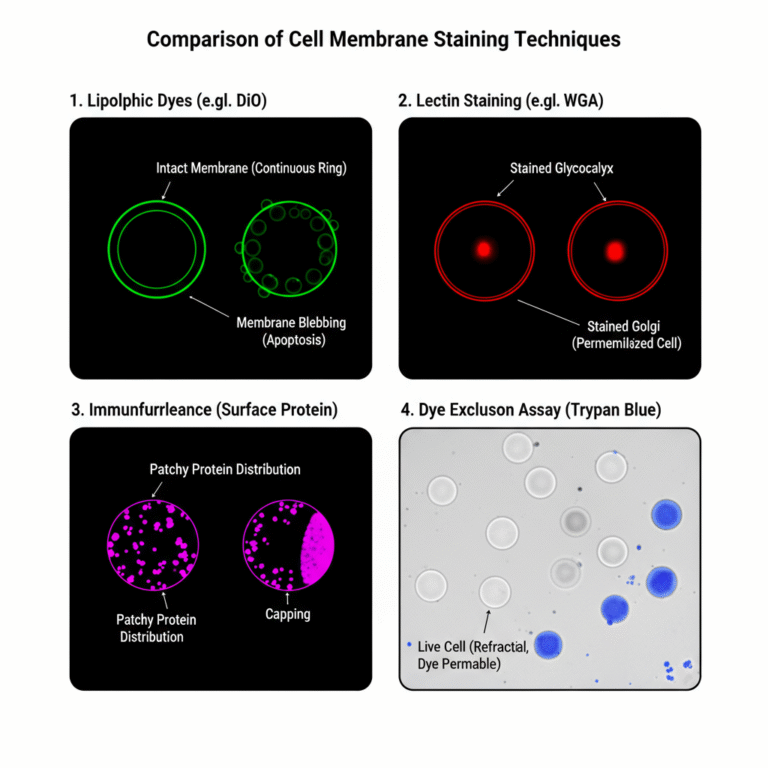
Cell membrane staining is the process in which the plasma membrane of a cell is visualized with the help of dyes or fluorescent probes. It is the thin outer covering of the cell, and it is normally transparent, so it cannot be seen clearly under a simple light microscope. It is the process where special
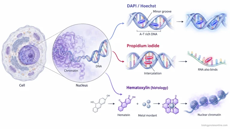
Nuclear staining labels cell nuclei for microscopy and analysis. Learn its principle, common stains such as DAPI and Hoechst, protocol, uses, and limitations.
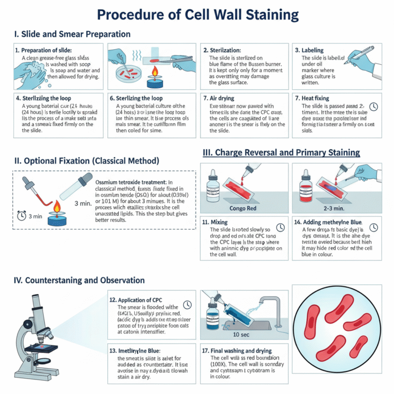
The composition of cell wall varies from species to species, it has been reported that the main constituents of cell wall is chitin, hemicellulose, and cellulose. In addition, prokaryotic cell wall contains peptidoglycan (also known as murein and mucopeptide). Peptidoglycan is mainly composed of sugar, amino acids (peptide; amino acids + glycan; sugar).
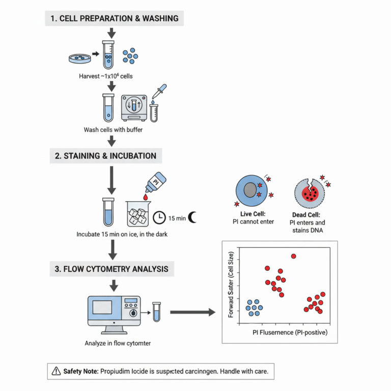
Cell viability analysis using Propidium Iodide is a simple method in which the dye is used to distinguish between living and dead cells. It is the process where Propidium Iodide (PI) is added to a cell suspension and the stained cells is then examined usually by flow cytometer. PI is a red-fluorescent dye that binds
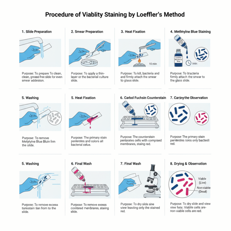
Loeffler’s methylene blue staining is a simple staining method used mainly for observing the morphology of Corynebacterium diphtheriae. It is the process where Loeffler’s alkaline methylene blue solution is applied on a fixed smear, and this stain contains a small amount of potassium hydroxide (KOH) which makes the solution alkaline. It is the alkalinity that
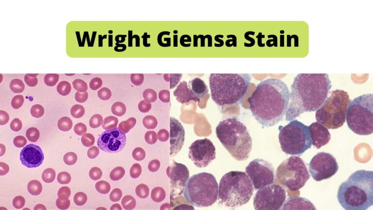
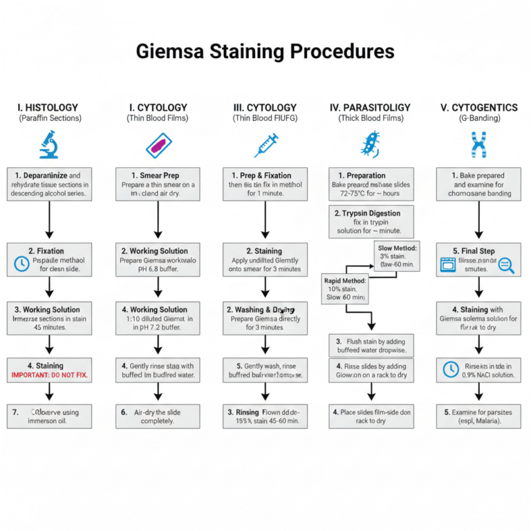
Giemsa stain is a polychromatic nucleic acid stain that is used mainly for demonstrating different blood cells and parasites in stained blood films. It is named after the German chemist Gustav Giemsa. It belongs to a group of stains known as Romanowsky stains. These are neutral stains made from a mixture of oxidized methylene blue,