

Endospore staining is commonly carried out using the Schaeffer–Fulton method in microbiology laboratories. In this method, malachite green is used as the primary stain. The bacterial smear is stained with malachite green and heated with steam. It is then washed with water and counterstained with safranin. Under the microscope, bacterial endospores appear green, while the vegetative cells appear pink or red.
In conventional Gram-stained preparations, bacterial endospores may appear as clear, refractile areas within the cells. Gram staining alone cannot reliably demonstrate the presence of endospores, so a special staining method is used for their examination.
What is Endospore Staining?
Endospore staining is a specialized differential staining technique used to visualize bacterial endospores and distinguish them from vegetative cells. It is also known as spore staining.
Bacterial endospores are dormant survival structures formed within certain bacterial cells. They help the bacteria survive under unfavourable environmental conditions. These are not reproductive spores, and their formation does not increase the number of bacterial cells.
Endospores present within vegetative cells are known as intracellular endospores. When the mother cell breaks down, the endospore is released and is called a free spore. Both intracellular endospores and free spores can be observed by endospore staining.
Purpose of Endospore Staining
The following are the purposes of endospore staining:
- It is used to detect the presence of endospores in a bacterial preparation.
- It helps to differentiate bacterial endospores from vegetative cells.
- It helps to distinguish spore-forming bacteria from non-spore-forming bacteria.
- It is used to observe the shape and position of endospores within bacterial cells. These characteristics help in the identification of bacteria.
Principle of Endospore Staining
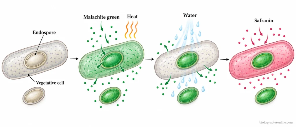
The principle of endospore staining is based on the difference in the staining properties of bacterial endospores and vegetative cells. Mature endospores have protective layers that are less permeable to ordinary stains. This makes the endospores difficult to stain.
In the Schaeffer–Fulton method, heat is used to help malachite green enter the endospores. Heat assists in the penetration of the stain and does not act as a chemical mordant. Once the endospores are stained, they retain the malachite green even after washing with water.
Malachite green is not strongly retained by the vegetative cells and is removed by washing with water. The vegetative cells then take up safranin and appear pink or red, while the endospores remain green.
Different Methods of Endospore Staining
The following are the different methods of endospore staining:
- Schaeffer–Fulton Method (Wirtz–Conklin Method) – It is the most commonly used method of endospore staining. Malachite green is used as the primary stain and safranin as the counterstain. The endospores appear green, while the vegetative cells appear pink or red.
- Dorner Method – In this method, carbol fuchsin is used to stain the endospores and nigrosin is used to provide a dark background. The endospores appear red, while the vegetative cells remain colourless.
- Moeller’s Method – The bacterial smear is treated with chromic acid before staining with heated carbol fuchsin. Methylene blue is used as the counterstain. The endospores appear red and the vegetative cells appear blue.
- Modified Ziehl–Neelsen Method – In this method, carbol fuchsin is used as the primary stain. The smear is decolorized and counterstained with methylene blue. The endospores appear red, while the vegetative cells appear blue.
Materials and Reagents Required for Endospore Staining
The following are the materials and reagents required for endospore staining by the Schaeffer–Fulton method:
Materials Required
- Bacterial Culture – A suitable culture of endospore-forming bacteria is used for preparing the smear.
- Clean Glass Slides – Used for the preparation of bacterial smears.
- Inoculating Loop – It is used to transfer the bacterial sample onto the slide.
- Bunsen Burner or Hot Plate – Used for heating the water to produce steam.
- Beaker and Slide Rack – The beaker contains water, and the rack supports the slide during steaming.
- Absorbent Paper – It is placed over the bacterial smear to hold the malachite green stain during heating.
- Forceps or Slide Holder – Used for handling the slide and removing the absorbent paper.
- Staining Tray and Wash Bottle – Used for washing the stained slide with water.
- Bibulous Paper – It is used to dry the slide after staining.
- Light Microscope and Immersion Oil – Used for examining the stained preparation under the oil-immersion objective.
Reagents Required
- Malachite Green Solution – It is used as the primary stain for bacterial endospores.
- Safranin Solution – It is used as the counterstain for vegetative cells.
- Distilled or Deionized Water – It is used to remove malachite green from vegetative cells and to wash off excess stain. Water also acts as the decolorizing agent.
Procedure of Endospore Staining
Procedure of Schaeffer–Fulton Method (Wirtz–Conklin Method)
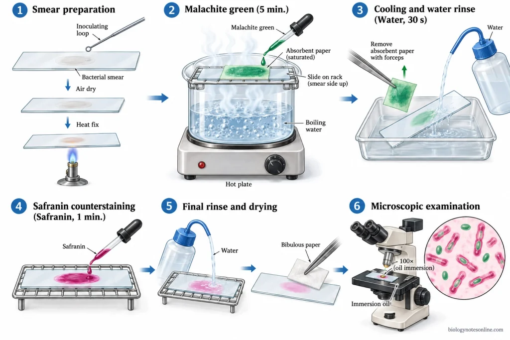
The procedure of the Schaeffer–Fulton method of endospore staining is given below:
- Take a clean glass slide and prepare a thin smear of the bacterial culture.
- Allow the smear to air dry completely and heat-fix it.
- Place the heat-fixed slide over a beaker of boiling water.
- Cover the bacterial smear with a small piece of absorbent paper.
- Add malachite green solution to the absorbent paper until it is completely soaked.
- Steam the slide for about 5 minutes. Keep the paper moist by adding more malachite green when required. Do not allow the stain to dry.
- Remove the absorbent paper using forceps and allow the slide to cool.
- Wash the slide gently with water for about 30 seconds to remove the excess malachite green.
- Cover the smear with safranin for 1 minute.
- Wash the slide gently with water to remove the excess safranin.
- Blot the slide dry using bibulous paper.
- Examine the stained smear under the oil immersion objective (100×) of a light microscope.
Procedure of Dorner Method
The procedure of Dorner method of endospore staining is given below:
- Take a clean glass slide and prepare a thin smear of the bacterial culture.
- Allow the smear to air dry completely and heat-fix it.
- Cover the bacterial smear with a small piece of blotting paper.
- Add carbol fuchsin solution to the blotting paper until it is completely soaked.
- Steam the slide for 5–10 minutes. Keep the paper moist by adding more carbol fuchsin when required.
- Remove the blotting paper using forceps.
- Decolorize the smear with acid-alcohol for 1 minute.
- Wash the slide gently with water and blot it dry.
- Apply saturated aqueous nigrosin solution and spread it to form a thin, even film.
- Allow the slide to air dry.
- Examine the stained smear under the oil immersion objective (100×) of a light microscope.
Procedure of Moeller’s Method
The procedure of Moeller’s method of endospore staining is given below:
- A thin smear of the bacterial culture is prepared on a clean glass slide.
- Allow the smear to air dry completely and heat-fix it.
- The fixed slide is immersed in absolute alcohol for 2 minutes and then in chloroform for 2 minutes.
- Wash the slide thoroughly with water.
- The bacterial smear is treated with 5% aqueous chromic acid for 1 minute.
- Wash the slide with water to remove the chromic acid.
- Cover the smear with Ziehl’s carbol fuchsin and heat gently until steam rises. Keep the stain on the smear for 10 minutes. Do not allow it to dry.
- The excess stain is removed by washing the slide with water.
- Decolorize the smear with 5% sulphuric acid for 5 seconds.
- Wash the slide thoroughly with water.
- The smear is counterstained with Kühne’s carbol methylene blue for 1–2 minutes.
- Wash the slide with water and allow it to dry.
- Examine the stained smear under the oil immersion objective (100×) of a light microscope. The endospores appear red, while the vegetative cells appear blue.
Procedure of Modified Ziehl–Neelsen Method
The procedure of modified Ziehl–Neelsen method of endospore staining is given below:
- Take a clean glass slide and prepare a thin smear of the bacterial culture using normal saline.
- Allow the smear to air dry completely and heat-fix it.
- Cover the bacterial smear with concentrated carbol fuchsin.
- Heat the slide gently until steam rises. Continue heating for 3–4 minutes. Do not allow the stain to boil or dry.
- Wash the slide with water to remove the excess carbol fuchsin.
- Decolorize the smear with 0.5% sulphuric acid.
- Wash the slide thoroughly with water.
- The smear is counterstained with Loeffler’s alkaline methylene blue for 2 minutes.
- Wash the slide with water and blot it dry.
- Examine the stained smear under the oil immersion objective (100×) of a light microscope. The endospores appear red, while the vegetative cells appear blue.
Results and Interpretation of Endospore Staining
The following are the results and interpretation of endospore staining by the Schaeffer–Fulton method:
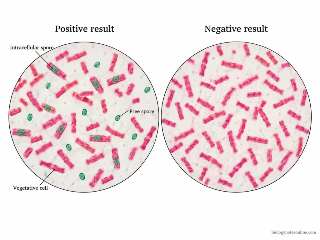
Positive Endospore Staining Result
- The endospores appear green, while the vegetative cells appear pink or red.
- Green endospores may be present within the pink or red vegetative cells. These are called intracellular endospores.
- Free endospores appear as separate green structures outside the vegetative cells.
- The presence of green endospores indicates a positive endospore staining result.
Negative Endospore Staining Result
- In a negative result, only pink or red vegetative cells are observed. No green endospores are seen.
- A negative result does not always mean that the bacteria cannot form endospores. Young cultures may not produce endospores if the conditions required for sporulation are not present.
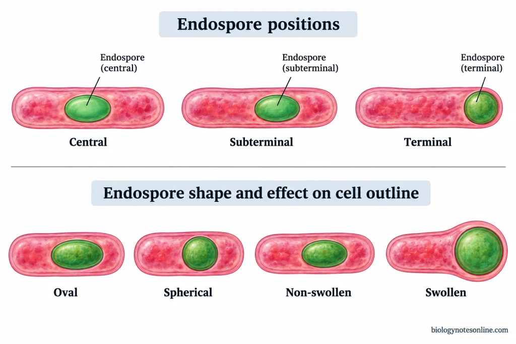
Endospore Shape and Position
- Endospores may be oval or spherical in shape.
- The endospores may be present in the central, subterminal or terminal position of the bacterial cell.
- In some bacteria, the endospore causes swelling of the vegetative cell. In others, the endospore does not cause swelling.
- The shape, position and swelling of endospores help in the identification of bacteria. These characteristics alone are not sufficient for the identification of a bacterial species.
Examples of Endospore Staining
The following are some examples of bacteria used in endospore staining by the Schaeffer–Fulton method:
- Bacillus subtilis – It is an endospore-forming bacterium that produces oval endospores. The endospores appear green within the pink or red vegetative cells. Free green spores may also be present.
- Bacillus megaterium – This bacterium produces endospores that appear green within the pink or red rod-shaped cells. Free endospores may also be seen in older cultures.
- Clostridium sporogenes – It produces oval endospores that are commonly found near the ends of the bacterial cells. The endospores appear green, while the vegetative cells appear pink or red.
- Escherichia coli – It is a non-endospore-forming bacterium. Only pink or red vegetative cells are observed after staining. No green endospores are present.
Comparison of Endospore Staining Results
| Bacterial Organism | Endospore Formation | Expected Staining Result |
|---|---|---|
| Bacillus subtilis | Positive | Green endospores within pink/red cells or as free spores |
| Bacillus megaterium | Positive | Green endospores within pink/red rods or as free spores |
| Clostridium sporogenes | Positive | Green endospores, commonly near the ends of pink/red cells |
| Escherichia coli | Negative | Pink/red vegetative cells without green endospores |
The formation of endospores depends upon the age of the bacterial culture and the growth conditions. In spore-forming bacteria, endospore formation usually takes place when nutrients become limited or the environmental conditions are unfavourable. Young cultures may contain only vegetative cells, and endospores may not be observed even in spore-forming bacteria.
The shape and position of endospores help in the identification of bacteria. However, these characteristics alone are not sufficient for the identification of a bacterial species.
Applications of Endospore Staining
The following are the applications of endospore staining:
- Endospore staining is used in microbiology laboratories for the examination of spore-forming bacteria such as Bacillus and Clostridium.
- The shape and position of endospores help in the preliminary identification of bacteria. Other tests are required for the identification of the bacterial species.
- In clinical microbiology, it is used to examine suspected spore-forming bacteria isolated from clinical samples.
- It is used in food microbiology to detect the presence of endospores in bacterial isolates.
- Spore-forming bacteria isolated from soil and other environmental samples are studied by endospore staining in environmental microbiology.
- It is used to study endospore formation in bacterial cultures grown under different conditions.
- In teaching laboratories, the difference between endospores and vegetative cells can be observed under the microscope by endospore staining.
Advantages of Endospore Staining
The following are the advantages of endospore staining:
- Endospores can be distinguished from vegetative cells by their colour. In the Schaeffer–Fulton method, the endospores appear green, while the vegetative cells are pink or red.
- Detection of endospores that are not clearly visible after Gram staining.
- Both free endospores and those present inside the bacterial cells can be observed under a light microscope.
- The presence of endospores in a positive staining result helps to differentiate spore-forming bacteria from non-spore-forming bacteria.
- Examining the shape and position of endospores for the preliminary identification of spore-forming bacteria.
- The method requires only common laboratory reagents and a light microscope.
- To study the formation of endospores in bacterial cultures grown under different conditions.
Limitations of Endospore Staining
The following are the limitations of endospore staining:
- Endospore staining cannot identify the bacterial species. The shape and position of endospores can only help in the preliminary identification of bacteria.
- Endospores may not be present in young bacterial cultures or under conditions that do not favour sporulation. A negative result does not always mean that the bacteria cannot produce endospores.
- The staining result depends upon proper heating of the smear. Insufficient heating may prevent malachite green from entering the endospores.
- The malachite green must not be allowed to dry during heating. Drying of the stain may affect the staining result.
- It cannot determine whether the stained endospores are viable or non-viable. Other methods are required to test their viability.
- The method does not provide an accurate count of endospores present in a bacterial sample.
- Immature and damaged endospores may not be easily distinguished from mature endospores by this staining method.
- Endospore staining does not determine the resistance of bacterial endospores to heat or disinfectants.
Precautions and Quality Control of Endospore Staining
Precautions of Endospore Staining
The following are the precautions of endospore staining:
- A thin smear of the bacterial culture should be prepared on a clean glass slide. Thick smears may make microscopic examination difficult.
- Allow the smear to air dry completely before heat fixation. Excessive heating should be avoided.
- Heat the slide gently during staining. Do not allow the malachite green to boil.
- Keep the absorbent paper moist by adding more malachite green when required. Do not allow the stain to dry during steaming.
- Allow the slide to cool before washing. Wash gently with water to prevent the removal of the bacterial smear.
- The safranin should be applied for the required time. Wash the excess stain with water.
- The glass slide should be free from dirt and stain deposits. These may be mistaken for endospores during microscopic examination.
- Use forceps to handle hot slides. Gloves and protective eyewear should be worn when handling staining reagents.
Quality Control of Endospore Staining
The following are the quality control measures of endospore staining:
- A known spore-forming bacterium such as Bacillus subtilis is used as a positive control. The culture should contain endospores.
- In the positive control, the endospores should appear green, while the vegetative cells appear pink or red.
- Escherichia coli is used as a negative control. Only pink or red vegetative cells should be observed.
- Both positive and negative control slides should be stained along with the test sample.
- The staining reagents should be of the correct concentration and within their expiry dates.
- If the control slides do not give the expected results, the reagents and staining procedure should be checked and the staining repeated.
- The absence of endospores in a test culture does not always indicate a non-spore-forming bacterium. The age of the culture and growth conditions should also be considered.
References
- Bartholomew, J. W., & Mittwer, T. (1950). A simplified bacterial spore stain. Stain Technology, 25(3), 153–156. https://doi.org/10.3109/10520295009110979
- Brown, A. E., & Smith, H. R. (2017). Benson’s microbiological applications: Laboratory manual in general microbiology (Concise version, 14th ed.). McGraw-Hill Education. https://www.mheducation.co.uk/looseleaf-benson-s-microbiological-applications-laboratory-manual-concise-version-9781259705236-emea
- Eyre, J. W. H. (1902). The elements of bacteriological technique: A laboratory guide for the medical, dental, and technical student. W. B. Saunders. https://books.google.com/books?id=D778v2N8U_0C
- Parker, N., Schneegurt, M., Tu, A.-H. T., Lister, P., & Forster, B. M. (2016). Microbiology. OpenStax. https://openstax.org/books/microbiology/pages/1-introduction
- Reynolds, J., Moyes, R., & Breakwell, D. P. (2009). Differential staining of bacteria: Endospore stain. Current Protocols in Microbiology, 15(1), A.3J.1–A.3J.5. https://doi.org/10.1002/9780471729259.mca03js15