

In capsule staining, a bacterial smear is prepared on a glass slide without heat fixation. The smear is stained with suitable staining reagents and then examined under a light microscope. The staining reagents and preparation of the smear depend upon the method used.
The bacterial capsule may not be clearly visible by simple staining or Gram staining. Heat fixation may cause shrinkage or distortion of the capsule. Special care is required during the preparation and staining of the smear.
What is Capsule Staining?
Capsule staining, also known as capsular staining, is a specialized microscopic staining technique used to demonstrate the extracellular capsules surrounding certain bacterial cells.
The bacterial capsule is a well-organized layer present outside the cell envelope and is closely attached to the cell surface. It is mainly composed of polysaccharides, but some bacterial capsules are made up of polypeptides. A slime layer is less organized and loosely attached to the cell surface.
In many staining preparations, the capsule appears as a clear halo around the bacterial cell. Similar staining techniques are also used to demonstrate the capsules of certain yeasts.
Objectives of Capsule Staining
The following are the objectives of capsule staining:
- To detect the presence of capsules around bacterial cells.
- To differentiate between encapsulated and non-encapsulated bacteria.
- To observe the size and position of the bacterial capsule under a light microscope.
- To prepare and stain bacterial smears using a suitable capsule staining method.
- To examine and interpret the microscopic results of capsule staining.
- To help in the identification of capsule-producing bacteria.
Principle of Capsule Staining
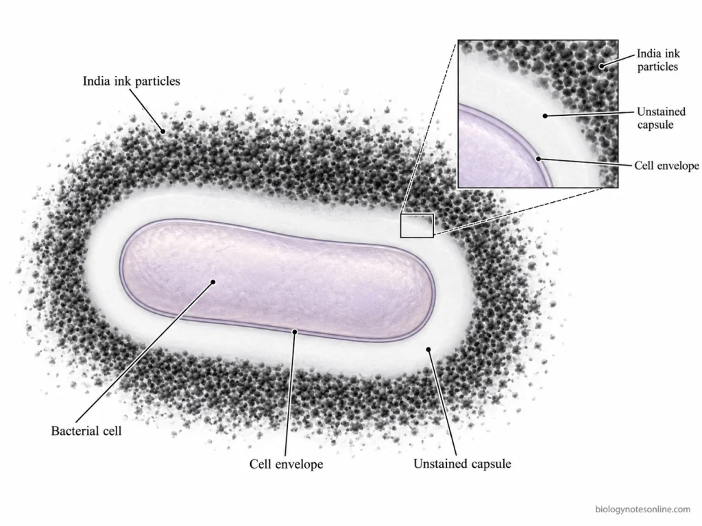
The bacterial capsule contains a large amount of water. Many conventional dyes have a low affinity for the capsular material and do not bind strongly to it. Capsule staining depends upon the difference in the staining of the bacterial cells, capsules and the surrounding background. The bacterial cells, background or both are stained, while the capsule remains unstained or takes up very little colour.
India ink is used for negative staining of the background. The ink particles cannot penetrate the capsule and remain outside it, forming a dark background around the bacterial cells. The unstained capsule is seen as a clear zone between the bacterial cell and the dark background.
In Anthony’s method, crystal violet is used to stain the bacterial cells and the protein-containing background. Copper sulphate acts as a mordant and decolorizing agent. It removes the crystal violet loosely attached to the capsule, but the stain remains in the bacterial cells. These cells appear purple. The capsule may appear clear or pale blue.
Congo red is used for staining the background in Maneval’s method. Acid fuchsin stains the bacterial cells. The acetic acid present in Maneval’s stain lowers the pH and changes the Congo red background from red to blue. It also acts as a mordant and helps in stabilizing the capsule. The unstained capsule is seen as a clear halo around the stained bacterial cell.
Reagents and Materials Required for Capsule Staining
The following are the materials and reagents required for capsule staining. The staining reagents depend upon the method used.
Materials and Equipment
- Glass Slides and Coverslips – Clean glass slides are required for the preparation of bacterial smears. Coverslips are used in certain wet-mount methods.
- Bacterial Culture – A suitable bacterial culture grown in an appropriate culture medium.
- Inoculating Loop – Used to transfer the bacterial culture onto the glass slide.
- Staining Rack and Tray – The slides are placed on a staining rack during staining, and the excess staining solution is collected in the tray.
- Light Microscope and Immersion Oil – The stained bacterial cells are examined under a light microscope. Immersion oil is used with the oil immersion objective.
- Protective Equipment – Laboratory coat, disposable gloves and safety goggles.
Staining Reagents
- Crystal Violet – In Anthony’s method, crystal violet stains the bacterial cells and the protein-containing background purple. It is used as the primary stain.
- Copper Sulfate Solution – Copper sulfate is used with crystal violet in Anthony’s method. It acts as a mordant and decolorizing agent. The capsule may appear pale blue.
- Congo Red – It is used for staining the background in Maneval’s method. The red background changes to blue under acidic conditions.
- Maneval’s Staining Solution – This solution contains acid fuchsin and acetic acid. Acid fuchsin stains the bacterial cells red. The acetic acid lowers the pH and helps in stabilizing the capsule.
- India Ink or Nigrosin – These are used for negative staining. They form a dark background around the bacterial cells, while the capsule remains unstained.
- Serum or Protein-containing Medium – Serum protein, milk broth or litmus milk is used in Anthony’s method to provide a protein-containing background for staining with crystal violet.
Methods of Capsule Staining
Capsule staining can be carried out by negative staining, Anthony’s method, Maneval’s method and Hiss staining. The staining reagents are different in these methods. The appearance of the bacterial cells and capsules also depends upon the method used.
1. Negative Staining Method (India Ink or Nigrosin)
India ink or nigrosin stains the background in this method. The stain particles remain outside the capsule and form a dark background around the bacterial cells. The cells are colourless or may appear pale.
The capsule remains unstained. It is visible as a clear halo between the cell and the dark background.
Negative staining is used to demonstrate capsules in bacterial cultures. It is also used for certain encapsulated yeasts.
2. Anthony’s Capsule Staining Method
Anthony’s method uses crystal violet as the primary stain and copper sulphate as a mordant and decolorizing agent. Crystal violet stains the bacterial cells and the protein-containing background purple.
Milk broth, litmus milk or serum protein may be used to provide the protein-containing background. The crystal violet loosely attached to the capsule is removed by copper sulphate. It does not remove the stain from the bacterial cells.
The cells are purple, whereas the capsule may appear clear or pale blue.
3. Maneval’s Capsule Staining Method
Maneval’s method uses Congo red for staining the background. The bacterial cells are stained red or pink by acid fuchsin present in Maneval’s solution. This solution also contains acetic acid.
The pH is lowered by acetic acid, which changes the Congo red background from red to blue. Acetic acid also acts as a mordant and helps in stabilizing the capsule.
The capsule does not take up the stain and is visible as a clear or white halo around the bacterial cells. The background is blue.
4. Hiss Capsule Staining Method
Hiss staining is used for staining bacterial capsules and has different modifications. In some commonly used modifications, gentian violet or crystal violet is used along with copper sulphate solution.
The bacterial cells are dark violet. The capsule may be pale blue or light violet. The background colour varies and may appear light blue.
Serum or other protein-containing material is used in some methods. The historical and modified methods may differ in the reagents used, preparation and microscopic appearance.
Difference Between Capsule Staining Methods
| Method | Principal Reagents | Staining Approach | Bacterial Cell | Background | Capsule |
|---|---|---|---|---|---|
| Negative staining | India ink or nigrosin | Staining of the background | Colourless or pale | Dark | Clear halo |
| Anthony’s method | Crystal violet and copper sulphate | Staining of bacterial cells and protein-containing background | Purple | Purple | Clear or pale blue |
| Maneval’s method | Congo red and Maneval’s solution | Staining of bacterial cells and background | Red or pink | Blue | Clear or white halo |
| Hiss method | Gentian violet or crystal violet and copper sulphate | Positive staining followed by copper sulphate treatment | Dark violet | Variable, often light blue | Pale blue or light violet |
Procedure of Capsule Staining
The procedure of capsule staining by different methods is given below:
Procedure of Anthony’s Capsule Staining Method
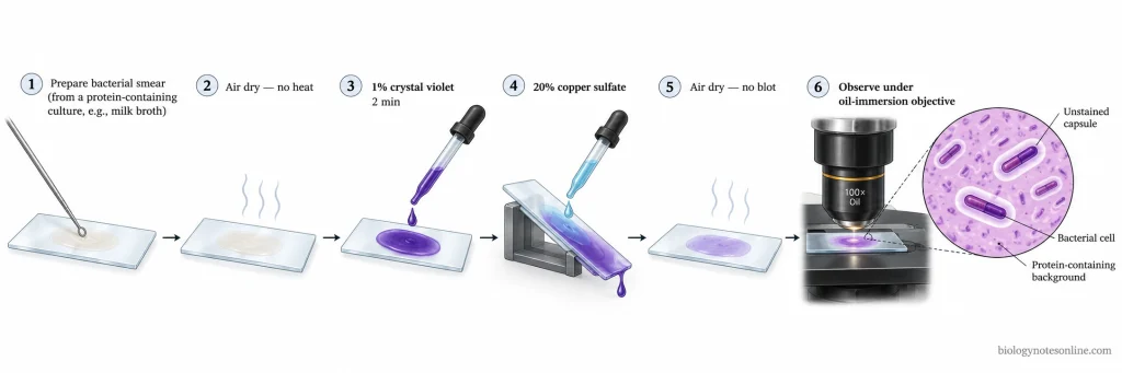
- Take a clean glass slide and prepare a thin smear using a 12–18-hour bacterial culture grown in milk broth or litmus milk.
- If the culture is grown in another medium, serum protein may be added during smear preparation.
- Allow the smear to air dry completely. Do not heat fix the smear.
- Cover the smear with 1% crystal violet solution for 2 minutes.
- The excess stain is removed by gently washing with 20% copper sulphate solution. Water should not be used.
- Allow the slide to air dry. Do not blot the smear as the bacterial cells are not heat fixed.
- Examine the stained smear under the oil immersion objective of a light microscope.
- The bacterial cells and the protein-containing background appear purple. The capsule is seen as a clear zone around the cells.
Procedure of Maneval’s Capsule Staining Method
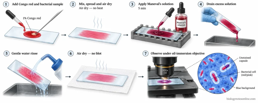
- Take a clean glass slide and place a few drops of 1% aqueous Congo red solution on it.
- Transfer a small amount of bacterial culture into the Congo red solution and prepare a thin smear without adding water.
- The smear is allowed to air dry at room temperature. Heating is not required.
- Place the slide on a staining rack and cover the smear with Maneval’s staining solution for 5 minutes.
- Remove the excess staining solution by gently tilting the slide.
- Wash the slide carefully with water. The water should flow gently across the slide to prevent the removal of the unfixed smear.
- Place the slide on absorbent paper and allow it to air dry. Do not blot the smear.
- Examine the slide under the oil immersion objective at 1000× magnification.
- The bacterial cells appear red or pink against a blue background. The unstained capsule is seen as a clear halo around the cells.
Procedure of India Ink Negative Staining
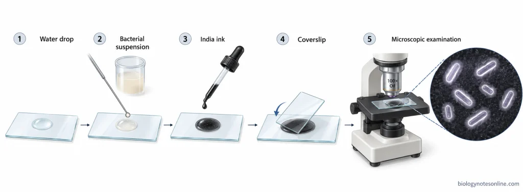
- Take a clean glass slide and place a small drop of water on it.
- Transfer a small amount of bacterial growth from an agar culture into the drop of water and prepare a suspension.
- Add a drop of filtered India ink to the bacterial suspension and mix gently.
- Place a clean coverslip over the preparation. Air bubbles should be avoided.
- Examine the wet mount immediately under a light microscope. Start with the 10× objective, followed by the 40× objective. The 100× oil immersion objective may be used if required.
- The bacterial cells appear colourless or pale against a dark background. The capsule is seen as a clear zone around the cells.
- Air-drying, heat fixation and washing are not required in this method. After examination, the slide and coverslip are discarded in a suitable disinfectant.
Results of Capsule Staining
The result of capsule staining depends upon the presence or absence of a capsule around the bacterial cell. The colour of the bacterial cells, capsule and background depends upon the staining method used.
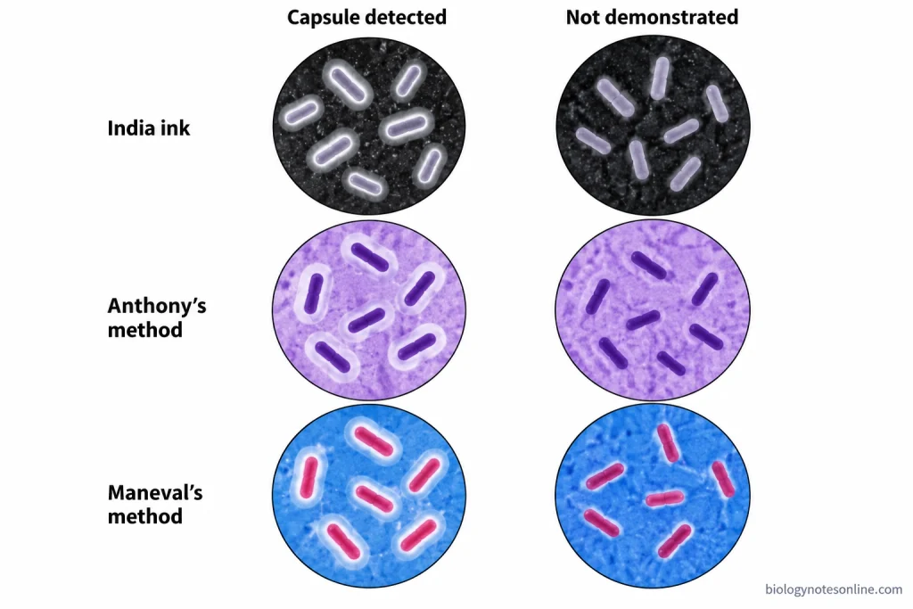
Positive Capsule Staining Result
In a positive capsule stain, the capsule is seen as a clear or lightly coloured zone around the bacterial cell. The bacterial cell is present in the centre, while the stained background is seen outside the capsule.
In negative staining with India ink or nigrosin, the bacterial cells appear colourless or pale against a dark background. The capsule remains unstained and appears as a transparent halo.
In Anthony’s method, the bacterial cells and the protein-containing background appear purple. The capsule is seen as a clear or pale blue zone around the cells.
In Maneval’s method, the bacterial cells appear red or pink against a blue background. The capsule remains colourless and is seen as a clear halo between the bacterial cell and the background.
Negative Capsule Staining Result
In a negative capsule stain, no clear halo is seen around the bacterial cells. The cell boundary appears directly against the stained background without a separate capsule zone.
This result indicates that the capsule is not demonstrated in the stained preparation.
A negative capsule staining result is different from the negative staining method. India ink or nigrosin is used in negative staining, which may give either a positive or negative capsule result.
Microscopic Appearance of Capsule Staining
The bacterial cell, capsule and background are the three main parts shown in a capsule staining diagram. In a positive result, the capsule forms a clear or pale zone between the cell and the background. In a negative result, this zone is absent.
| Method | Bacterial Cell Colour | Background Colour | Capsule Appearance | Interpretation |
|---|---|---|---|---|
| India ink or nigrosin – Positive | Colourless or pale | Dark | Clear halo around the cell | Capsule present |
| India ink or nigrosin – Negative | Colourless or pale | Dark | No clear halo | Capsule not demonstrated |
| Anthony’s method – Positive | Purple | Purple | Clear or pale blue zone | Capsule present |
| Anthony’s method – Negative | Purple | Purple | No separate capsule zone | Capsule not demonstrated |
| Maneval’s method – Positive | Red or pink | Blue | Clear or white halo | Capsule present |
| Maneval’s method – Negative | Red or pink | Blue | No clear halo | Capsule not demonstrated |
Examples of Capsule-Forming Bacteria
The following are some examples of bacteria that produce capsules:
- Klebsiella pneumoniae – It is a Gram-negative, rod-shaped bacterium having a polysaccharide capsule. In capsule staining, the capsule appears as a clear zone around the bacterial cells. The amount of capsule produced differs between strains and also depends upon the culture conditions.
- Streptococcus pneumoniae – It is a Gram-positive bacterium that produces a polysaccharide capsule. The composition of the capsule differs among different serotypes. The encapsulated cells are seen as pairs of cocci surrounded by a clear halo. Some strains may produce less capsule or may lose capsule production.
- Bacillus anthracis – The capsule of B. anthracis is composed of poly-γ-D-glutamic acid. It is a polypeptide capsule, whereas most bacterial capsules are composed of polysaccharides. The capsule is seen as a clear zone around the bacterial cells. Capsule production depends upon the presence of capsule genes and suitable growth conditions.
- Escherichia coli – Some strains of E. coli produce a polysaccharide capsule known as the K antigen. The K1 capsule is composed of polysialic acid. In capsule staining, the encapsulated cells may appear with a clear halo around them. This halo is absent in non-encapsulated strains. The production of certain capsules, including K1 and K5, also depends upon temperature and culture conditions.
Applications of Capsule Staining
The following are the applications of capsule staining:
- It is used to detect the presence of capsules around bacterial cells.
- It helps to differentiate between encapsulated and non-encapsulated bacteria.
- It is used to observe the size and position of the capsule around bacterial cells under a light microscope.
- Capsule staining helps in the preliminary identification of encapsulated bacteria such as Klebsiella pneumoniae and Streptococcus pneumoniae. Other tests are required for confirmation.
- It is used to study capsule production in different bacterial strains and under different culture conditions.
- It helps in studying the capsule as a virulence factor in pathogenic bacteria. The capsule protects certain bacteria from phagocytosis.
- India ink staining is used for the demonstration of the capsule of Cryptococcus neoformans in clinical specimens. Other diagnostic tests are required for confirmation.
Precautions of Capsule Staining
The following are the precautions of capsule staining:
- Use clean glass slides free from grease for the preparation of bacterial smears.
- The bacterial culture selected for staining should be suitable. Capsule production depends upon the culture medium and growth conditions.
- A thin smear is prepared using a small amount of bacterial culture. Thick smears should not be used.
- The capsular material may be removed from the bacterial cells if the culture is shaken vigorously.
- The smear is allowed to air dry completely. Heat fixation may cause shrinkage or distortion of the capsule and should be avoided.
- In Anthony’s method, the excess crystal violet is removed with 20% copper sulphate solution. Water is not used for washing the smear.
- The Congo red smear in Maneval’s method is prepared without adding water. After staining, it is washed gently with water.
- Blotting or rubbing the stained smear may remove the bacterial cells from the slide, as the smear is not heat fixed.
- Place the coverslip carefully on the India ink wet mount to avoid air bubbles. The preparation is examined immediately. Drying or heating is not required.
- The staining reagents should be used in the required concentrations, and the specified staining time should be maintained.
- A laboratory coat, gloves and safety goggles should be worn while handling bacterial cultures and staining reagents. Maneval’s solution containing phenol and glacial acetic acid should be prepared in a fume hood.
- Used slides, coverslips and bacterial cultures are discarded according to the laboratory biosafety procedures.
Limitations of Capsule Staining
The following are the limitations of capsule staining:
- Capsule production depends upon the bacterial strain, culture medium and growth conditions. A negative result does not always indicate that the bacterium cannot produce a capsule.
- Some bacterial capsules are very thin and may not be clearly visible under a light microscope.
- Heat fixation may cause shrinkage or distortion of the capsule. The shrinking of bacterial cells may also produce a false clear zone around them.
- In methods where the smear is not heat fixed, bacterial cells may be removed from the slide during washing.
- Thick smears or excess India ink may make microscopic examination difficult.
- Air bubbles and other particles present in the preparation may be mistaken for encapsulated cells and give false-positive results.
- Drying of certain stains may produce a clear zone around the bacterial cells. This may be mistaken for a capsule.
- The staining result depends upon the preparation of the smear and the staining method used. Improper staining may give a false-negative result.
- Capsule staining cannot determine the chemical composition or type of the bacterial capsule.
- It cannot be used alone for the identification of bacterial species. Other laboratory tests are required for confirmation.
Advantages of Capsule Staining
The following are the advantages of capsule staining:
- It is a simple and rapid method used for the detection of bacterial capsules.
- It helps to differentiate between encapsulated and non-encapsulated bacteria.
- The bacterial capsule is seen as a clear or pale zone around the cells, which is difficult to observe by ordinary staining methods.
- It does not require expensive instruments. A light microscope and commonly available staining reagents are sufficient.
- In India ink negative staining, heat fixation is not required. This helps to prevent the distortion of bacterial cells and capsules.
- A small amount of bacterial culture is required for staining.
- It is used to observe the size and position of the capsule around bacterial cells.
- Capsule staining helps in the preliminary identification of certain capsule-producing bacteria.
- It can also be used to study capsule production in different bacterial cultures and under different growth conditions.
References
- Aldawood, E., & Roberts, I. S. (2022). Regulation of Escherichia coli group 2 capsule gene expression: A mini review and update. Frontiers in Microbiology, 13, Article 858767. https://doi.org/10.3389/fmicb.2022.858767
- Anthony, E. E., Jr. (1931). A note on capsule staining. Science, 73(1890), 319–320. https://doi.org/10.1126/science.73.1890.319
- Breakwell, D. P., Moyes, R. B., & Reynolds, J. (2009). Differential staining of bacteria: Capsule stain. Current Protocols in Microbiology, 15(1), A.3I.1–A.3I.4. https://doi.org/10.1002/9780471729259.mca03is15
- Duguid, J. P. (1951). The demonstration of bacterial capsules and slime. Journal of Pathology and Bacteriology, 63(4), 673–685. https://doi.org/10.1002/path.1700630413
- Ezzell, J. W., Jr., & Welkos, S. L. (1999). The capsule of Bacillus anthracis, a review. Journal of Applied Microbiology, 87(2), 250. https://doi.org/10.1046/j.1365-2672.1999.00881.x
- Moat, A. G., Foster, J. W., & Spector, M. P. (2002). Microbial physiology (4th ed.). Wiley-Liss. https://doi.org/10.1002/0471223867
- Parker, N., Schneegurt, M., Tu, A.-H. T., Lister, P., & Forster, B. M. (2016). Microbiology. OpenStax. https://openstax.org/books/microbiology/pages/1-introduction
- Paton, J. C., & Trappetti, C. (2019). Streptococcus pneumoniae capsular polysaccharide. Microbiology Spectrum, 7(2), GPP3-0019-2018. https://doi.org/10.1128/microbiolspec.GPP3-0019-2018
- Walker, K. A., & Miller, V. L. (2020). The intersection of capsule gene expression, hypermucoviscosity and hypervirulence in Klebsiella pneumoniae. Current Opinion in Microbiology, 54, 95–102. https://doi.org/10.1016/j.mib.2020.01.006
- Whitfield, C., & Roberts, I. S. (1999). Structure, assembly and regulation of expression of capsules in Escherichia coli. Molecular Microbiology, 31(5), 1307–1319. https://doi.org/10.1046/j.1365-2958.1999.01276.x