

In Giemsa staining, a blood smear is prepared on a clean glass slide and stained with a diluted Giemsa staining solution. The slide is then rinsed, allowed to dry and examined under a microscope.
For malaria microscopy, two types of blood films are used: thin blood films and thick blood films. These two types of blood films are prepared differently and have different fixation requirements.
What is Giemsa Stain?
Giemsa stain is a Romanowsky-type differential stain containing a mixture of basic and acidic dyes. It is used for staining blood films and other cellular preparations. The stain contains more than one dye and produces different staining of cellular components, unlike a simple stain that uses only one dye.
Giemsa stain is named after Gustav Giemsa, a German chemist who developed this staining method. It is also known as Giemsa’s stain. Romanowsky stain refers to a group of related stains, which includes Giemsa, Wright and Leishman stains. It is not another name for Giemsa stain.
Objectives of Giemsa Stain
The following are the objectives of Giemsa staining:
- To stain and differentiate different types of blood cells.
- To examine the nucleus, cytoplasm and granules of blood cells.
- To detect malaria parasites in thick and thin blood films.
- To identify different species of Plasmodium by examining their morphology in thin blood films.
- To estimate the number of malaria parasites present in a blood sample.
- To detect other parasites such as Trypanosoma in blood and Leishmania in tissue samples.
- To study the banding patterns of chromosomes during cytogenetic analysis.
Principle of Giemsa Staining
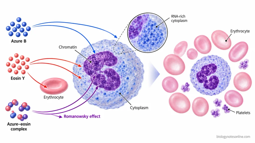
The principle of Giemsa staining is based on the binding of basic and acidic dyes to different cellular components. The basic dyes, mainly azure B and other methylene blue derivatives, are positively charged and bind to negatively charged substances such as DNA and RNA. Eosin Y is an acidic dye that binds to basic components of cellular proteins.
Azure B also combines with eosin Y in certain cellular structures and produces a characteristic purple colour. This is known as the Romanowsky–Giemsa effect. The interaction of these dyes produces different staining colours in chromatin, cytoplasm, cellular granules and erythrocytes. In malaria blood films, the chromatin of the parasites also reacts with these dyes.
The staining reaction depends upon the pH of the buffer, concentration of the dyes and fixation of the specimen. Changes in these conditions can affect the staining colours and differentiation of cellular structures.
Composition of Giemsa Stain
Giemsa stain consists of a mixture of basic and acidic dyes. The main colouring components are methylene blue, azure dyes and eosin Y. Methanol and glycerol are also used in the preparation of Giemsa stock solution.
The following are the components of Giemsa stain:
- Methylene Blue – It is a basic dye that stains acidic cellular components such as nucleic acids.
- Azure Dyes – These are basic dyes derived from methylene blue. Azure B combines with eosin Y to produce the characteristic purple staining.
- Eosin Y – It is an acidic dye that stains basic components of cells, mainly proteins.
- Methanol – It is used as a solvent for dissolving the dyes in the stock solution.
- Glycerol – It is used as a solvent and helps in maintaining the stability of the stock solution.
Giemsa stain is available in the form of powder and stock solution. The powder contains the staining dyes, while the stock solution is prepared by dissolving the powder in methanol and glycerol. The stock solution is diluted with a suitable buffer to prepare the working staining solution.
The proportion of dyes and solvents may differ among commercial and laboratory preparations. Giemsa stain is a mixture of different chemical substances and does not have a single molecular formula.
Materials and Reagents Required for Giemsa Staining
The following are the materials and reagents required for Giemsa staining. The staining reagents depend upon the method used.
Materials and Equipment
- Clean Glass Slides – Used for preparing thin and thick blood films.
- Spreading Slide – A clean glass slide used to prepare a thin blood smear.
- Blood Sample – Fresh capillary blood or venous blood collected in an EDTA tube.
- Sterile Lancet and Collection Materials – Required for collecting blood by finger prick.
- Staining Rack or Coplin Jar – Used to hold the slides during staining.
- Pipettes and Measuring Cylinder – Required for measuring and diluting the staining solutions.
- Timer – Used to measure the staining time.
- Slide Drying Rack – Used for drying the prepared and stained blood films.
- Light Microscope – A compound microscope with an oil immersion objective for examining the stained blood films.
- Immersion Oil – Required for microscopic examination under the oil immersion objective.
- Protective Equipment – Laboratory coat, gloves and other protective materials.
- Disposal Materials – Sharps container and biohazard waste container for used laboratory materials.
Staining Reagents
- Giemsa Stock Solution – A ready-made Giemsa stain solution used for preparing the working staining solution. Certified Giemsa stain powder may also be used for preparing the stock solution.
- Phosphate-Buffered Water – Used for diluting Giemsa stock solution and rinsing the stained slides. A buffer of pH 7.2 is used for malaria microscopy.
- Absolute Methanol – Used for fixing thin blood films before staining. Thick blood films are not fixed with methanol.
- Glycerol and Methanol – Required for preparing Giemsa stock solution from the stain powder. These are not required for diluting a ready-made stock solution.
- Distilled or Deionized Water – Used for preparing phosphate-buffered water.
- Additional Reagents – Certain staining methods require additional reagents, such as Triton X-100 in the rinsing buffer.
Preparation of Giemsa Stain
Giemsa stain is prepared by dissolving Giemsa powder in methanol and glycerol. The stock solution is diluted with phosphate-buffered water before staining. The preparation of Giemsa stain for malaria microscopy follows the WHO methods MM-SOP-02, MM-SOP-03a and MM-SOP-04.
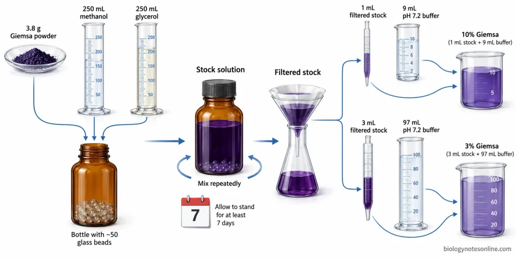
Preparation of Giemsa Stock Solution
For the preparation of approximately 500 mL of Giemsa stock solution, the following materials are required:
- Giemsa stain powder – 3.8 g
- Absolute methanol (acetone-free) – 250 mL
- Glycerol – 250 mL
- Glass beads – About 50 beads of 3–5 mm diameter.
The steps involved in the preparation of Giemsa stock solution are given below:
- Take a clean, dry amber glass bottle with a screw cap. Place about 50 glass beads cleaned with methanol in the bottle.
- Weigh 3.8 g of Giemsa powder and transfer it into the bottle through a funnel.
- Pour 100 mL of absolute methanol into the bottle and close the cap. Shake the bottle in a circular motion for 2–3 minutes.
- Add 250 mL of glycerol to the mixture and shake again for 3–5 minutes.
- The remaining 150 mL of methanol is then added to the bottle. Close the cap tightly.
- During the first day, shake the bottle about six times for 2–3 minutes each time.
- Continue shaking the bottle six times daily for at least 7 days.
- Label the bottle with the name of the stain, batch number, preparation date and expiry date. Keep the bottle tightly closed in a cool, dry place away from direct sunlight.
The stock solution should be left without shaking for at least 24 hours before use. A small amount of the stain is filtered through Whatman No. 1 filter paper into a clean, dry container for preparing the working solution.
Water should not be added to the stock solution because it causes deterioration of the stain.
Preparation of Phosphate Buffer
Phosphate-buffered water of pH 7.2 is required for staining malaria blood films with Giemsa stain.
The following reagents are used for preparing 1 litre of phosphate buffer:
- Potassium dihydrogen phosphate (KH₂PO₄), anhydrous – 0.7 g
- Disodium hydrogen phosphate (Na₂HPO₄), anhydrous – 1.0 g
- Distilled or deionized water – To make 1 litre.
The procedure for preparing phosphate buffer is given below:
- Take 0.7 g of potassium dihydrogen phosphate in a clean beaker.
- Add about 150 mL of distilled water and stir the mixture until the salt dissolves.
- Weigh 1.0 g of disodium hydrogen phosphate and dissolve it in the same solution.
- Transfer the solution into a 1-litre volumetric flask.
- Add distilled water to make up the volume to 1 litre.
- Measure the pH of the solution using a calibrated pH meter.
- If the pH is below 7.2, add a small amount of 2% disodium hydrogen phosphate solution. If the pH is above 7.2, add 2% potassium dihydrogen phosphate solution. These solutions are prepared separately by dissolving 2 g of the respective salt in 100 mL of water.
- Pour the prepared buffer into a clean bottle and label it properly.
The phosphate buffer can be stored for a maximum of 7 days in a tightly closed bottle in a cool place away from sunlight.
The pH of the buffer should be checked before staining. An incorrect pH affects the staining colours and may interfere with the identification of malaria parasites.
Preparation of Giemsa Working Solution
Giemsa working solution is obtained by diluting the filtered stock solution with phosphate-buffered water of pH 7.2. In the WHO malaria microscopy method, 10% Giemsa is used for rapid staining and 3% Giemsa for slow staining.
1. Preparation of 10% Giemsa Working Solution
The 10% Giemsa solution is used when a small number of blood films are to be stained.
- Take 9 mL of phosphate-buffered water of pH 7.2 in a clean beaker.
- Using a clean, dry pipette, add 1 mL of filtered Giemsa stock solution.
- Mix the solution gently to obtain 10 mL of 10% Giemsa working solution.
The blood films are stained with this solution for about 8–10 minutes.
2. Preparation of 3% Giemsa Working Solution
The 3% Giemsa solution is used for staining larger batches of blood films.
- Measure 97 mL of phosphate-buffered water of pH 7.2 in a clean measuring cylinder.
- Add 3 mL of filtered Giemsa stock solution and mix gently.
- This gives 100 mL of 3% Giemsa working solution.
The staining time for this method is about 45–60 minutes.
For preparing different volumes of Giemsa working solution, the amount of stock solution can be calculated using the following formula:
Volume of stock solution = (Required concentration × Final volume) / 100
The remaining volume is made up with phosphate-buffered water.
The working solutions should be prepared immediately before staining. Both the 10% and 3% solutions should be used within 15 minutes of preparation. Any remaining diluted stain is discarded after use.
Other laboratory methods may use different concentrations of Giemsa stain. The CDC staining method uses a 2.5% Giemsa working solution.
Procedure of Giemsa Staining
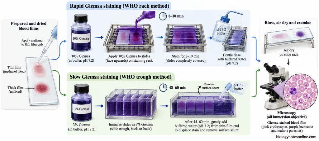
Giemsa staining of blood films can be carried out by two methods. The 10% Giemsa solution is used for rapid staining, while the 3% solution is used for slow staining. The procedure for Giemsa staining of malaria blood films is given below.
Preparation of Thin and Thick Blood Smears
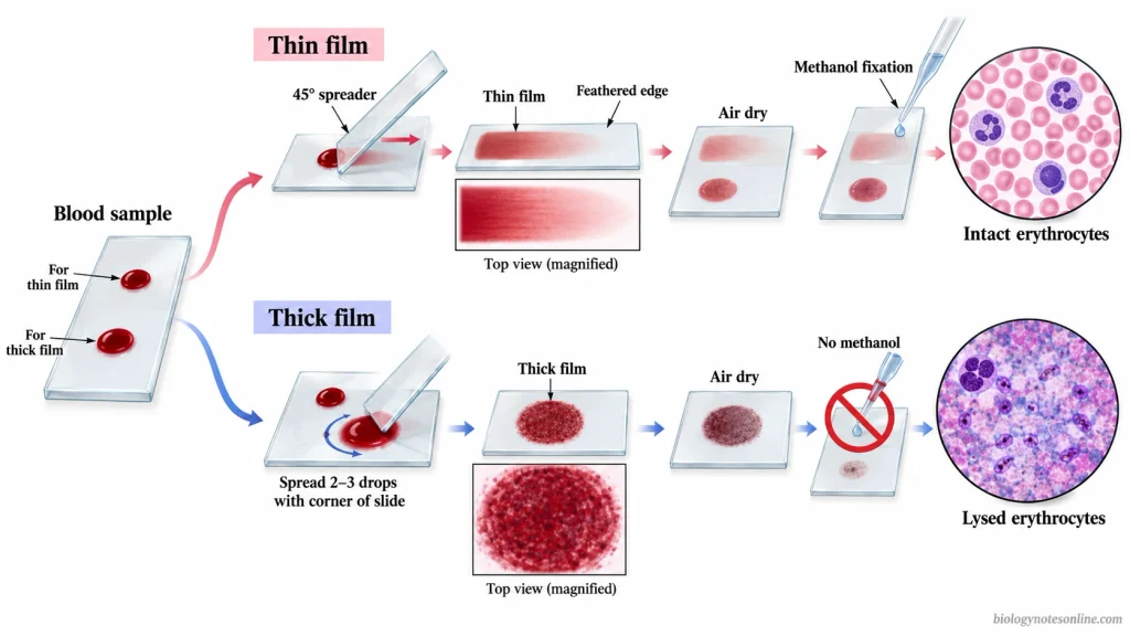
The following are the steps involved in the preparation of thin and thick blood smears:
- Take a clean glass slide free from grease and label it with the sample details.
- Collect fresh blood by finger prick using a sterile lancet. Blood collected in an EDTA tube can also be used, but the smears should be prepared soon after collection.
- Place a small drop of blood near the centre of the slide for preparing the thin blood film.
- Take another clean slide and hold it at an angle of about 45°. Bring its edge in contact with the blood drop and push it forward to prepare a thin smear with a feathered edge.
- For the thick blood film, place two or three drops of blood about 1 cm away from the thin film.
- Spread the blood into a circle of about 1 cm in diameter using the corner of the spreading slide.
- Keep the slide in a horizontal position and allow both blood films to dry completely in air. The films should be protected from dust and excessive heat.
- Dip only the thin blood film in absolute methanol for about 2 seconds. Remove the slide and allow the fixed film to dry in air for approximately 2 minutes.
The thick blood film is not fixed with methanol. During staining, the red blood cells in the thick film are lysed, but the parasites and other blood elements remain on the slide. Methanol fixation of the thin blood film helps in preserving the morphology of red blood cells.
Rapid Giemsa Staining Procedure (10% Solution)
The rapid Giemsa staining method uses a 10% working solution. It is used when a small number of blood films are to be stained.
The procedure of rapid Giemsa staining is given below:
- Take 9 mL of phosphate-buffered water of pH 7.2 and add 1 mL of filtered Giemsa stock solution to prepare a fresh 10% working solution.
- The thin blood film should be fixed with absolute methanol and dried before staining. The thick blood film is left unfixed.
- Place the prepared slides on a staining rack with the blood films facing upwards. A curved staining tray may also be used with the films facing downwards.
- Pour the Giemsa solution gently over the slides to cover the blood films. About 3 mL of stain is required for each slide.
- Allow the stain to remain on the blood films for 8–10 minutes.
- Add phosphate-buffered water of pH 7.2 gently to wash off the excess stain. Do not pour the stain directly off the slide, as the scum present on the surface may remain on the blood film.
- Place the slides in a drying rack with the thick films facing downwards. Allow the films to dry in air without touching the thick films against the rack.
- Discard the remaining Giemsa working solution after staining.
The staining time depends upon the quality and concentration of the stock stain. The stock stain should be tested to determine the optimum staining time before use.
In the WHO rapid staining method, the staining time is about 8–10 minutes. For individual slides stained on a rack, the WHO procedure also describes a staining time of 10–15 minutes using 10% Giemsa solution.
Slow Giemsa Staining Procedure (3% Solution)
The slow Giemsa staining method uses a 3% working solution and is used for staining a larger number of blood films.
The following are the steps involved in slow Giemsa staining:
- Prepare a fresh 3% Giemsa solution by adding 3 mL of filtered stock solution to 97 mL of phosphate-buffered water of pH 7.2.
- Take the completely dried thin and thick blood films. If the thin films are not fixed earlier, fix them with absolute methanol. The thick films should remain unfixed.
- Place the slides back-to-back in a staining trough with the thick films together at one end.
- Pour the Giemsa solution slowly into the trough until the blood films are covered. The stain should not be poured directly onto the thick blood films.
- Leave the slides in the staining solution for 45–60 minutes. The staining time depends upon the stock stain used.
- After staining, add phosphate-buffered water gently into the trough from the end containing the thin films. This removes the metallic green scum present on the surface of the stain.
- Pour off the remaining stain carefully and wash the slides with phosphate-buffered water of pH 7.2.
- Take out the slides one by one and place them in a drying rack with the blood films facing downwards. The thick films should not touch the edges of the rack.
- Allow the blood films to dry completely in air. Discard the remaining 3% Giemsa working solution.
Microscopic Examination of Stained Blood Films
The stained blood films are examined under a compound light microscope. The procedure for microscopic examination is given below:
- Take the completely dried stained slide and place it on the microscope stage.
- First examine the thick blood film using the 10× objective and select a suitable area for examination.
- Place a small drop of immersion oil on the selected area and examine the film under the 100× oil immersion objective.
- Adjust the fine focus and examine the thick blood film systematically for malaria parasites. At least 100 oil immersion fields should be examined before reporting that no malaria parasites were seen.
- If parasites are found, examine another 100 fields to check for the presence of mixed infections.
- Examine the thin blood film under the 100× oil immersion objective. Select an area near the feathered edge where the red blood cells are present without much overlapping.
- The thin blood film is used to study the morphology of malaria parasites and identify different species of Plasmodium.
- Record the presence of parasites along with the species and stages identified during microscopic examination.
Results and Interpretation of Giemsa Staining
Giemsa stain is used to examine blood cells and malaria parasites present in blood films. It stains the nucleus, cytoplasm and granules of cells with different colours. The staining results depend upon the type of cells, staining time and pH of the buffer used.
Staining Results of Blood Cells
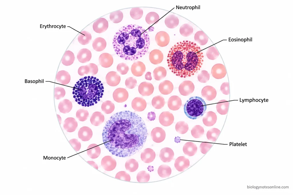
The following are the staining results of different blood cells:
| Blood Cells or Structures | Staining Colour | Microscopic Appearance |
|---|---|---|
| Red Blood Cells (Erythrocytes) | Pink to pinkish-grey | The cells are pink with a lighter central region. |
| White Blood Cell Nuclei | Blue-purple to deep purple | The nuclei are clearly visible and differ in shape among leukocytes. |
| Lymphocytes | Light blue cytoplasm and dark purple nucleus | They have a large, round nucleus with a narrow region of cytoplasm. |
| Monocytes | Grey-blue cytoplasm and purple nucleus | The nucleus is usually kidney-shaped. Fine granules may be present in the cytoplasm. |
| Neutrophils | Pale pink cytoplasm with fine purple or lilac granules | The nucleus is divided into several lobes. |
| Eosinophils | Orange-red to purple-red granules | Large granules are present in the cytoplasm. The nucleus is usually bilobed. |
| Basophils | Dark blue to purple granules | The cytoplasm contains large, dark granules which may cover the nucleus. |
| Platelets | Pink to purple with darker granules | Small cell fragments are present between the red blood cells. |
The nucleus, cytoplasm and granules are examined to identify different types of white blood cells. Their size and nuclear shape also help in identification.
Staining Results of Malaria Parasites
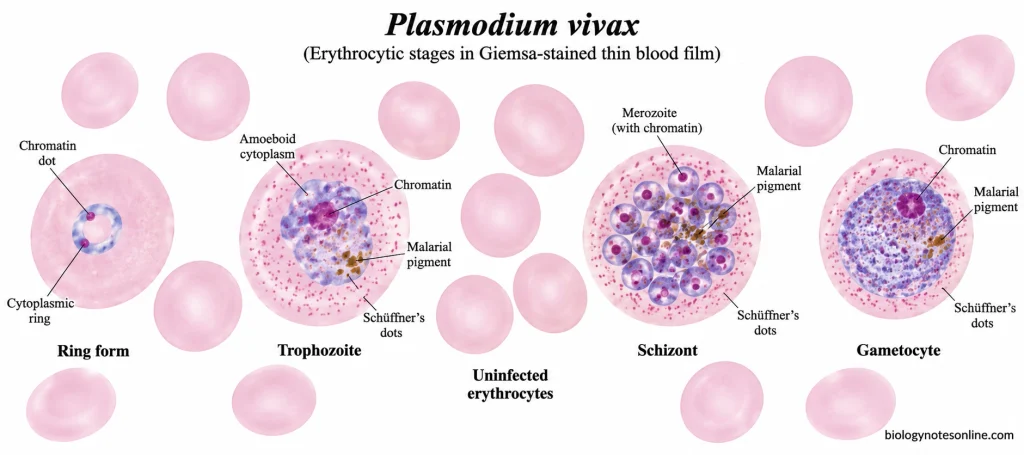
The cytoplasm of malaria parasites stains blue, while the nuclear chromatin appears red or pink after Giemsa staining.
In the early ring stage, the parasite is seen as a blue ring with a red chromatin dot inside the red blood cell. Other stages of the parasite, such as trophozoites, schizonts and gametocytes, can also be observed. Dark brown or black malarial pigment may be present in some stages.
Certain changes are also seen in the infected red blood cells. Schüffner’s dots are present in red blood cells infected with Plasmodium vivax and Plasmodium ovale. Maurer’s clefts may be seen in Plasmodium falciparum infection.
The thick blood film is used for the detection of malaria parasites. During staining, the red blood cells are lysed and the parasites remain concentrated in the film.
The thin blood film is used to study the morphology of malaria parasites and identify different species of Plasmodium. The identification depends upon the shape, size, staining and developmental stages of the parasites. Proper microscopic examination and experience are required for species identification.
Interpretation of Staining Quality
Proper Giemsa staining gives a pink colour to the red blood cells and a purple colour to the nuclei of white blood cells in thin blood films. The cytoplasmic granules are clearly visible and the background is clear.
In thick blood films, the red blood cells are lysed and the background is usually pale grey.
The following are the changes observed due to improper Giemsa staining:
- The blood cells appear pale when the staining time is insufficient, the stain concentration is low or the slide is washed excessively. The nuclei and granules are not clearly visible.
- Excessive staining produces darkly stained blood cells, making the cellular structures difficult to identify.
- An acidic buffer gives a reddish-pink colour to the blood film, whereas an alkaline buffer produces an excessively blue colour.
- Stain precipitates form dark blue or purple particles on the slide. These particles may be confused with malaria parasites.
- Platelets present over red blood cells may be mistaken for malaria parasites. The blue cytoplasm and red chromatin dot help in the identification of the parasites.
- Fixation of thick blood films with methanol or heat may prevent proper haemolysis. The red blood cells remain intact.
- Improper fixation of thin blood films may result in partial lysis of red blood cells and changes in their morphology.
- Different batches of Giemsa stain may give slightly different staining colours. The staining time and quality of the buffer should be checked if the staining results are not satisfactory.
Applications of Giemsa Staining
The following are the applications of Giemsa staining:
- It is used for the detection of malaria parasites of the genus Plasmodium in thick and thin blood films.
- The stained thin blood film is used to identify different species of Plasmodium and examine their developmental stages.
- The number of malaria parasites present in the blood film can be counted to determine the parasite density.
- It is used to detect other blood parasites such as Babesia and Trypanosoma species.
- Giemsa staining helps in the detection of microfilariae of Wuchereria bancrofti and Brugia malayi in blood samples.
- It is used to detect Leishmania amastigotes in bone marrow and splenic aspirates.
- Examination of the morphology of red blood cells, white blood cells and platelets in peripheral blood smears.
- It helps in the identification and differential counting of white blood cells based on their nuclear shape and cytoplasmic granules.
- Giemsa-based stains are used to examine bone marrow cells and study their morphology.
- It is used in chromosome G-banding to identify individual chromosomes and detect structural abnormalities.
- Modified Giemsa staining is used for the detection of Helicobacter pylori in gastric biopsy sections.
Advantages of Giemsa Staining
The following are the advantages of Giemsa staining:
- Giemsa staining is a simple and inexpensive method used for the microscopic examination of blood films.
- It can be used for staining both thick and thin blood films.
- The thick blood film helps in the detection of malaria parasites even when they are present in small numbers.
- The thin blood film is used to study the morphology of malaria parasites and identify different species of Plasmodium.
- It helps in the identification of different stages of malaria parasites, such as trophozoites, schizonts and gametocytes.
- The number of malaria parasites present in a blood sample can be counted to determine the parasite density.
- Giemsa stain produces different colours in the nucleus, cytoplasm and granules of blood cells. This helps in the examination of red blood cells and identification of different types of white blood cells.
- Other blood parasites, such as Babesia and Trypanosoma, can also be detected using Giemsa staining.
- The Giemsa stock solution can be stored for a long period when prepared and stored properly.
Limitations of Giemsa Staining
The following are the limitations of Giemsa staining:
- The staining results depend upon the pH of the buffer, stain concentration and staining time. Improper staining may affect the identification of cells and parasites.
- Giemsa staining requires proper preparation of blood smears. Thick or uneven smears may make microscopic examination difficult.
- Malaria parasites present in very small numbers may not be detected in a single blood film. A negative result does not always exclude malaria infection.
- Stain precipitates and platelets may be mistaken for malaria parasites during microscopic examination.
- The thick blood film does not preserve the morphology of red blood cells. This makes the identification of different species of Plasmodium difficult.
- Some species of Plasmodium have similar morphological features, making them difficult to distinguish. Mixed infections may also be missed during examination.
- Delayed preparation of blood smears may cause changes in the morphology and staining characteristics of parasites.
- Fixation of thick blood films with methanol or heat may prevent proper haemolysis of red blood cells and affect parasite detection.
- It requires a microscope and trained personnel for the correct identification of blood cells and parasites.
- The slow Giemsa staining method requires about 45–60 minutes. Microscopic examination of the stained films also takes time.
Precautions of Giemsa Staining
The following are the precautions of Giemsa staining:
- Use clean glass slides free from grease and dust for preparing blood smears. The staining equipment should be cleaned and dried before use.
- Blood smears should be prepared soon after the collection of blood. Delay in preparation may change the morphology of blood cells and parasites.
- Allow the thick blood film to dry completely in air. Excessive heat should be avoided as it may cause fixation of the blood film.
- Fix the thin blood film with absolute methanol. The thick film should not be fixed or exposed to methanol vapours, as the red blood cells may not undergo proper haemolysis.
- Prepare a fresh Giemsa working solution before staining. The stock solution should be stored in a tightly closed bottle to prevent moisture from entering.
- Use phosphate-buffered water of pH 7.2 for the preparation of Giemsa working solution. A change in pH affects the staining colour of blood cells and parasites.
- The staining time depends upon the concentration of Giemsa stain. Staining for a longer or shorter period than required may affect the examination of blood films.
- Avoid pouring the staining solution directly onto the thick blood film. The film may get detached from the slide.
- Wash the stained slides gently with phosphate-buffered water of pH 7.2. Do not use a strong stream of water, especially on the thick blood film.
- The metallic green scum present on the surface of the stain should be removed during washing. If it remains on the slide, it may form deposits that interfere with microscopic examination.
- Dry the stained blood films completely in air. The thick film should not come in contact with the edges of the drying rack.
- Wear laboratory gloves, protective clothing and safety glasses while working with blood samples and staining reagents.
- Methanol is toxic and highly flammable. Avoid direct contact with methanol and inhalation of its vapours. It should be kept away from flames and other sources of ignition.
- Discard the remaining Giemsa working solution after staining. Wash the staining equipment properly to remove any remaining stain and detergent.
Differences Between Giemsa and Other Romanowsky Stains
Giemsa, Leishman, Wright, Wright–Giemsa and May–Grünwald–Giemsa belong to the Romanowsky group of stains. These stains contain acidic and basic dyes, mainly eosin and methylene blue derivatives. They differ in their composition, preparation, fixation and staining methods.
The following table shows the differences between these Romanowsky stains:
| Stain | Composition | Fixation and Staining Method | Common Applications |
|---|---|---|---|
| Giemsa | Eosin, methylene blue and azure dyes. The conventional stock solution contains methanol and glycerol. | Thin blood films are fixed with methanol and stained with diluted Giemsa. Thick blood films are stained without methanol fixation. | Detection and identification of blood parasites, especially malaria parasites. |
| Leishman | Polychromed methylene blue and eosin in methanol. | Undiluted stain can fix the dried blood film. Buffered water is then added for staining. | Peripheral blood smear examination, differential leukocyte counting and malaria parasite detection. |
| Wright | Eosin and oxidised methylene blue derivatives in methanol. | Methanol in the staining solution can act as a fixative. The stain is then diluted with buffer. | Routine examination of blood cells and bone marrow smears. |
| Wright–Giemsa | A combined preparation containing eosin and thiazine dyes, including methylene blue and azure dyes. | Usually performed with a methanol-based staining solution and buffer. Fixation depends upon the method used. | Blood cell differentiation, bone marrow examination and detection of certain blood parasites. |
| May–Grünwald–Giemsa (MGG) | Separate May–Grünwald and Giemsa staining solutions. | Air-dried smears are fixed and stained first with May–Grünwald, followed by Giemsa. | Hematology, bone marrow examination and cytological examination of air-dried smears. |
Giemsa Stain vs Leishman Stain
Giemsa stain contains eosin, methylene blue and azure dyes, usually dissolved in methanol and glycerol. Leishman stain is prepared using polychromed methylene blue and eosin in methanol.
In Leishman staining, the undiluted stain is applied to the dried thin blood film. The methanol present in the stain helps in fixation. Buffered water is then added for staining. Separate methanol fixation may also be used in certain procedures.
In Giemsa staining, the thin blood film is fixed with methanol before staining. The film is then stained with Giemsa working solution prepared using phosphate-buffered water. Thick blood films are not fixed with methanol.
Leishman staining generally requires less staining time than the conventional slow Giemsa method.
Both stains are used for the examination of blood cells and malaria parasites. Leishman staining may give better nuclear chromatin detail and cytoplasmic colour contrast in blood cells. Giemsa staining is commonly preferred for the identification of malaria parasites and their characteristic morphological features.
Giemsa Stain vs Wright and Wright–Giemsa Stains
Wright stain contains eosin and polychromed methylene blue in methanol. Wright–Giemsa is a combined Romanowsky stain containing eosin, methylene blue and azure dyes. The proportions of these dyes differ from those in the individual Wright and Giemsa preparations.
Wright stain is commonly used for staining peripheral blood smears. The methanol present in the stain can fix the dried blood film. Buffered water is then added to develop the staining colours.
Wright–Giemsa staining is used in routine hematology and bone marrow examination. It is also used in automated staining methods. The staining procedure and fixation depend upon the reagent preparation used.
Wright and Wright–Giemsa stains give characteristic colours to red blood cells, white blood cell nuclei and cytoplasmic granules. These stains are used for differential white blood cell counting and examination of blood cell morphology.
Wright–Giemsa staining can detect malaria parasites, but it is not optimal for their identification. Certain features, such as Schüffner’s dots, are not demonstrated as reliably as with Giemsa staining.
For confirmation of blood parasites, Giemsa staining is recommended. A Giemsa-stained blood film prepared with buffer of pH 7.2 helps in the identification of malaria species and their characteristic staining features.
Giemsa Stain vs May–Grünwald–Giemsa (MGG) Stain
May–Grünwald–Giemsa (MGG) is a combined staining method in which May–Grünwald and Giemsa solutions are used successively.
May–Grünwald stain contains eosin and methylene blue derivatives in methanol. Giemsa stain contains additional azure dyes responsible for the characteristic purple staining of certain cellular structures.
In MGG staining, the air-dried smear is first fixed with methanol or the methanolic May–Grünwald solution, depending upon the method used. The smear is stained with May–Grünwald solution, followed by diluted Giemsa stain.
The staining time and buffer pH depend upon the procedure used. A buffer of pH 6.8 is commonly used for routine hematological and cytological preparations.
MGG staining gives pink to orange-red colour to erythrocytes and purple colour to the nuclei of leukocytes. The cytoplasmic granules and other cellular components are also clearly visible.
MGG staining is used for the examination of peripheral blood smears, bone marrow aspirates, fine-needle aspiration smears, lymph node imprints and other air-dried cytological preparations.
Unlike Giemsa staining alone, MGG uses two staining solutions in sequence. The method is commonly used for studying the morphology of blood cells and other cells present in cytological specimens.
References
- Bain, B. J., Bates, I., & Laffan, M. A. (2017). Dacie and Lewis practical haematology (12th ed.). Elsevier. https://www.sciencedirect.com/book/9780702066962/dacie-and-lewis-practical-haematology
- Mathison, B. A., & Pritt, B. S. (2017). Update on malaria diagnostics and test utilization. Journal of Clinical Microbiology, 55(7), 2009–2017. https://doi.org/10.1128/JCM.02562-16
- Miller, J. M., Binnicker, M. J., Campbell, S., Carroll, K. C., Chapin, K. C., Gonzalez, M. D., Harrington, A., Jerris, R. C., Kehl, S. C., Leal, S. M., Jr., Patel, R., Pritt, B. S., Richter, S. S., Robinson-Dunn, B., Snyder, J. W., Telford, S., III, Theel, E. S., Thomson, R. B., Jr., Weinstein, M. P., & Yao, J. D. (2024). Guide to utilization of the microbiology laboratory for diagnosis of infectious diseases: 2024 update by the Infectious Diseases Society of America (IDSA) and the American Society for Microbiology (ASM). Clinical Infectious Diseases, Article ciae104. https://doi.org/10.1093/cid/ciae104
- Piaton, E., Fabre, M., Goubin-Versini, I., Bretz-Grenier, M.-F., Courtade-Saïdi, M., Vincent, S., Belleannée, G., Thivolet, F., Boutonnat, J., Debaque, H., Fleury-Feith, J., Vielh, P., Egelé, C., Bellocq, J.-P., Michiels, J.-F., & Cochand-Priollet, B. (2016). Guidelines for May-Grünwald–Giemsa staining in haematology and non-gynaecological cytopathology: Recommendations of the French Society of Clinical Cytology (SFCC) and of the French Association for Quality Assurance in Anatomic and Cytologic Pathology (AFAQAP). Cytopathology, 27(5), 359–368. https://doi.org/10.1111/cyt.12323
- Sathpathi, S., Mohanty, A. K., Satpathi, P., Mishra, S. K., Behera, P. K., Patel, G., & Dondorp, A. M. (2014). Comparing Leishman and Giemsa staining for the assessment of peripheral blood smear preparations in a malaria-endemic region in India. Malaria Journal, 13, Article 512. https://doi.org/10.1186/1475-2875-13-512