Romanowsky staining, also known as Romanowsky–Giemsa staining, is a classic staining technique that paved the way for several different but similar stains widely used in haematology (the study of blood) and cytopathology (the study of cells) (the study of diseased cells). Romanowsky stains are used to distinguish cells for microscopic study in pathological materials, particularly blood and bone marrow films, and to detect blood parasites such as malaria. In addition to the Romanowsky-type stains, the Giemsa, Jenner, Wright, Field, May–Grünwald, and Leishman stains are linked to or derived from the Romanowsky-type stains. The technique is named after Russian physician Dmitri Leonidovich Romanowsky (1861–1921), who was among the first to realise its potential as a blood stain.
Contents
Principle of Romanowsky Stains
The dyes, which include Eosin Y and oxidised methylene blue (azure), produce neutral stains. The azures are blue-purple because they are basic dyes that bond to the acid nucleus. Eosin, an acid dye, combines with the highly basic cytoplasm to produce a red hue.
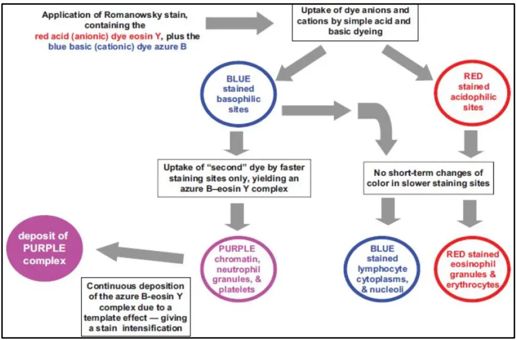
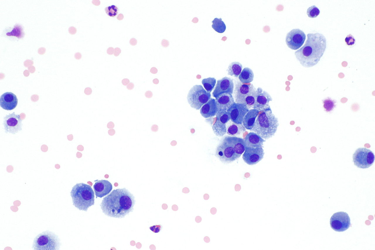
Romanowsky staining is effective because it generates a range of colours, each of which can be used to identify a specific subset of cells. The Romanowsky effect, sometimes known as metachromasia, describes this talent. Romanowsky defined the Romanowsky effect, also known as the Romanowsky–Giemsa effect, which describes the production of purple hues in the chromatins of the nucleus and the granules in the cytoplasm of some white blood cells when active eosin Y and active methylene blue are mixed.
And by the action of oxidative demethylation, oxidised unmethylated methylene blue can be used to create stains of the Romanowsky type. Methylene blue disintegrates into several coloured stains as a result, and some of these stains are responsible for the Romanowsky phenomenon. Oxidatively demethylated methylene blue, also known as polychrome methylene blue, contains roughly 11 different dyes, including azure A, azure B, azure C, methylene blue, methylene violet Bernthesen, methyl thionoline, and thionoline.
Types of Romanowsky Stains
Romanowsky stains are a group of staining techniques used in hematology, pathology and histology to visualize and differentiate blood cells, tissue structures, and microorganisms. The most common Romanowsky stains include:
- Wright-Giemsa stain: This is one of the most widely used Romanowsky stains. It is used to stain peripheral blood smears and bone marrow aspirates, and it can be used to differentiate leukocytes, erythrocytes, platelets and microorganisms. Wright’s stain is versatile; it can be used on its own or in tandem with the Giemsa stain to create the Wright-Giemsa stain. Wright’s stain gets its name from James Homer Wright, who in 1902 reported a method for making polychromed methylene blue by combining it with eosin Y and applying heat. To make eosinate, polychromed methylene blue is mixed with eosin, allowed to precipitate, and then redissolved in methanol. The “reddish-purple” colour of the cytoplasmic granules is become more noticeable when Giemsa is added to Wright’s stain. U.S. pathologists frequently use Romanowsky-type stains like Wright’s and Wright-Giemsa for staining blood and bone marrow films.
- May-Grünwald stain: This is a modified Wright-Giemsa stain and is used for the same purposes as Wright-Giemsa stain. Staining with the May-Grünwald stain, which does not generate the Romanowsky effect, is the first stage of the May-Grünwald-Giemsa stain, which entails a second staining phase with the Giemsa stain, which does cause the Romanowsky effect.
- Leishman stain: This stain is used to stain blood smears, and it is especially useful for the visualization of parasites such as Plasmodium and Trypanosoma. In 1901, William Leishman created a stain comparable to Louis Jenner’s, but with polychromed methylene blue instead of pure methylene blue. The eosinate of polychromed methylene blue and eosin Y are dissolved in methanol to create Leishman’s stain.
- Giemsa stain: The Giemsa stain consists of “Azure II” and eosin Y, with methanol and glycerol serving as solvents. “Azure II” is believed to be a combination of azure B (which Giemsa referred to as “azure I”) and methylene blue, while the precise composition of “azure I” is a trade secret. Comparable formulations with well-established colours have been published and are commercially available. Giemsa stain is regarded as the gold standard for detecting and identifying the malaria parasite.
- Field stain: This stain is a rapid staining technique used for peripheral blood smears.
- Supravital stains: These are vital dyes used to stain blood cells, especially red blood cells, for in vitro examinations. Some of the common supravital stains include eosin, acridine orange, and ethidium bromide.
- Diff-Quik stain: This is a quick and easy staining method used to differentiate cells and tissues in smears and cytologic specimens.
Each of these Romanowsky stains has its own specific protocol, staining properties and applications, and the choice of stain often depends on the type of specimen being examined and the specific information needed.
Factors affecting staining
- Composition of solvent – purple hue with high quantities of lesser alcohols that is unstable (methanol content).
- The azure B to Eosin Y ratio.
- Stain purity >80%
- pH value of phosphate buffer is crucial – optimum range 6.8 to 7.2
- Timing of staining dependent on specimen thickness.
- Cell count
- Fixative – formalin stops nuclear chromatin from becoming purple.
Troubleshooting of Romanowsky Stains
| Too PINK | • Improper azure B/eosin Y ratio • Impure dye • Low pH |
| Too BLUE | • Improper azure B/eosin Y ratio • Stock stain exposed to light • Excess staining time • Thick film • Inadequate time in buffer solution |
| Too PALE | • Old solution • Weak/Impure dyes • High temperature |
| Nuclei too dark | • Stain too concentrated • Incorrect staining time |
Artefacts
Blood smear abnormalities can be either pathological or artefactual. Any result of a method that is caused by the procedure and not the entity being studied. It was presented in 1992. It is crucial to accurately identify artefacts so they are not misinterpreted as pathogenic processes. Can lead to confusion and diagnostic difficulties. Possibly because of…
- Fixation
- Storage
- Heat artefact
- Poor spreading techniques
- Poorly or irregularly spread blood films, often with poor morphology.
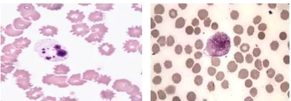
Fixation artefact
The presence of water in the methanol used to attach the blood film – The presence of refractile rings in red cells makes it impossible to evaluate red cell morphology. Excessive water on the slide or in the stain causes erythrocytes to have refractile edges. Inadequate or delayed fixation, sluggish drying in moist conditions

Storage artefact
White cells become brittle and may develop into smudge/smear cells. Similar to NRBC, neutrophil nuclei form spherical, homogenous masses or a single mass. Crenation or echinocytic alteration occurs in red cells.
- Pseudo toxic changes
- Pseudoechinocytosis and
- Platelet degranulation

Heat artefact
- Red cells bud off vesicles.
- Microspherocytes seen.
- White cells disintegrate.
- Proteins coagulate, producing weakly basophilic particles, similar in size to platelets.
Applications of Romanowsky Stains
The Romanowsky stains have a wide range of applications in different fields, including:
- Hematology: Romanowsky stains are used in the diagnosis and characterization of blood disorders such as anemia, leukemias, and lymphomas. They can be used to differentiate different types of leukocytes, erythrocytes, and platelets, and to assess their morphological features.
- Pathology: In pathology, Romanowsky stains are used to diagnose and evaluate the progress of diseases and conditions affecting different organs and tissues, including cancer, infectious diseases, and autoimmune disorders.
- Microbiology: Romanowsky stains are used in microbiology to identify and differentiate different types of microorganisms, including bacteria, fungi, and parasites. They can be used to evaluate the morphological features of microorganisms, such as cell shape, size, and staining characteristics, which can aid in the identification of different species.
- Histology: In histology, Romanowsky stains are used to visualize and differentiate different types of cells and tissues in tissue sections. They can be used to highlight specific cellular structures, such as the nucleus, cytoplasm, and organelles, and to evaluate the morphological features of cells and tissues.
- Research: Romanowsky stains are also used in research to evaluate the morphological and staining properties of cells and tissues, and to study the molecular and cellular mechanisms underlying various biological processes.
- Detection of malaria and other parasites: The Giemsa stain is the most essential of the Romanowsky-type stains for detecting and identifying malaria parasites in blood samples. Alternatives to the staining and microscopic analysis of blood films for the identification of malaria include antigen detection techniques.
- Blood and bone marrow pathology: Blood, in the form of blood films, and bone marrow biopsies and aspirate smears are frequently examined with Romanowsky-type stains. Blood and bone marrow examinations can be useful in the diagnosis of a range of blood illnesses. In the United States, the Wright and Wright-Giemsa varieties of the Romanowsky-type stains are extensively employed, although in Europe, the Giemsa stain is the most prevalent.
Overall, Romanowsky stains play a crucial role in the diagnosis, treatment, and research of various diseases and conditions, and they provide valuable information that can be used to improve patient outcomes.
Advantages of Romanowsky Stains
- The stains are widely accessible.
- They are straightforward to create, maintain, and employ.
Disadvantages of Romanowsky Stains
- The cell’s morphological characteristics may be distorted.
- It is essential to use the necessary pH buffer to preserve the dye colour.
FAQ
What are Romanowsky stains give example?
Romanowsky stains are a group of staining techniques used in hematology, pathology, and histology to visualize and differentiate cells, tissues, and microorganisms. They work by using dyes that interact with specific cellular structures, resulting in the selective staining of different structures based on their staining properties.
Here is an example of a commonly used Romanowsky stain:
Wright-Giemsa stain: This is a commonly used Romanowsky stain that is used to stain peripheral blood smears and bone marrow aspirates. It is made up of a mixture of dyes, including methylene blue and eosin, that interact with different cellular structures. The Wright-Giemsa stain can be used to differentiate leukocytes, erythrocytes, platelets, and microorganisms based on their morphological features and staining characteristics. For example, the nucleus of a leukocyte is typically stained blue by the methylene blue component of the stain, while the cytoplasm is stained pink by the eosin component. This allows for the clear visualization and differentiation of different types of cells in the blood.
What are Romanowsky stains?
Romanowsky stains are a group of staining techniques used in hematology, pathology, and histology to visualize and differentiate cells, tissues, and microorganisms.
How do Romanowsky stains work?
Romanowsky stains work by using dyes that interact with specific cellular structures, resulting in the selective staining of different structures based on their staining properties.
What are the most common Romanowsky stains?
The most common Romanowsky stains include Wright-Giemsa stain, May-Grünwald stain, Leishman stain, Field stain, Supravital stains, and Diff-Quik stain.
What are the applications of Romanowsky stains?
Romanowsky stains are used in the diagnosis and characterization of blood disorders, the diagnosis of diseases and conditions affecting different organs and tissues, the identification of microorganisms, the visualization and differentiation of cells and tissues in histology, and in research to study biological processes.
How are Romanowsky stains prepared?
Romanowsky stains are typically prepared as a mixture of different dyes, which can be combined in different ratios to produce the desired staining properties.
How are Romanowsky stains used?
Romanowsky stains are used by first preparing a sample of the tissue or fluid being examined, such as a peripheral blood smear or a tissue section. The sample is then stained with the Romanowsky stain and examined under a microscope to visualize and differentiate different structures.
What are the benefits of using Romanowsky stains?
Romanowsky stains provide valuable information about the morphological and staining properties of cells and tissues, which can be used to diagnose and evaluate diseases and conditions, to identify microorganisms, and to study biological processes.
What are the limitations of using Romanowsky stains?
Romanowsky stains are limited by the quality of the sample being examined, as well as the limitations of the staining technique itself. For example, some stains may not provide clear differentiation of certain structures, or they may be subject to variability based on the preparation and staining procedure.
What are the differences between Romanowsky stains and other staining techniques?
Romanowsky stains differ from other staining techniques, such as H&E stains and special stains, in the dyes used, the specific staining properties, and the applications for which they are used.
How can the quality of Romanowsky stains be improved?
The quality of Romanowsky stains can be improved by optimizing the staining protocol, using high-quality samples, and ensuring the proper storage and handling of the stains. In addition, advances in technology, such as the use of automated staining systems, can also improve the accuracy and reproducibility of Romanowsky stains.
References
- Bentley SA, Marshall PN, Trobaugh FE Jr. Standardization of the Romanowsky staining procedure: an overview. Anal Quant Cytol. 1980 Mar-Apr;2(1):15-8. PMID: 6155098.
- https://tmc.gov.in/tmh/PDF/Hemato%20Pathology%20Course/Romanowsky%20stain%20Dr%20Archana.pdf
- https://www.diseasefix.com/page/diagnosis-and-tests-for-urinary-tract-infection/3479/
- http://histology-world.com/stains/stains.htm
- https://en.m.wikipedia.org/wiki/Leishman_stain
- https://www.newhealthadvisor.org/Types-of-White-Blood-Cells.html
- https://wikimili.com/en/Romanowsky_stain
- https://en.wikipedia.org/wiki/Romanowsky_stain


