Bacterial cells contain nuclear material, made of single-stranded circular DNA, in contrast to eukaryotes. This genetic material is present within the membrane-bounded structure known as the nucleus.
In case of prokaryotes the nuclear material is present within the nucleoid which lacks the nuclear membrane and does not follow the mitosis or miosis cell division. Some bacteria carry their genetic information in plasmid. It is a circular, small fragment of DNA, which replicates independently.
Nucleus involves in different vital activities such as growth, metabolic activities, multiplication, and transfer of hereditary characters.
Objective of Nuclear Staining
The main purpose of this staining method is to stain the nuclear material and study the detailed morphology of the nucleus.
Contents
Principle of Nuclear Staining
In this method, the cytoplasm which contains a high affinity for most stains and interferes with the observation of the nuclear material is first hydrolyzed with HCL and later stained with the Giemsa stain. Nuclear bodies purple color.
Giemsa’s stain contains both acidic and basic properties because it is a mixture of two different dyes called methylene blue ( basic dye) and eosin( acidic dye). This Giemsa’s stain reacts with the chromatids, which are acidic in nature, as a result of this reaction it gives a reddish-purple color to DNA.
Before staining, the cells are treated with 1 N HCl solution to remove the RNA material from the cells. This step is also known as the acid hydrolysis step. In this step the cells are treated with 1 N HCl solution in a water bath of about 60°C temperature for 5 minutes, as result, the double bonds in RNA become week and break.
While DNA contains triple bonds in between some base pairs, therefore they don’t get hydrolyzed and get stain, and cytoplasm remains colorless.
Requirement
- Bacterial Culture
- 1N hydrochloric Acid
- Giemsa stain
- Clean grease-free glass slide
- Waterbath
- Inoculating loop
- Beaker
- Blotting paper
- Bunsen burner
- Microscope
Nuclear Staining Procedure
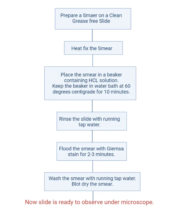
- Take a clean slide and make it grease-free by washing it with soap and water.
- Allow to air dry.
- Take an inoculating loop and sterilize it by holding it on bunsen burner flame until it becomes hot red. Allow cooling the loop.
- Transfer the bacterial culture on the slide by using this sterile cooled loop.
- Spread the culture on the slide.
- Heat fix the smear, by passing the slide through the flame of bunsen burner for 2-3 times. It will fix the cell at the surface of slide.
- Place the smear in a beaker containing HCL solution.
- Keep the beaker in water bath at 60 degrees centigrade for 10 minutes.
- Rinse the slide with running tap water.
- Flood the smear with Giemsa stain for 2-3 minutes.
- Wash the smear with running tap water.
- Blot dry the smear.
- Now the slide is ready to observe under a microscope.
Nuclear Staining Flow Chart
Result of Nuclear Staining
- Cytoplasm: Appear colorless.
- Nuclear Material: appear purple in color.
Reference Book
Experiments in Microbiology, Plant Pathology and Biotechnology by K.R. Aneja


