

Loeffler’s method (Löffler’s method) is a classical staining method used for the demonstration of bacterial flagella. Bacterial flagella are very thin, thread-like structures that are difficult to observe under an ordinary light microscope. The method uses a mordant followed by staining with a dye to make the flagella visible under a light microscope.
Flagella are present in many motile bacteria and help in their movement. Loeffler’s method is used to observe the presence and arrangement of bacterial flagella, which helps in the identification of bacteria. However, the staining method does not show the actual movement of bacteria. Bacterial motility can be examined by the hanging drop method or other motility tests.
The method was developed by Friedrich Loeffler in 1889. Several modifications of the original method have been introduced using different mordants and staining dyes.
Methylene blue was also used in some early flagella-staining methods. However, Loeffler’s alkaline methylene blue stain is a separate staining preparation used for staining bacterial cells. Loeffler’s serum medium is a culture medium and is not used as part of the flagella-staining procedure.
Objectives of Loeffler’s Method
The following are the objectives of Loeffler’s method:
- To demonstrate the presence of bacterial flagella under a light microscope.
- To determine the number and arrangement of flagella on bacterial cells.
- To differentiate between flagellated and non-flagellated bacteria.
- To help in the identification and classification of bacteria based on their flagellar characteristics.
Principle of Loeffler’s Method
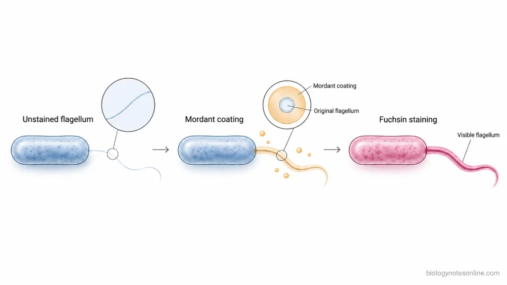
The principle of Loeffler’s method is based on increasing the apparent thickness of bacterial flagella by using a mordant. Bacterial flagella are extremely thin and cannot be seen clearly under an ordinary light microscope.
The mordant used in the classical method contains tannic acid, ferrous sulphate and a small amount of fuchsin. Tannic acid and ferrous sulphate help in the formation of a coating around the flagella. This coating increases the apparent thickness of the flagella and helps in retaining the stain.
The aniline-water fuchsin staining solution contains a basic dye which binds to the coated flagella. The dye gives colour to the flagella and makes them visible under a light microscope.
Reagents and Materials Required for Loeffler’s Method
The following are the materials and reagents required for Loeffler’s method of flagella staining.
Materials and Equipment
- Bacterial Culture– A suitable culture of a flagellated bacterium.
- Glass Slides or Coverslips– Clean and free from grease.
- Inoculating Loop or Needle– For transferring the bacterial culture.
- Dropper or Pipette– For adding staining reagents.
- Staining Rack– For holding the slides during staining.
- Wash Bottle– For washing the slides with distilled water.
- Bunsen Burner or Spirit Lamp– For gentle heating in the classical method.
- Filter Paper– For filtering the mordant and staining solutions.
- Light Microscope with Oil Immersion Objective– For microscopic examination of stained flagella.
Staining Reagents
The following reagents are used in the fuchsin-containing formulation of Loeffler’s method:
- Loeffler’s Mordant– It contains 10 mL of 20% aqueous tannic acid solution, 5 mL of cold saturated aqueous ferrous sulphate solution and 1 mL of saturated alcoholic basic fuchsin solution.
- Aniline-Water Fuchsin Stain– A staining solution containing basic fuchsin dissolved in aniline water.
- Distilled Water– Used for the preparation of staining solutions and washing the smear.
Procedure of Loeffler’s Method
The procedure of Loeffler’s method using the historical heat-assisted method with the fuchsin-containing mordant is given below:
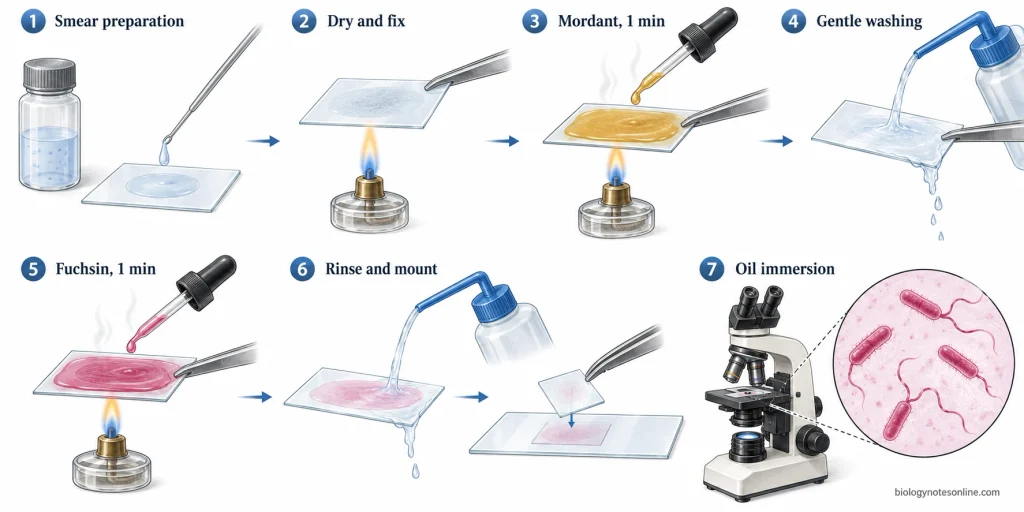
- Take a fresh agar culture of a suitable flagellated bacterium.
- Take a clean, grease-free coverslip. If the coverslip is cleaned by flaming, allow it to cool before use.
- Transfer a small amount of bacterial culture into a few millilitres of distilled water. Mix gently to prepare a dilute bacterial suspension.
- Place a drop of the suspension on the coverslip and spread it into a thin film without rubbing.
- Allow the film to dry completely in air, protected from dust.
- Fix the dried film by passing the coverslip three times through a Bunsen flame.
- Cover the film with the prepared mordant. Warm gently over a flame until steam rises and allow the mordant to act for 1 minute, keeping it gently steaming.
- Wash the coverslip thoroughly with distilled water until no more colour is removed.
- Add a few drops of filtered aniline-water fuchsin stain to the film. Warm gently until steam rises and allow the stain to act for 1 minute.
- Wash the film thoroughly with distilled water to remove the excess stain.
- Allow the stained preparation to dry and mount the coverslip on a clean glass slide.
- Place a drop of immersion oil on the mounted preparation and examine under the oil-immersion objective. Observe the stained bacterial cells and their attached flagella.
In a later modification of Loeffler’s method, heating is not required during mordanting or staining. In Kulp’s modification, both the mordant and the stain are allowed to act for 15 minutes without heating.
Results and Interpretation of Loeffler’s Method
After staining by Loeffler’s method, the bacterial cells and flagella can be observed under the oil-immersion objective. The flagella appear as thin, thread-like structures extending from the surface of bacterial cells. They may be straight, curved or wavy. The cells and flagella usually appear pink to red when aniline-water fuchsin is used. The colour depends upon the staining dye.
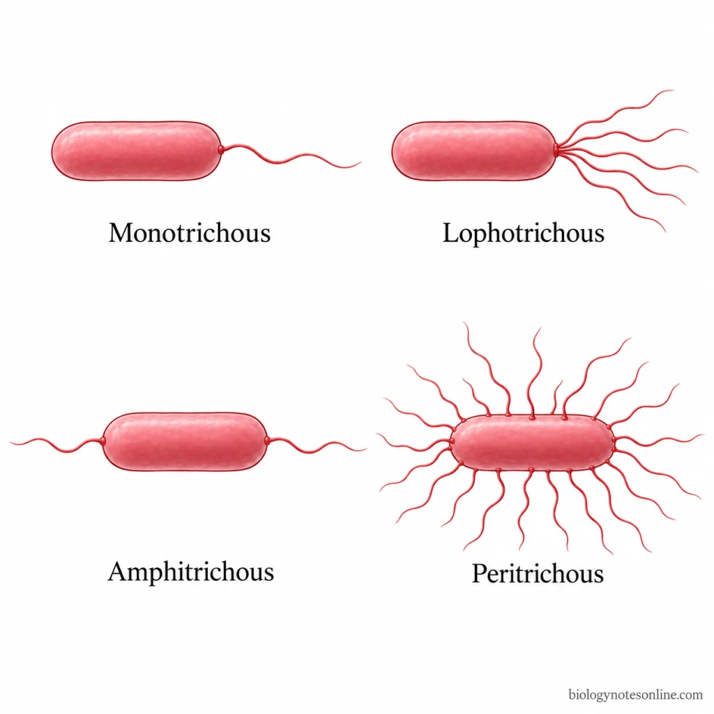
The number and arrangement of flagella may differ among bacteria. The following flagellar arrangements can be observed:
- Monotrichous– A single flagellum is present at one end of the bacterial cell.
- Lophotrichous– A tuft of flagella is present at one end of the bacterial cell.
- Amphitrichous– Flagella are present at both ends of the bacterial cell.
- Peritrichous– Flagella are distributed all around the surface of the bacterial cell.
The number and arrangement of flagella are used for the morphological characterization and identification of bacteria.
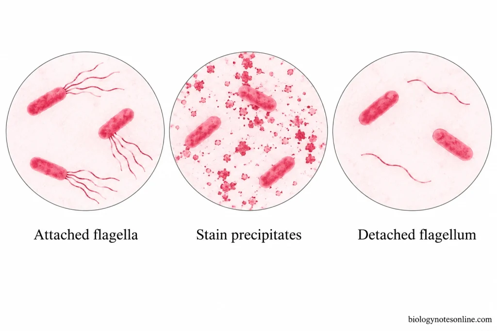
Staining precipitates may appear as irregular particles or masses in the preparation. These should not be confused with bacterial flagella. The flagella appear as fine filaments attached to the bacterial surface, whereas staining precipitates may be present separately in the background. Broken or detached flagella and other staining artefacts may also affect the microscopic observation.
The absence of visible flagella does not always indicate that the bacteria are non-flagellated. Flagella may be damaged or removed during the preparation of the smear.
Applications of Loeffler’s Method
The following are the applications of Loeffler’s method:
- It is used to detect the presence of flagella in bacterial cells.
- To determine the number and arrangement of flagella on the bacterial surface.
- It helps in the identification and classification of bacteria based on their flagellar arrangement.
- Used in microbiological studies to compare the flagellar structures of different bacteria.
- It is used in teaching laboratories for the microscopic examination of bacterial flagella.
Limitations of Loeffler’s Method
The following are the limitations of Loeffler’s method:
- The method requires a properly prepared bacterial smear and suitable staining reagents.
- Bacterial flagella are very delicate and can break or get detached from the cells during smear preparation and washing.
- Excessive heating may damage the flagella. Improper staining also affects the staining results.
- Staining precipitates and other deposits may be present in the smear. These can be confused with bacterial flagella under the microscope.
- The age and growth conditions of the bacterial culture affect the staining results. Older cultures may lose their flagella.
- The flagella of some bacterial species are difficult to stain by this method.
- Flagella may be present on the bacterial cells but may not be visible in the stained smear.
- The method is used to observe bacterial flagella but cannot show their actual movement. A separate motility test is required to examine bacterial movement.
Advantages of Loeffler’s Method
The following are the advantages of Loeffler’s method:
- The mordant increases the thickness of bacterial flagella. These can then be observed under a light microscope.
- The number of flagella present on a bacterial cell can be determined.
- It helps in studying the arrangement of flagella on bacterial cells.
- It is used to identify and differentiate bacteria based on their flagellar arrangement.
- The stained preparation can be mounted for further microscopic examination.
Precautions of Loeffler’s Method
The following are the precautions of Loeffler’s method:
- The glass slides and coverslips should be clean and free from grease and dust.
- Use a fresh culture of motile bacteria for staining.
- A thin smear should be prepared using a small amount of bacterial culture.
- Do not rub the smear during preparation. The bacterial flagella are delicate and may get detached from the cells.
- Allow the smear to dry in air before fixation. Excessive heating may damage the flagella.
- Filter the mordant and staining solutions before use to remove any precipitates.
- Heat the mordant and stain gently. The solutions should not be boiled.
- Wash the smear carefully with water. A strong stream of water may remove the flagella.
- The stained preparation should be examined carefully, as stain precipitates may be mistaken for bacterial flagella.
References
- Eyre, J. W. H. (1913). The elements of bacteriological technique: A laboratory guide for medical, dental, and technical students (2nd ed.). W. B. Saunders. https://www.gutenberg.org/ebooks/27713
- Kulp, W. L. (1926). A method for the staining of bacterial flagella. Stain Technology, 1(2), 60–62. https://doi.org/10.3109/10520292609115619
- Parker, N., Schneegurt, M., Tu, A.-H. T., Lister, P., & Forster, B. M. (2016). Microbiology. OpenStax. https://openstax.org/books/microbiology/pages/1-introduction