

Propidium iodide staining is a fluorescent staining technique used to examine cell viability and DNA content. Propidium iodide (PI) is a fluorescent nucleic acid stain. It is commonly used in flow cytometry and fluorescence microscopy.
In cell viability studies, intact and unfixed cells are stained with PI. Cells with intact plasma membranes generally exclude the dye, while cells with damaged membranes may take up PI. The uptake of PI indicates a loss of membrane integrity, but does not always mean that the cells are dead.
For DNA content and cell cycle analysis, cells are fixed or permeabilized before staining. PI can then enter the cells, and its fluorescence is measured to determine their DNA content. The interpretation of PI fluorescence depends upon the biological sample, preparation of cells and purpose of the experiment.
Objectives of Propidium Iodide Staining
The following are the objectives of propidium iodide staining:
- To detect cells with damaged plasma membranes.
- To determine the percentage of PI-positive cells in a cell population.
- To identify and exclude membrane-compromised cells during flow cytometry analysis.
- To measure the DNA content of fixed or permeabilized cells.
- To determine the distribution of cells in different phases of the cell cycle.
- To detect changes in cellular DNA content after experimental treatments.
Principle of Propidium Iodide Staining
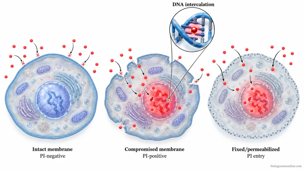
Propidium iodide (PI) is a fluorescent dye used for staining DNA and RNA. It binds to double-stranded DNA by intercalation, in which the dye enters between the base pairs. PI also binds to RNA. Its fluorescence increases on binding to nucleic acids. The bound dye produces red fluorescence when exposed to suitable excitation light.
PI does not readily enter cells through an intact plasma membrane. It enters cells with damaged plasma membranes and cells made permeable by laboratory treatment.
In viability staining, the cells are not fixed or permeabilized before adding PI. Cells with intact plasma membranes remain unstained, whereas cells with damaged membranes are stained. PI staining is used to detect the loss of membrane integrity. Dead cells and cells in the late stages of apoptosis commonly stain with PI. Early apoptotic cells may remain unstained as their plasma membranes are still intact.
For DNA-content staining, the cells are fixed and permeabilized or made permeable by another treatment. This allows PI to enter the cells. RNA is removed by RNase treatment to prevent RNA staining by PI. Under suitable staining conditions, the fluorescence intensity is directly proportional to the amount of DNA present in each cell. DNA content is measured by flow cytometry to identify cells in G₀/G₁, S and G₂/M phases of the cell cycle. The original membrane integrity of the cells cannot be determined after fixation by PI staining.
Fluorescence Properties of Propidium Iodide
Propidium iodide (PI) is a fluorescent dye that gives red fluorescence when bound to nucleic acids. In aqueous solution, unbound PI has an excitation maximum of about 493 nm and an emission maximum of about 636 nm.
The fluorescence of PI increases about 20–30 times after binding to DNA or RNA. The excitation maximum of DNA-bound PI is approximately 535 nm, while the emission maximum is about 617 nm.
In flow cytometry, PI is commonly excited using a 488 nm blue laser. It can also be excited using 532 nm or 561 nm lasers. The excitation maximum of 535 nm is different from the laser wavelength used in the instrument.
PI fluorescence is detected using suitable fluorescence channels and filters, such as a 610/20 nm or 620/15 nm bandpass filter. Some flow cytometers use the FL2 or FL3 channel for PI detection. The selection of the channel and filter depends upon the instrument.
In fluorescence microscopy, PI can be observed using a suitable red fluorescence filter set, such as Texas Red. The excitation and emission filters are selected according to the light source and filter system of the microscope.
Materials and Reagents Required for Propidium Iodide Staining
The following are the materials and reagents required for propidium iodide staining. The materials and reagents depend upon the staining method used.
Materials and Equipment
- Cell Sample – A cell suspension or prepared cells for staining.
- Pipettes and Pipette Tips – Used for transferring cell samples and staining solutions.
- Centrifuge – Used for collecting and washing cells when required.
- Centrifuge Tubes – For preparation and washing of cell suspensions.
- Flow Cytometry Tubes – Used for holding cell suspensions during flow cytometry analysis.
- Flow Cytometer – Used to measure the fluorescence of PI-stained cells.
- Glass Slides and Coverslips – Required for the microscopic examination of stained cells.
- Fluorescence Microscope – Used to observe PI fluorescence in stained cells.
- Mounting Equipment – Required for preparing slides for fluorescence microscopy.
- Aluminium Foil – Used to protect the staining solution and stained samples from light.
Reagents
- Propidium Iodide (PI) – The fluorescent dye used for staining. It can be used in the form of powder, stock solution or prepared working solution.
- Phosphate-Buffered Saline (PBS) – Used for washing cells and preparing the staining solution. Other suitable staining buffers may also be used.
- Distilled Water – Used for preparing the PI stock solution from powder.
- RNase A – Added to remove RNA during DNA content and cell cycle analysis. It is not generally required for cell viability staining.
- 70% Ethanol – Used for fixation and permeabilization of cells before DNA content analysis. It is not used for membrane-integrity staining of intact, unfixed cells.
- Triton X-100 – Used for permeabilization of cells in certain PI staining methods.
- Mounting Medium – Used for preparing microscopic slides. An antifade mounting medium may be used for fluorescence microscopy.
Preparation of Propidium Iodide Staining Solution
Propidium iodide (PI) staining solution is prepared by diluting the stock solution with a suitable buffer. The concentration of PI depends upon the staining method used.
Preparation of Propidium Iodide Stock Solution
- Take 10 mg of propidium iodide powder.
- The powder is dissolved in deionized or double-distilled water and the volume is made up to 10 mL. This gives a 1 mg/mL PI stock solution, which is equal to 1,000 µg/mL.
- Transfer the solution to a suitable container and label it with the reagent name, concentration, solvent and date of preparation.
- The container is covered with aluminium foil to protect the solution from light. A light-resistant container may also be used.
- Store the aqueous stock solution at 2–6°C. The solution prepared in this way can be stored for at least six months when protected from light. For a purchased PI solution, use the storage conditions given with the product.
Preparation of Propidium Iodide Working Solution
The PI working solution is prepared by diluting the stock solution with PBS or a suitable staining buffer. The required volume of stock solution is calculated using the formula C₁V₁ = C₂V₂.
The following are examples of PI working solution preparation:
- For cell viability staining, a 10 µg/mL PI working solution can be prepared by adding 100 µL of the 1 mg/mL stock solution to 9.9 mL of 1× PBS.
- Add one volume of this working solution to nine volumes of unfixed cell suspension to obtain a final PI concentration of 1 µg/mL.
- For cell cycle analysis, 500 µL of the 1 mg/mL PI stock solution is diluted with a PBS-based staining buffer to a final volume of 10 mL. This gives a PI concentration of 50 µg/mL.
- The staining solution used for fixed or permeabilized cells also contains RNase A to remove RNA before DNA content analysis.
- The working solution should be protected from light. The cell cycle staining solution containing RNase is generally prepared fresh, while a PI viability staining solution may be stored at 4°C in the dark according to the protocol.
The final staining concentration is not the same for all PI staining methods. Cell viability staining generally uses a lower PI concentration than fixed-cell DNA content analysis.
Procedure of Propidium Iodide Staining
Propidium iodide (PI) staining can be carried out by different methods depending upon the purpose of staining. The procedures for cell viability staining, cell cycle analysis and fluorescence microscopy are given below.
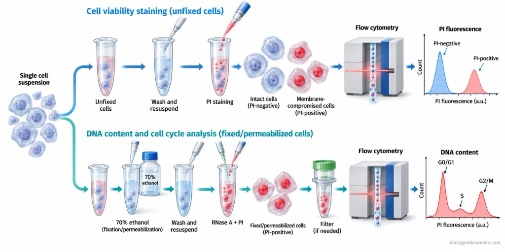
Propidium Iodide Staining for Cell Viability by Flow Cytometry
The following are the steps involved in PI staining for cell viability analysis:
- Collect the cells and prepare a single-cell suspension. Adherent cells may be detached before collection.
- Wash the cells once or twice with flow cytometry staining buffer.
- Resuspend the cells in 100 µL of staining buffer and transfer them to a flow cytometry tube.
- Use a ready-to-use PI staining solution prepared for flow cytometry.
- Add 5 µL of PI staining solution to 100 µL of cell suspension and mix gently.
- Incubate for 5–15 minutes on ice or at room temperature. Protect the samples from light.
- Prepare an unstained control and a suitable positive control containing cells with damaged plasma membranes.
- Do not wash the cells after PI staining. The dye is kept in the suspension during flow cytometry analysis.
- Analyze the samples using a flow cytometer with a suitable laser, such as a 488 nm laser, and a detector for red fluorescence. The controls are used to identify PI-positive and PI-negative cells.
- The samples should be analyzed within 4 hours after staining. If analysis is delayed, keep the samples at 2–8°C and protect them from light.
The cells are not fixed or permeabilized before staining in this method, because PI staining is used to examine the integrity of the plasma membrane.
Propidium Iodide Staining for Cell Cycle Analysis by Flow Cytometry
The following are the steps involved in PI staining for cell cycle analysis:
- Harvest the cells and count them. Wash the cells with phosphate-buffered saline (PBS) and prepare a single-cell suspension.
- Centrifuge the cell suspension and remove the supernatant.
- Add 1 mL of ice-cold 70% ethanol dropwise to the cell pellet while vortexing to prevent clumping.
- Keep the cells overnight at 4°C for fixation and permeabilization.
- Prepare the PI staining solution by mixing 8.5 mL of staining buffer containing 0.1% bovine serum albumin (BSA) in PBS, 1 mL of 1% RNase A stock solution and 0.5 mL of 1 mg/mL PI stock solution. The resulting solution contains 50 µg/mL PI and 1 mg/mL RNase A.
- Centrifuge the fixed cells and remove the ethanol. In this method, not more than 0.2 mL of ethanol is left in the tube.
- Resuspend the cells in 0.5–1.0 mL of PI staining solution containing RNase A.
- Incubate for at least 30 minutes at room temperature in the dark. RNase A removes RNA which can also bind to PI.
- Transfer the stained cells to flow cytometry tubes. If cell aggregates are present, pass the suspension through a suitable cell filter.
- Analyze the cells using a flow cytometer with a suitable PI fluorescence channel. A 488 nm laser can be used for excitation. The DNA fluorescence intensity is measured on a linear scale.
- Exclude cell doublets during analysis using fluorescence pulse area and height or width measurements.
Propidium Iodide Staining for Fluorescence Microscopy
PI staining can be used for microscopic examination of cells with damaged plasma membranes and for staining DNA in fixed or permeabilized specimens.
The following are the steps involved in PI staining for fluorescence microscopy:
- Prepare the cultured cells in a suitable culture vessel or prepare the specimen on a microscope slide.
- For cell viability staining, use unfixed cells without permeabilization.
- In a two-colour live/dead staining method, add 2 drops each of NucBlue Live reagent and PI reagent from the ReadyProbes Blue/Red kit per mL of culture medium. This method uses PI along with NucBlue Live reagent.
- Incubate the cells for 5–30 minutes.
- Examine the cells using a fluorescence microscope. A DAPI filter is used for NucBlue fluorescence, while an RFP or TRITC filter is used for the red fluorescence of PI.
- For DNA counterstaining, use fixed and permeabilized specimens. In one method used for prepared plant ovules, the specimens are stained with 10 µg/mL PI in PBS for 15 minutes at room temperature.
- Wash the fixed specimens with PBS for 15 minutes after staining.
- Mount the specimens using an antifade mounting medium. In this method, the mounting medium also contains 10 µg/mL PI.
- Place the prepared slide on the fluorescence microscope and observe the stained nuclei using a suitable red fluorescence filter set.
Results and Interpretation of Propidium Iodide Staining
The result of propidium iodide (PI) staining depends upon the condition of the cells and the method used for staining.
Interpretation of PI-Positive and PI-Negative Cells
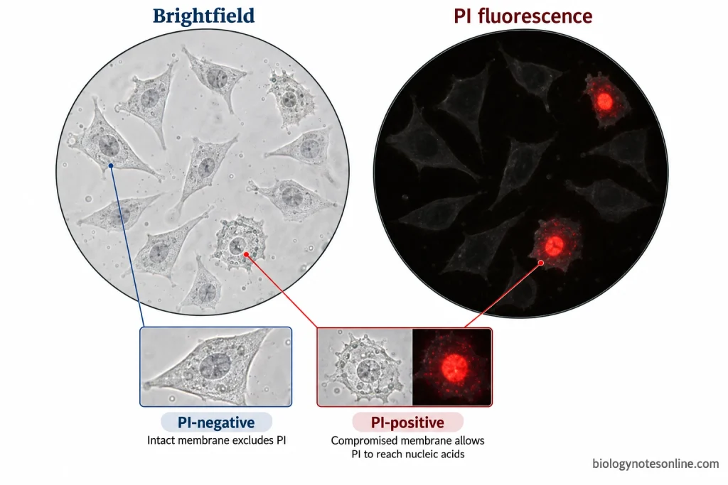
In cell viability staining, PI is added to unfixed cells. The intact plasma membrane prevents the entry of PI. These cells are called PI-negative cells and show little or no red fluorescence. When the plasma membrane is damaged, PI enters the cell and gives red fluorescence. Such cells are known as PI-positive cells.
PI-positive cells are generally considered nonviable, while PI-negative cells are considered viable in cell viability assays. However, the entry of PI only indicates that the plasma membrane has lost its integrity. It does not necessarily mean that the cell is irreversibly dead. Similarly, a PI-negative result does not confirm that all the cellular functions are normal.
The result is different when cells are fixed or permeabilized before staining. PI can enter these cells and bind to the nucleic acids present inside them. Therefore, PI fluorescence in fixed or permeabilized cells does not indicate cell death.
Under a fluorescence microscope, red fluorescence is observed mainly in the nuclei of PI-positive cells. The stained cells are counted and compared with the total number of cells. Brightfield images or a suitable counterstain can be used to determine the total cell number.
An unstained sample is used to measure background fluorescence. A sample containing cells with damaged plasma membranes is used as a positive control. In flow cytometry, cell debris is excluded by gating before calculating the percentage of PI-positive cells. Single-stained controls are also required for fluorescence compensation when other fluorescent dyes are used.
Interpretation of Cell Cycle Histograms
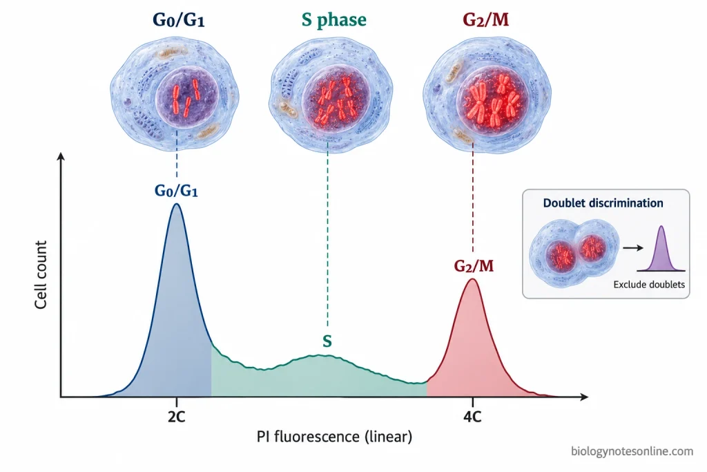
For cell cycle analysis, cells are fixed or permeabilized before PI staining. The dye enters the cells and binds to DNA. RNase is used to remove RNA, which can also contribute to PI fluorescence.
The amount of fluorescence depends upon the DNA content of each cell. The fluorescence intensity is measured on a linear scale and the results are represented in the form of a DNA-content histogram.
In a diploid cell population, the following regions are observed in the histogram:
- G0/G1 Phase: Cells containing approximately 2N DNA form the first peak of the histogram. These cells are present in the G0 or G1 phase.
- S Phase: DNA replication takes place during the S phase. The DNA content increases from 2N to 4N. These cells give intermediate fluorescence and are present between the two peaks.
- G2/M Phase: Cells in the G2 and M phases contain approximately 4N DNA and form the second peak. Their fluorescence intensity is about twice that of G0/G1 cells.
The percentage of cells in each region is calculated to determine the distribution of cells in different phases of the cell cycle.
Cell doublets may produce fluorescence similar to a single cell containing twice the amount of DNA. These cells are excluded by gating using the area and height or width of the fluorescence pulse. If the doublets are not removed, the G2/M population may be overestimated.
PI staining measures DNA content but cannot distinguish G0 from G1 or G2 from M. Additional markers are required for the separate identification of these phases.
Applications of Propidium Iodide Staining
The following are the applications of propidium iodide (PI) staining:
- It is used in cell viability assays to identify cells with damaged plasma membranes. These cells take up PI and give red fluorescence.
- PI staining is used in flow cytometry to exclude dead cells from the cell population during analysis.
- It is used to measure the DNA content of fixed or permeabilized cells and determine the distribution of cells in different phases of the cell cycle.
- The DNA content is used to identify cells in the G0/G1, S and G2/M phases.
- PI staining is used to estimate the DNA ploidy of cells by comparing their fluorescence intensity with a reference cell population.
- It is used along with Annexin V staining to study apoptosis and necrosis. The two stains help to distinguish cells based on phosphatidylserine exposure and plasma membrane integrity.
- Used in drug testing to determine the percentage of cells with damaged membranes after treatment with drugs or other toxic substances.
- PI is used in fluorescence microscopy to stain the nuclei of fixed or permeabilized cells. The stained nuclei give red fluorescence.
- It can be used with live-cell dyes such as calcein AM for the identification of live and dead cells in the same sample.
- PI staining is also used to detect cells with reduced DNA content in the sub-G1 region during apoptosis studies. However, the presence of a sub-G1 population alone does not confirm apoptosis.
Limitations of Propidium Iodide Staining
The following are the limitations of propidium iodide staining:
- PI staining is based on plasma membrane integrity. It does not directly measure metabolic activity or the ability of a cell to reproduce.
- Early apoptotic cells may remain unstained because their plasma membranes are still intact. These cells cannot be distinguished from viable cells by PI alone.
- PI alone cannot distinguish necrotic cells from late apoptotic cells. Both may take up the dye after the loss of membrane integrity.
- The original viability of cells cannot be determined by PI staining after fixation or permeabilization. These treatments allow the dye to enter the cells.
- PI binds to RNA as well as DNA. RNA staining adds to the fluorescence measured during DNA-content analysis, and RNase treatment is required to remove RNA.
- Cell doublets may be counted as cells with a higher DNA content. Two G₁ cells passing together through the flow cytometer can give a signal similar to that of a single G₂ cell.
- Extracellular nucleic acids can also bind PI. In bacterial biofilms, this staining may give a higher dead-cell count than the actual number of dead cells.
Advantages of Propidium Iodide Staining
The following are the advantages of propidium iodide staining:
- PI is used to detect cells with damaged plasma membranes. Fixation and permeabilization are not required for membrane-integrity-based viability staining.
- The fluorescence of PI increases on binding to nucleic acids. The stained cells give a strong red fluorescence.
- DNA content can be measured in fixed or permeabilized cells after RNase treatment. Under suitable staining conditions, the fluorescence intensity is directly proportional to the amount of DNA present in each cell.
- PI staining can be examined by fluorescence microscopy or measured by flow cytometry. Confocal microscopy can also be used.
- PI can be excited by a 488 nm laser, which is commonly used in flow cytometers.
- PI can be used along with Annexin V to identify viable, early apoptotic and late apoptotic or dead-cell populations.
- The dye is economical and stable under suitable storage conditions.
Precautions of Propidium Iodide Staining
The following are the precautions of propidium iodide staining:
- Wear protective gloves, a laboratory coat and safety glasses while handling propidium iodide. The dye is suspected of causing genetic damage.
- Keep the propidium iodide solution protected from light. The prepared solution should be stored at the recommended temperature.
- Use the correct concentration of propidium iodide for staining. Excess dye may increase the background fluorescence.
- For cell viability staining, use unfixed cells. Fixation or permeabilization allows propidium iodide to enter cells through the damaged or permeabilized plasma membrane.
- Add propidium iodide shortly before the examination of cells by flow cytometry. Do not wash the cells after staining when PI is used for cell viability analysis.
- Avoid damage to the cell membrane during sample preparation and washing. Damaged cells take up propidium iodide and give positive fluorescence.
- Maintain the same staining time and concentration of propidium iodide for all samples being compared.
- Use a properly prepared single-cell suspension. Cell clumps may interfere with the flow cytometric analysis.
- For cell cycle analysis, the cells should be fixed properly before staining. Cold ethanol should be added slowly while mixing to prevent cell clumping.
- Treat the fixed cells with RNase A before staining for cell cycle analysis. Propidium iodide also binds to RNA, which may affect the measurement of DNA content.
- Use unstained and suitable positive controls to check the staining results.
- Select the appropriate fluorescence channel for propidium iodide. Its fluorescence may overlap with other fluorescent dyes used in the same sample.
- Dispose of propidium iodide solutions and contaminated materials according to the laboratory chemical waste disposal procedures.
References
- Crowley, L. C., Scott, A. P., Marfell, B. J., Boughaba, J. A., Chojnowski, G., & Waterhouse, N. J. (2016). Measuring cell death by propidium iodide uptake and flow cytometry. Cold Spring Harbor Protocols, 2016(7), pdb.prot087163. https://doi.org/10.1101/pdb.prot087163
- Davey, H. M., & Hexley, P. (2011). Red but not dead? Membranes of stressed Saccharomyces cerevisiae are permeable to propidium iodide. Environmental Microbiology, 13(1), 163–171. https://doi.org/10.1111/j.1462-2920.2010.02317.x
- Fried, J., Perez, A. G., & Clarkson, B. D. (1976). Flow cytofluorometric analysis of cell cycle distributions using propidium iodide: Properties of the method and mathematical analysis of the data. The Journal of Cell Biology, 71(1), 172–181. https://doi.org/10.1083/jcb.71.1.172
- Krämer, C. E. M., Wiechert, W., & Kohlheyer, D. (2016). Time-resolved, single-cell analysis of induced and programmed cell death via non-invasive propidium iodide and counterstain perfusion. Scientific Reports, 6, Article 32104. https://doi.org/10.1038/srep32104
- Krishan, A. (1975). Rapid flow cytofluorometric analysis of mammalian cell cycle by propidium iodide staining. The Journal of Cell Biology, 66(1), 188–193. https://doi.org/10.1083/jcb.66.1.188
- Nicoletti, I., Migliorati, G., Pagliacci, M. C., Grignani, F., & Riccardi, C. (1991). A rapid and simple method for measuring thymocyte apoptosis by propidium iodide staining and flow cytometry. Journal of Immunological Methods, 139(2), 271–279. https://doi.org/10.1016/0022-1759(91)90198-O
- Riccardi, C., & Nicoletti, I. (2006). Analysis of apoptosis by propidium iodide staining and flow cytometry. Nature Protocols, 1(3), 1458–1461. https://doi.org/10.1038/nprot.2006.238
- Rieger, A. M., Hall, B. E., Luong, L. T., Schang, L. M., & Barreda, D. R. (2010). Conventional apoptosis assays using propidium iodide generate a significant number of false positives that prevent accurate assessment of cell death. Journal of Immunological Methods, 358(1–2), 81–92. https://doi.org/10.1016/j.jim.2010.03.019
- Rieger, A. M., Nelson, K. L., Konowalchuk, J. D., & Barreda, D. R. (2011). Modified Annexin V/propidium iodide apoptosis assay for accurate assessment of cell death. Journal of Visualized Experiments, 50, Article 2597. https://doi.org/10.3791/2597
- Shapiro, H. M. (2003). Practical flow cytometry (4th ed.). Wiley-Liss. https://doi.org/10.1002/0471722731
- Vermes, I., Haanen, C., Steffens-Nakken, H., & Reutelingsperger, C. (1995). A novel assay for apoptosis: Flow cytometric detection of phosphatidylserine expression on early apoptotic cells using fluorescein labelled Annexin V. Journal of Immunological Methods, 184(1), 39–51. https://doi.org/10.1016/0022-1759(95)00072-I
- Zhao, H., Oczos, J., Janowski, P., Trembecka, D., Dobrucki, J., Darzynkiewicz, Z., & Wlodkowic, D. (2010). Rationale for the real-time and dynamic cell death assays using propidium iodide. Cytometry Part A, 77A(4), 399–405. https://doi.org/10.1002/cyto.a.20867