

Cell wall staining is required because the bacterial cell wall is very thin and cannot be seen clearly in an ordinary stained smear. The bacterial cell wall is present outside the cytoplasmic membrane and helps to maintain the shape of the bacterial cell.
In bright-field microscopy, ordinary staining can show the shape and arrangement of bacterial cells, but the cell wall cannot be clearly distinguished from the cytoplasm.
What is Cell Wall Staining?
Cell wall staining is a special staining technique used in microbiology to demonstrate the bacterial cell wall separately from the other components of the cell. In this technique, the cell wall may appear as a coloured outline surrounding the cell contents. The cytoplasm may remain colourless or appear in a different colour.
Gram staining is a differential staining technique in which bacterial cells are distinguished by their retention of the primary stain. It does not specifically show the cell wall as a separate outline.
Chance’s method, Ringen et al.’s method and Dyar’s method are some of the methods used for cell wall staining.
Principle of Cell Wall Staining
Cell wall staining is based on the different staining reactions of the bacterial cell wall and cytoplasm. The bacterial cell surface is usually negatively charged. Basic dyes are positively charged and bind to the negatively charged components of the cell wall and cytoplasm.
Acidic dyes are negatively charged and are normally repelled by the bacterial surface. In some methods, an acidic dye is used to remove the basic stain from the cytoplasm, while the cell wall retains the stain. Certain methods use a mordant to increase the affinity of the dye for the cell wall.
In other methods, a cationic surface-active agent acts as a mordant and gives the bacterial cell wall a positive charge. The acidic dye then binds to the treated cell wall. The stained cell wall appears as a coloured outline around the cytoplasm, which may remain colourless or be stained with a contrasting dye under the light microscope.
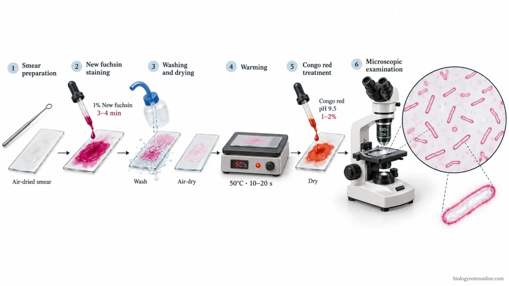
Cell Wall Staining by Chance’s Method
Chance’s method is a bacterial cell wall staining method developed by H. L. Chance in 1953. This method uses new fuchsin and Congo red to stain the bacterial cell wall separately from the cytoplasm. It was described particularly for Escherichia coli and Bacillus cereus.
Principle of Chance’s Method
Chance’s method is based on the selective retention of new fuchsin by the bacterial cell wall. New fuchsin is a basic dye which stains both the cell wall and cytoplasm. The cell wall retains the stain more strongly than the cytoplasm.
Congo red is an acidic dye used as a selective decolourising agent. It removes much of the new fuchsin from the cytoplasm, while the cell wall retains the stain. In the original method, Congo red is used at an alkaline pH of about 9.5.
The bacterial cell wall appears pink or reddish, while the cytoplasm remains colourless or lightly stained.
Reagents and Materials Required
The following are the materials and reagents required for cell wall staining by Chance’s method:
Materials and Equipment
- Bacterial culture – A suitable culture of Escherichia coli or Bacillus cereus for the preparation of bacterial smear.
- Clean glass slides – The bacterial smear is prepared on a clean glass slide.
- Inoculating loop – For transferring the bacterial cells from the culture onto the glass slide.
- Dropper or pipette – To add the staining solutions to the smear.
- Staining rack – The slides are placed on the rack during staining and washing.
- Warm plate – A warm plate maintained at 50°C.
- Timer – For measuring the staining and warming time.
- Light microscope – A microscope with an oil immersion objective.
- Immersion oil – Required for examining the stained smear under the oil immersion objective.
- pH meter or pH paper – For checking the pH of the Congo red solution.
Staining Reagents
- New fuchsin solution – 1% aqueous solution of new fuchsin. It is used as the primary stain.
- Congo red solution – 1–2% Congo red solution with a pH of approximately 9.5.
- Distilled water – Used for the preparation of bacterial smear and washing off the excess stain.
Procedure of Chance’s Method
The procedure of cell wall staining by Chance’s method is given below:
- Take a clean glass slide and prepare a thin smear of bacterial culture using water.
- Allow the smear to air dry completely.
- Cover the dried smear with 1% aqueous new fuchsin solution for 3–4 minutes.
- Wash the smear with water until no more stain comes off.
- Allow the smear to air dry.
- Place the slide on a warm plate maintained at 50°C for 10–20 seconds.
- Cover the smear with a thin film of 1–2% Congo red solution at a pH of about 9.5.
- Allow the Congo red film to dry.
- The dried smear can be examined directly or washed with water before examination.
- Examine the stained smear under the oil immersion objective of a light microscope.
Observation and Result
The bacterial smear is examined under the oil immersion objective of a light microscope. The observations of Chance’s method are as follows:
- The bacterial cell wall is stained pink or reddish.
- The cytoplasm is either colourless or lightly stained.
- A coloured outline is seen around the bacterial cell due to the staining of the cell wall.
- The cross septa may show faint staining.
The cell wall is identified by the pink or reddish outline present around the colourless or lightly stained cytoplasm. If the outline cannot be seen clearly, the cell wall is recorded as not clearly demonstrated in the stained smear.
Cell Wall Staining by Ringen et al. Method
Ringen et al. method is a special staining method used to demonstrate the bacterial cell wall separately from the cytoplasm. The method uses tannic acid, crystal violet and Congo red for staining the bacterial cell wall.
In this method, tannic acid is used as a mordant which increases the affinity of the cell wall for crystal violet. It may also increase the apparent thickness of the stained cell wall. Crystal violet stains both the cell wall and cytoplasm. Congo red is used to remove the stain mainly from the cytoplasm, while the cell wall retains the violet colour.
Under the light microscope, the bacterial cell wall appears violet and the cytoplasm remains colourless or lightly stained.
Principle of Ringen et al. Method
Ringen et al. method is based on the selective staining of the bacterial cell wall with crystal violet. Tannic acid is used as a mordant which increases the affinity of the cell wall for crystal violet. It also increases the apparent thickness of the cell wall.
Crystal violet is a basic dye which stains both the cell wall and cytoplasm. Congo red is used as a selective decolourising agent. It removes much of the crystal violet stain from the cytoplasm, while the cell wall retains the violet colour.
The bacterial cell wall appears violet under the light microscope. The cytoplasm remains colourless or lightly stained, but may appear pinkish in some preparations.
Reagents and Materials Required
The following are the materials and reagents required for cell wall staining by Ringen et al. method:
Materials and Equipment
- Bacterial culture – A suitable Gram-positive or Gram-negative bacterial culture for preparing the smear.
- Clean glass slides – For preparing the bacterial smear.
- Inoculating loop – To transfer the bacterial cells onto the glass slide.
- Bunsen burner or spirit lamp – For heat fixation of the bacterial smear.
- Staining rack – The slides are placed on the rack during staining and washing.
- Dropper or pipette – To add the staining solutions to the smear.
- Timer – For measuring the staining time.
- Blotting paper – To remove excess water from the slide after washing.
- Light microscope – A microscope with an oil immersion objective.
- Immersion oil – For examining the stained bacterial smear under the oil immersion objective.
Staining Reagents
- Tannic acid solution – 5% aqueous solution of tannic acid. It is used as a mordant.
- Crystal violet solution – 0.5% aqueous solution of crystal violet chloride, used as the primary stain.
- Congo red solution – 0.5% aqueous solution of Congo red. It is used as a selective decolourising agent.
- Distilled water – For preparing the bacterial smear and washing off the excess stain.
Procedure of Ringen et al. Method
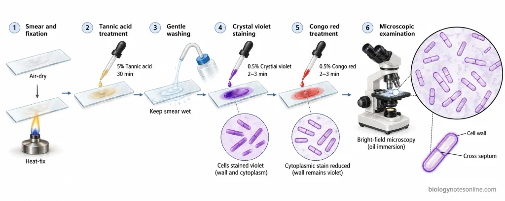
The procedure of Ringen et al. method of cell wall staining is given below:
- Take a clean glass slide and prepare a thin smear of the bacterial culture.
- Allow the smear to air dry and heat fix it.
- Cover the smear with 5% aqueous tannic acid solution for 30 minutes.
- Wash the slide with water. Do not dry the smear.
- Stain the wet smear with 0.5% aqueous crystal violet chloride solution for 2–3 minutes.
- Wash off the crystal violet stain with water.
- Cover the smear with 0.5% aqueous Congo red solution for 2–3 minutes.
- Wash the slide with water and allow it to dry. Excess water may be removed gently with blotting paper.
- Examine the stained smear under the oil immersion objective of a light microscope.
Observation and Result
The bacterial smear is examined under the oil immersion objective of a light microscope. The observations of Ringen et al. method are as follows:
- The bacterial cell wall is stained violet.
- The cytoplasm is either colourless or lightly stained.
- A violet outline is seen around the bacterial cell due to the staining of the cell wall.
- The cell wall may appear slightly thicker due to the treatment with tannic acid.
The cell wall is identified by the violet outline present around the colourless or lightly stained cytoplasm. If the outline cannot be seen clearly, the cell wall is recorded as not clearly demonstrated in the stained smear.
Cell Wall Staining by Dyar’s Method
Cell wall staining by Dyar’s method is a staining technique used to demonstrate the bacterial cell wall separately from the cytoplasm. The method was developed by M. T. Dyar in 1947. It uses cetylpyridinium chloride, Congo red and methylene blue for staining the bacterial cells.
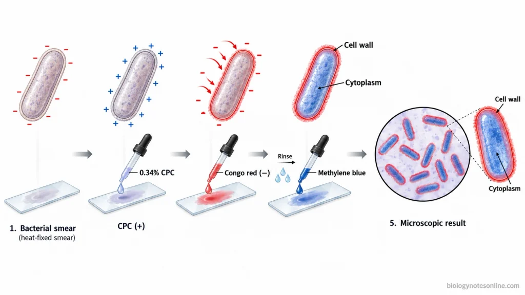
Cetylpyridinium chloride (CPC) is a cationic surface-active agent used as a mordant. It gives the bacterial cell wall a positive charge. Congo red is an acidic dye which stains the treated cell wall red. Methylene blue is used as a counterstain for the cytoplasm.
Under the light microscope, the bacterial cell wall appears red and the cytoplasm appears blue. The method has also been used for staining the cell walls of yeasts such as Saccharomyces and Schizosaccharomyces.
Principle of Dyar’s Method
The bacterial cell wall normally carries a negative surface charge. Cetylpyridinium chloride (CPC) is a cationic surface-active agent which is used as a mordant. It binds to the bacterial cell wall and makes the surface positively charged.
Congo red is an acidic dye which has a negative charge. It binds to the positively charged CPC present on the cell wall and stains the wall red. A red precipitate of Congo red and CPC may also form on the surface of the cell wall.
Methylene blue is a basic dye used as a counterstain. It stains the cytoplasm blue, while the cell wall remains red.
Reagents and Materials Required
The following are the materials and reagents required for cell wall staining by Dyar’s method:
Materials and Equipment
- Bacterial culture – A suitable culture of Bacillus cereus or Escherichia coli for the preparation of bacterial smear.
- Clean glass slides – A thin bacterial smear is prepared on a clean glass slide.
- Inoculating loop – For transferring bacterial cells from the culture onto the glass slide.
- Bunsen burner or spirit lamp – The dried bacterial smear is heat fixed using a flame.
- Dropper or pipette – To add the staining solutions to the bacterial smear.
- Staining rack – The slides are placed on the rack during staining and washing.
- Wash bottle – A wash bottle containing distilled water for washing the stained smear.
- Glass coverslips – The stained smear is covered with a coverslip after adding a drop of water.
- Light microscope – A microscope fitted with an oil immersion objective.
- Immersion oil – Required for examining the stained bacterial cells under the oil immersion objective.
Staining Reagents
- Cetylpyridinium chloride (CPC) solution – 0.34% aqueous solution (M/100) of cetylpyridinium chloride. It is used as a mordant.
- Congo red solution – A saturated aqueous solution of Congo red for staining the bacterial cell wall.
- Methylene blue solution – Used as a counterstain for the cytoplasm.
- Distilled water – For the preparation of bacterial smear and washing off the excess stain.
Procedure of Dyar’s Method
The procedure of cell wall staining by Dyar’s method is given below:
- Take a clean glass slide and prepare a thin smear of bacterial culture.
- Allow the smear to air dry completely and heat fix it.
- Add 3 drops of M/100 (0.34%) aqueous cetylpyridinium chloride solution to the smear.
- Add 1 drop of saturated aqueous Congo red solution and mix the two solutions on the slide.
- Wash the smear with water to remove the excess stain.
- Counterstain the smear with methylene blue for a few seconds.
- Wash the slide again with water.
- Examine the stained smear in a drop of water under a coverslip or directly in oil using a light microscope. Side illumination may be used for better observation of the cell wall.
Observation and Result
The stained bacterial smear is examined under the oil immersion objective of a light microscope. The observations of Dyar’s method are as follows:
- The bacterial cell wall is stained red.
- The cytoplasm is stained blue with methylene blue.
- A red outline is seen around the bacterial cell due to the staining of the cell wall.
- In some bacterial cells examined in water, the cytoplasm may appear separated from the cell wall.
- The cell wall may appear contracted when the stained smear is examined dry in oil.
The bacterial cell wall is identified by the red outline present around the blue cytoplasm. If the red outline cannot be seen clearly, the cell wall is recorded as not clearly demonstrated in the stained smear.
Comparison of Cell Wall Staining Methods
Chance’s method, Ringen et al. method and Dyar’s method are used for staining the bacterial cell wall. These methods differ in the staining reagents, mordants, fixation and colour of the stained cells.
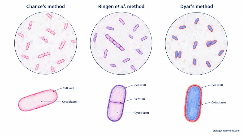
The differences between the three cell wall staining methods are given below:
| Comparison Parameter | Chance’s Method | Ringen et al. Method | Dyar’s Method |
|---|---|---|---|
| Primary staining agent | 1% aqueous new fuchsin | 0.5% aqueous crystal violet chloride | Saturated aqueous Congo red |
| Mordant or surface-active agent | No separate mordant is used. | 5% aqueous tannic acid | 0.34% cetylpyridinium chloride (CPC) |
| Role of mordant | No mordant treatment is required. | Tannic acid increases the affinity of the cell wall for crystal violet. | CPC gives a positive charge to the bacterial cell wall. |
| Decolourising treatment | 1–2% Congo red solution at approximately pH 9.5 | 0.5% aqueous Congo red solution | No separate decolourising agent |
| Cytoplasmic counterstain | Not used | Not used | Methylene blue |
| Smear fixation | The smear is air-dried without heat fixation. | The smear is air-dried and heat-fixed. | The smear is air-dried and heat-fixed. |
| Additional treatment | The slide is warmed at 50°C before adding Congo red. | Tannic acid is applied before crystal violet. | CPC and Congo red are mixed on the smear. |
| Cell wall appearance | Pink or red | Violet or blue-violet | Red |
| Cytoplasm appearance | Colourless or lightly stained | Colourless or lightly stained | Blue |
| Cross septa | The cross septa may be faintly stained. | The cross septa may be stained more clearly. | The red outline of the cell wall is the main observation. |
| Microscopic examination | Dried smear under oil immersion | Dried smear under oil immersion | The stained cells may be examined in water under a coverslip or in oil. |
| Main difference | New fuchsin staining followed by Congo red treatment | Tannic acid mordanting followed by crystal violet staining | CPC-assisted staining with Congo red and methylene blue |
Differences in Laboratory Observations
In Chance’s method, the bacterial cell wall is stained pink or red, while the cytoplasm remains colourless or lightly stained. Ringen et al. method gives a violet cell wall and may show the transverse septa more clearly.
Dyar’s method stains the cell wall red and the cytoplasm blue. Congo red is used as the cell wall stain in this method, whereas it is used for selective decolourisation in Chance’s and Ringen et al. methods.
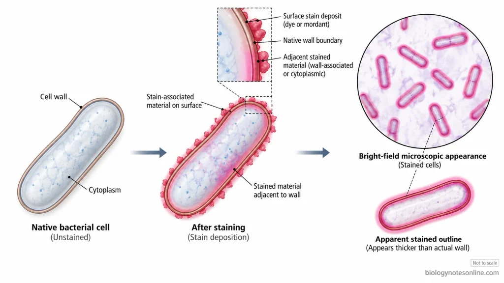
The apparent thickness of the stained cell wall may increase due to the deposition of mordant or dye precipitates. In Dyar’s method, the cell wall may also contract when the stained cells are examined dry in oil.
Precautions of Cell Wall Staining
The following are the precautions of cell wall staining:
- Use glass slides free from grease and dust.
- The bacterial smear should be thin. Thick smears may contain overlapping cells and give uneven staining.
- Take a small amount of bacterial material from a fresh culture.
- Allow the smear to air dry completely. Heat fixation is not required in Chance’s method, but it is required in the heat-fixed protocols of Ringen et al. and Dyar’s methods.
- Avoid overheating the smear during fixation, as it may change the shape of bacterial cells.
- Use the staining reagents at the required concentrations. The pH of Congo red solution in Chance’s method should be maintained at approximately 9.5.
- Keep the warm plate at 50°C in Chance’s method. Do not heat the slide longer than the required time.
- In Ringen et al. method, apply crystal violet to the wet smear after washing off the tannic acid. The smear should not be allowed to dry between these treatments.
- Do not stain the smear for longer than the specified time. Excess staining may make it difficult to differentiate the cell wall from the cytoplasm.
- Wash the smear gently with distilled water. Avoid using a strong stream of water.
- In Dyar’s method, use the required amounts of cetylpyridinium chloride and Congo red. Methylene blue is applied for a few seconds only.
- Do not dry the stained cells excessively in Dyar’s method. The cell wall may shrink or collapse when examined dry in oil.
- Wear laboratory gloves and protective clothing while handling bacterial cultures and staining reagents. Avoid direct contact with the staining solutions.
Applications of Cell Wall Staining
The following are the applications of cell wall staining:
- Cell wall staining is used to demonstrate the bacterial cell wall under a light microscope.
- It helps in studying the shape and outline of bacterial cells.
- The cell wall can be differentiated from the cytoplasm by staining the two structures with different colours.
- It is used for the observation of cross walls or septa formed during bacterial cell division.
- The staining reactions of the cell walls of Gram-positive and Gram-negative bacteria can be compared using different staining reagents.
- Cell wall staining is used in studies of bacterial cell division and the arrangement of cells in chains or groups.
- Dyar’s method can also be used for staining the cell walls of certain yeasts, such as Saccharomyces and Schizosaccharomyces.
- Cell wall staining may be combined with Gram staining to study the relationship between the Gram reaction and the bacterial cell wall.
Limitations of Cell Wall Staining
The following are the limitations of cell wall staining:
- The bacterial cell wall is very thin and cannot be seen in its actual thickness under an ordinary light microscope.
- The stained cell wall may appear thicker than its actual size due to the deposition of dye or mordant on the cell surface.
- Heat fixation and drying may change the shape of bacterial cells. In Dyar’s method, the stained cell wall may shrink or collapse when examined dry.
- The staining result depends upon the concentration of staining reagents, staining time and washing of the smear.
- Incomplete treatment with tannic acid may stain only the outer outline of the cell wall. The transverse septa may not be clearly visible.
- Excess staining or improper washing may stain the cytoplasm and make it difficult to differentiate the cell wall from the cytoplasm.
- Certain bacteria, such as mycobacteria and corynebacteria, are difficult to stain by some cell wall staining methods.
- The cell membrane cannot be clearly differentiated from the cell wall by these staining methods.
- A faint or absent stained outline does not always indicate the absence of a bacterial cell wall.
- Cell wall staining does not provide information about the exact chemical composition of the bacterial cell wall.
References
- Bartholomew, J. W., & Finkelstein, H. (1958). Relationship of cell wall staining to Gram differentiation. Journal of Bacteriology, 75(1), 77–84. https://doi.org/10.1128/jb.75.1.77-84.1958
- Chance, H. L. (1953). A bacterial cell wall stain. Stain Technology, 28(4), 205–207. https://doi.org/10.3109/10520295309105600
- Dyar, M. T. (1947). A cell wall stain employing a cationic surface-active agent as a mordant. Journal of Bacteriology, 53(4), 498. https://doi.org/10.1128/jb.53.4.498-498.1947
- Finkelstein, H., & Bartholomew, J. W. (1958). An interpretation of several cell wall stains applied to heat-fixed Bacillus subtilis on glass slides. Stain Technology, 33(4), 177–186. https://doi.org/10.3109/10520295809111845
- Hale, C. M. F., & Bisset, K. A. (1956). A comparison of the staining reactions of the cell walls of Azotobacter chroococcum and those of Gram-positive and Gram-negative bacteria. Journal of General Microbiology, 15(3), 423–427. https://doi.org/10.1099/00221287-15-3-423
- Lamanna, C., & Mallette, M. F. (1950). The relation of the Gram stain to the cell wall and the ribonucleic acid content of the cell. Journal of Bacteriology, 60(4), 499–505. https://doi.org/10.1128/jb.60.4.499-505.1950
- Mandal, S., Tomar, S. K., & Singh, R. (2012). Laboratory manual on microbial morphology and taxonomy. National Dairy Research Institute. https://studylib.net/doc/8185735/laboratory-manual-on-microbial-morphology-and-taxonomy