

The cell cycle is a regulated sequence of events through which a dividing eukaryotic cell grows, replicates its deoxyribonucleic acid (DNA) and divides.
It consists of two main stages, interphase and the mitotic phase (M phase). During interphase, the cell grows and prepares for division. In M phase, the duplicated chromosomes are separated by mitosis (nuclear division), and the cytoplasm is usually divided by cytokinesis to form two daughter cells. Cell division is only a part of the complete cell cycle.
- The four main phases of the cell cycle are G1 (first gap), S (DNA synthesis), G2 (second gap) and M. G1 and G2 are mainly involved in cell growth and preparation, while DNA replication takes place during S phase. M phase includes mitosis and usually cytokinesis.
- The progression of the cell cycle is controlled by cyclins, cyclin-dependent kinases (CDKs) and cell cycle checkpoints. These checkpoints can delay the cell cycle when DNA is damaged or certain events have not been completed.
- Interphase occupies most of the cell cycle in many dividing cells. It is often described as approximately 90% of the total cycle in introductory examples, but this duration varies with cell type and growth conditions.
- Some cells leave the active cell cycle and enter a non-dividing state called G0 phase. The cells may remain in this state temporarily or for a prolonged period, and G0 is not a compulsory fifth phase of the cell cycle.
Phases of the Cell Cycle
The cell cycle is divided into four main phases, G1 (first gap), S (synthesis), G2 (second gap) and M (mitotic phase). These phases occur in a definite sequence, G1 → S → G2 → M. The cell cycle has two major parts, interphase and mitotic phase.
Interphase consists of G1, S and G2 phases. During this period, the cell grows, carries out its normal metabolic activities and replicates its deoxyribonucleic acid (DNA). The M phase involves the division of the nucleus by mitosis, followed by the completion of cytoplasmic division (cytokinesis), which usually begins before mitosis is fully completed. Two daughter cells are formed after cytokinesis.
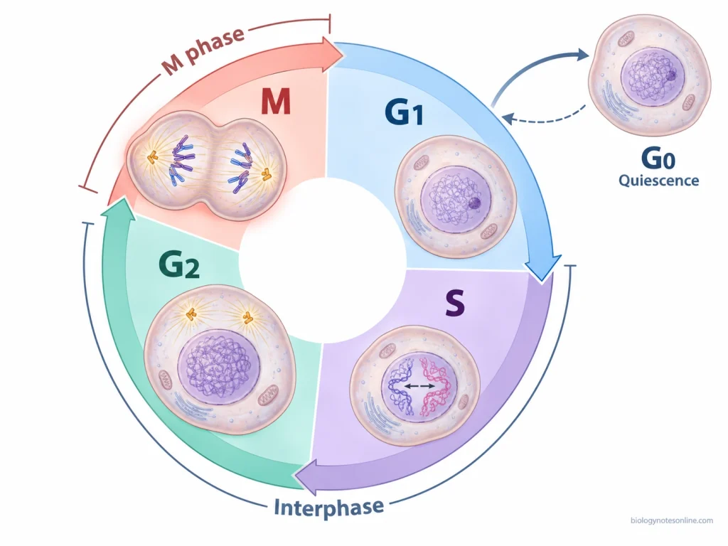
Four Main Phases of the Cell Cycle
The four phases and their major activities are given below-
| Phase | Main activity | Biological outcome |
|---|---|---|
| G1 (First gap) | Cell growth, synthesis of proteins and other cellular components. | Cell increases in size and prepares for DNA replication. |
| S (Synthesis) | Replication of DNA. | Each chromosome is duplicated into two sister chromatids. |
| G2 (Second gap) | Further cell growth and synthesis of proteins required for mitosis. | The cell completes its preparation for nuclear division. |
| M (Mitotic phase) | Mitosis and usually cytokinesis. | Duplicated chromosomes are separated, and two daughter cells are generally formed. |
Why Are There Four, Five or Seven Stages of the Cell Cycle?
The number of stages of the cell cycle varies in different educational resources. This is due to the different ways of counting the phases and their subdivisions.
The conventional cell cycle consists of four phases (G1, S, G2 and M). Some descriptions count cytokinesis separately from mitosis and refer to five stages. The G0 phase is also sometimes included as an additional stage. It is a non-dividing state in which certain cells remain temporarily or for a prolonged period. Not all cells enter G0.
In some textbooks, the mitotic phase is further divided into prophase, metaphase, anaphase and telophase. When these four stages are counted along with G1, S and G2, the total becomes seven. Other classifications also include prometaphase as a separate stage of mitosis or count cytokinesis separately.
A. Interphase
Interphase is the period of the cell cycle in which the cell grows, replicates its deoxyribonucleic acid (DNA) and prepares for division. It consists of three phases, G1, S and G2.
The cell remains metabolically active during interphase and carries out various activities required for its growth and division. The DNA within the nucleus is mainly present in the form of less condensed chromatin.
1. G1 Phase (First Gap Phase)
G1 phase (first gap phase) is the first stage of interphase, which occurs after cell division and before the S phase. In this phase, the cell grows in size and prepares for deoxyribonucleic acid (DNA) replication.
The major activities of G1 phase are as follows-
- The newly formed daughter cell increases in size. The amount of cytoplasm and other cellular components also increases during this period.
- The cell remains metabolically active and carries out various activities required for its normal functioning. Ribonucleic acid (RNA) and different types of proteins are synthesized in this phase.
- Some cellular organelles also increase in number during G1 phase.
- Various nucleotides, enzymes and other proteins required for DNA replication are produced and accumulated within the cell. Energy reserves are also built up for the next phase.
- The DNA remains unreplicated during G1 phase. Each chromosome consists of a single chromatid, and the amount of nuclear DNA has not yet doubled.
- The duration of G1 phase is not same in all cells. It varies depending on the type of cell, growth conditions and availability of external signals.
- Before entering S phase, the cell checks for sufficient nutrients, growth signals, cell size and any damage present in the DNA. This control takes place at the G1 checkpoint. If the required conditions are not met, the progression of the cell cycle may be delayed.
- In absence of suitable growth conditions or signals, some cells may leave G1 and enter into the non-dividing G0 phase. These cells can return to the active cell cycle when suitable signals become available.
2. S Phase (Synthesis Phase)
S phase (synthesis phase) is the second stage of interphase during which the deoxyribonucleic acid (DNA) of the cell undergoes replication. It occurs after G1 phase and before G2 phase.
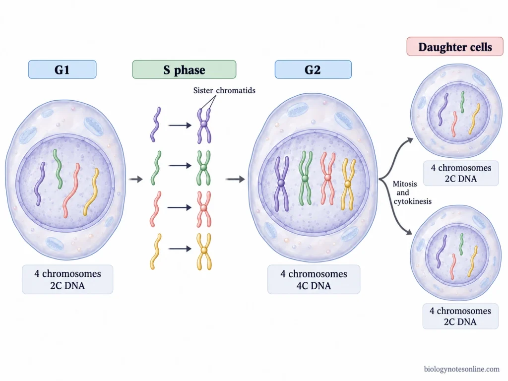
The major activities of S phase are as follows-
- The DNA present in the nucleus is replicated to produce two copies of each chromosome. During this process, the chromosomes remain in the form of less condensed chromatin.
- Each chromosome is duplicated to form two identical copies, known as sister chromatids. They remain attached to each other at the centromeric region after replication.
- The total amount of DNA becomes double by the end of S phase. But the chromosome number remains unchanged, as the two attached sister chromatids are still counted as one chromosome.
- DNA replication takes place by a semiconservative process, in which each newly formed DNA molecule contains one original strand and one newly synthesized strand.
- The synthesis of histone proteins also increases during S phase. These proteins are required for the packaging of newly synthesized DNA into chromatin.
- In typical animal cells, the centrosome also undergoes duplication during this phase. The two centrosomes are involved in the formation of the mitotic spindle, which is required for chromosome separation during mitosis.
- The DNA is normally replicated only once during each cell cycle. The replication process is regulated to prevent the same DNA region from being copied more than once in a single cycle.
- Accurate DNA replication is required for maintaining the genetic information of the cell. Errors occurring during DNA synthesis may produce mutations, which can be passed to the daughter cells.
- The duration of S phase is not same in all cells. It varies depending on the cell type and growth conditions. After the completion of DNA replication, the cell enters into G2 phase.
3. G2 Phase (Second Gap Phase)
G2 phase (second gap phase) is the last stage of interphase, which occurs after the completion of DNA replication in S phase. During this phase, the cell continues its growth and prepares for mitosis.
The major activities of G2 phase are as follows-
- The cell continues to grow in size after DNA replication. The amount of cytoplasm and other cellular components may also increase during this period.
- Various proteins required for mitosis are synthesized in G2 phase. The synthesis of ribonucleic acid (RNA) also continues. Some of these proteins are involved in the formation and functioning of the mitotic spindle, which is required for chromosome separation.
- The cell replenishes its energy reserves and produces various materials needed for mitotic division.
- Some cell organelles are also duplicated during this phase. The cytoskeleton undergoes changes in its organization as the cell prepares for chromosome movement and division.
- The deoxyribonucleic acid (DNA) has already been replicated during S phase. Each chromosome contains two sister chromatids, which remain attached to each other. The amount of DNA is double compared to G1 phase, but the chromosome number remains unchanged.
- DNA replication does not normally take place again during G2 phase.
- Before entering mitosis, the cell checks whether DNA replication has been completed and if there is any damage present in the DNA. This checking occurs at the G2 checkpoint. If any damaged DNA or incomplete replication is detected, the cell cycle may be temporarily stopped to allow DNA repair or completion of replication.
- The entry into mitosis is regulated by different cell cycle proteins, mainly cyclins and cyclin-dependent kinases. The activation of cyclin B-cyclin-dependent kinase 1 (CDK1) is involved in the transition from G2 to M phase.
- The duration of G2 phase is not same in all cells. It varies depending on the cell type and growth conditions. After the required preparations are completed, the cell enters into M phase.
B. Mitotic Phase (M Phase)
The mitotic phase (M phase) is the stage of the cell cycle during which the duplicated chromosomes are distributed into two daughter nuclei. It usually results in the formation of two daughter cells. The M phase involves two processes, mitosis and cytokinesis. Mitosis is the division of the nucleus, while the division of cytoplasm is carried out by cytokinesis.
Mitosis (Nuclear Division)
Mitosis is the process of nuclear division in which the duplicated chromosomes of a cell are separated into two daughter nuclei. It takes place during the M phase of the cell cycle. Each daughter nucleus normally receives the same number and types of chromosomes as the parent nucleus.
The chromosomes have already been duplicated during S phase. Each chromosome consists of two sister chromatids, which are separated during mitosis.
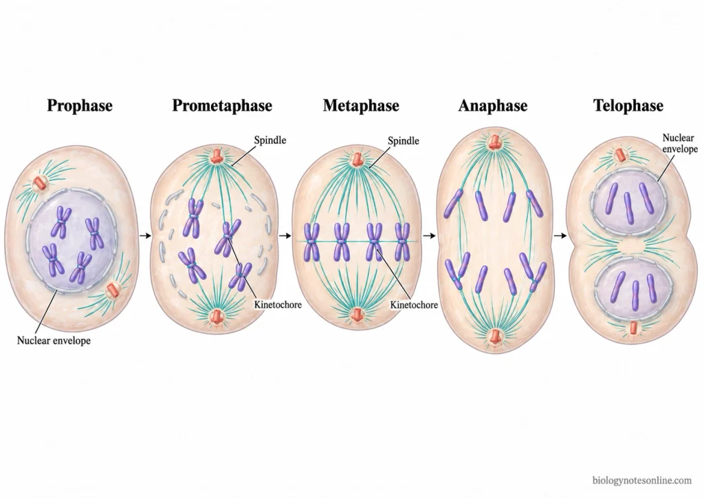
Mitosis is divided into five stages, which are as follows-
- During prophase, the chromatin starts to condense and forms distinct chromosomes. The mitotic spindle also begins to form, and in animal cells the duplicated centrosomes move towards the opposite poles of the cell.
- In prometaphase, the nuclear envelope breaks down in typical animal cells. The spindle microtubules attach to the chromosomes at their kinetochores, which are present in the centromeric region of each sister chromatid. The sister chromatids become attached to spindle microtubules from opposite poles.
- The chromosomes are arranged at the middle of the cell during metaphase, along an imaginary plane called the metaphase plate.
- Anaphase begins with the separation of sister chromatids. They are pulled towards the opposite poles of the cell by the spindle microtubules. After separation, each chromatid is considered an individual chromosome. The mitotic spindle also elongates during this period.
- In telophase, the separated chromosomes reach the opposite poles and start to decondense. A new nuclear envelope is formed around each group of chromosomes, forming two daughter nuclei. The mitotic spindle breaks down.
In some textbooks, prometaphase is included within prophase. In such cases, mitosis is divided into four stages instead of five.
The division of the cytoplasm is referred to as cytokinesis, which usually results in the formation of two separate daughter cells. It generally begins during the later stages of mitosis and may overlap with anaphase and telophase. Mitosis involves nuclear division, while cytokinesis is responsible for the physical separation of the cytoplasm.
Cytokinesis (Cytoplasmic Division)
Cytokinesis is the process of division of the cytoplasm, which usually results in the formation of two separate daughter cells. It takes place during the M phase of the cell cycle. While mitosis involves the division of the nucleus, cytokinesis separates the cytoplasm and other cellular components between the daughter cells.
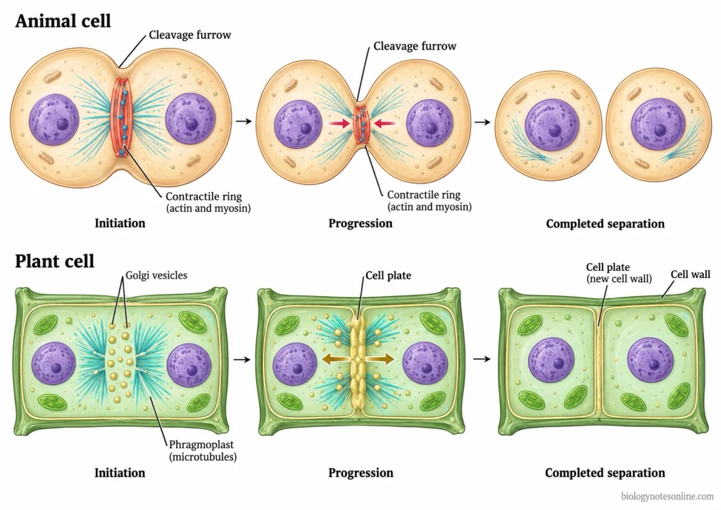
The major events of cytokinesis are as follows-
- Cytokinesis generally begins during the later stages of mitosis. In many cells, it starts during anaphase or telophase, before the nuclear division has been fully completed.
- In animal cells, cytokinesis takes place by the formation of a cleavage furrow. The plasma membrane starts to move inward at the middle region of the dividing cell, forming a small depression on the cell surface.
- A contractile ring made up of actin and myosin filaments is formed beneath the plasma membrane. The contraction of this ring pulls the membrane inward and causes the cleavage furrow to become deeper. The furrow gradually extends around the cell until the cytoplasm is separated into two daughter cells.
- The position of the cleavage furrow is determined by the mitotic spindle. It usually forms at the equatorial region of the cell, between the two separated groups of chromosomes.
- In plant cells, cytokinesis occurs by the formation of a cell plate instead of a cleavage furrow. The rigid cell wall does not allow the cell to divide by the inward contraction of the plasma membrane.
- The phragmoplast is formed between the two daughter nuclei during the later stages of mitosis. It contains microtubules and other cellular components which help in the formation of the cell plate. The vesicles, mainly derived from the Golgi apparatus, carry different materials required for the formation of the new cell wall.
- These vesicles accumulate at the middle of the cell and fuse together to form the cell plate. The cell plate gradually grows towards the existing cell wall by the addition of more vesicles. It finally joins with the parental plasma membrane and cell wall, forming a new partition between the two daughter cells. The membranes of the vesicles contribute to the formation of new plasma membranes on either side of the cell plate.
- Cytokinesis does not always occur after nuclear division. In some cells, mitosis may take place without cytokinesis, resulting in the formation of cells containing two or more nuclei.
C. G0 Phase (Quiescent Phase)
G0 phase (quiescent phase) is a non-dividing state of the cell cycle in which the cell stops its progression towards division. The cells usually enter into G0 from the G1 phase.
The major characteristics of G0 phase are as follows-
- In G0 phase, the cell remains outside the actively progressing cell cycle. It does not continue through the normal G1, S, G2 and M phases.
- The deoxyribonucleic acid (DNA) replication and cell division do not take place during this period. However, the cells remain metabolically active and carry out their normal cellular functions.
- Some cells enter into G0 in absence of sufficient nutrients, growth factors or other external signals required for cell division. High cell density can also cause the cells to leave the active cell cycle.
- The duration of G0 phase is not same in all cells. Some cells remain in this phase for a short period, while others may remain for a prolonged time.
- In many cells, G0 is a temporary state. When suitable growth conditions and signals become available, the cells can leave G0 and re-enter the G1 phase to start the cell cycle again.
- Certain lymphocytes and fibroblasts remain in a quiescent state until they receive suitable signals for cell division. These cells can return to the active cell cycle.
- The liver cells also have the ability to re-enter the cell cycle after liver damage. Under normal conditions, many liver cells remain in G0.
- Some adult stem cells, such as muscle stem cells, remain in a quiescent state for a prolonged period. They can become activated during tissue repair and start dividing again.
- In some cells, the withdrawal from the cell cycle is long-term or permanent. This is commonly found in terminally differentiated cells, which have lost most or all of their ability to divide. The mature neurons and skeletal muscle fibres are examples of cells having very limited proliferative capacity under normal conditions. Unlike temporary quiescence, terminal differentiation is generally associated with permanent withdrawal from the cell cycle.
Duration of the Cell Cycle
Duration of the cell cycle is the time required by a dividing cell to complete one cycle of growth, deoxyribonucleic acid (DNA) replication and cell division. The time taken is not same in all cells. Some cells may complete the cycle within a few minutes, while others take several hours or days.
The duration of the cell cycle varies in different cells and conditions, which are as follows-
- In certain human cells grown in culture, the complete cell cycle takes approximately 24 hours. This duration is commonly used as a textbook example. It is not applicable to all human cells.
- Most of the cell cycle is occupied by interphase. During this period, the cell grows, replicates its DNA and prepares for division. It consists of G1, S and G2 phases. The M phase generally takes a much shorter time compared to interphase.
- Interphase is often described as occupying about 90% of the cell cycle. However, this percentage is not fixed for all cells. In some cultured human cells having a 24-hour cycle, interphase may continue for about 23 hours, while the M phase takes approximately one hour. These are only general examples of cell cycle duration.
- The G1 phase is often the longest individual phase in many somatic cells. Its duration may vary considerably. In some cells, G1 lasts for only a few hours. Other cells may remain in this phase for a much longer period depending on the growth conditions and signals received by the cell.
- The duration of S and G2 phases also varies between cells. But in many mammalian cells, these phases show comparatively less variation than G1.
- Different types of cells do not divide at the same rate. Some cells undergo division regularly, whereas others divide only occasionally. Certain differentiated cells may remain in the non-dividing G0 phase for a prolonged period or permanently, without continuing through the active cell cycle.
- The duration of the cell cycle also differs among organisms and developmental stages. In early embryos, the cells may divide very rapidly. Some early embryonic cells do not have the normal G1 and G2 phases. They undergo repeated S and M phases without normal cell growth.
- The availability of nutrients, growth factors and other suitable conditions also affects the cell cycle duration. In absence of sufficient growth signals, the cells may delay their progression through G1. Some cells can also enter into the G0 phase instead of continuing the cell cycle.
Some examples of cell cycle duration under different conditions are given below-
| Cell type or organism | Approximate duration |
|---|---|
| Rapidly dividing human cells grown in culture | 24 hours |
| Budding yeast under suitable growth conditions | 90 minutes |
| Some rapidly dividing early embryonic cells | 30 minutes or less |
These durations are based on particular cells and growth conditions. The duration may differ in other cells of the same organism.
Cell Cycle Regulation
Cell cycle regulation is the control of cell division by different regulatory proteins and cellular signals. It controls the entry of the cell into division and its movement from one phase to another. In absence of suitable conditions, the cell cycle may be delayed or stopped.
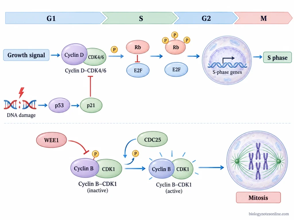
The important mechanisms of cell cycle regulation are as follows-
- Cyclins and CDKs- Cyclins are regulatory proteins whose amount increases and decreases during different phases of the cell cycle. They combine with cyclin-dependent kinases (CDKs) and control their activity. The cyclin-CDK complexes phosphorylate different proteins required for cell cycle progression. In many cells, the amount of CDKs remains comparatively stable. Different cyclin-CDK complexes become active at particular stages of the cycle.
- Growth Signals- The signals received by the cell from its surrounding environment can stimulate or inhibit cell division. During G1 phase, growth factors increase the production of certain cyclins and promote the activity of CDKs. In absence of sufficient nutrients or growth signals, the cell may delay its division or enter into G0 phase.
- G1 to S Phase Transition- The retinoblastoma protein (Rb) binds with E2F transcription factors and prevents the expression of certain genes required for deoxyribonucleic acid (DNA) replication. During G1 phase, Rb is phosphorylated by the cyclin-CDK complexes. The E2F transcription factors are then able to activate these genes. The cell can enter into S phase.
- S Phase Regulation- The S-phase cyclin-CDK complexes activate the machinery required for DNA synthesis. These complexes also help to prevent the same DNA region from undergoing replication more than once in a single cell cycle.
- CDK Inhibitors- Some proteins can bind with the cyclin-CDK complexes and reduce their activity. These are referred to as cyclin-dependent kinase inhibitors (CKIs). The p21 and p27 proteins are examples of these inhibitors. They can prevent the progression of the cell into the next phase when suitable conditions are not present.
- DNA Damage Response- When DNA damage is detected, the cell cycle may be stopped temporarily. DNA repair can take place during this period. The p53 protein is one of the major regulators involved in this process. It increases the production of p21, which inhibits cyclin-CDK activity and delays the progression from G1 to S phase. In case of severe DNA damage, p53 can also promote programmed cell death (apoptosis).
- G2 to M Phase Transition- The entry of cell into mitosis is mainly regulated by the cyclin B-cyclin-dependent kinase 1 (CDK1) complex. The WEE1 kinase adds inhibitory phosphate groups to CDK1 and keeps it in an inactive state. These phosphate groups are removed by CDC25 phosphatase. CDK1 becomes active. When DNA replication is incomplete or DNA damage is present, the checkpoint signals prevent the activation of this complex. The cell does not enter mitosis until the required conditions are met.
- Spindle Checkpoint- During mitosis, the separation of sister chromatids is controlled by the spindle checkpoint. It prevents the beginning of anaphase until the chromosomes are properly attached to the spindle microtubules. After the required attachments are formed, the inhibition of the anaphase-promoting complex/cyclosome (APC/C) is removed. The APC/C promotes the breakdown of securin. This allows separase to cleave the cohesin proteins holding the sister chromatids together.
- Protein Degradation- Certain cyclins and other regulatory proteins are broken down at particular stages of the cell cycle by the ubiquitin-proteasome system. During mitosis, the APC/C promotes the degradation of mitotic cyclins. The activity of CDK1 decreases, allowing the cell to exit mitosis.
Cell Cycle Checkpoints
Cell cycle checkpoints are the control points in the cell cycle where the cell checks whether the required conditions are fulfilled before entering the next phase. These checkpoints detect problems related to cell growth, deoxyribonucleic acid (DNA) replication and chromosome separation. In presence of any such problems, the cell cycle may be temporarily stopped.
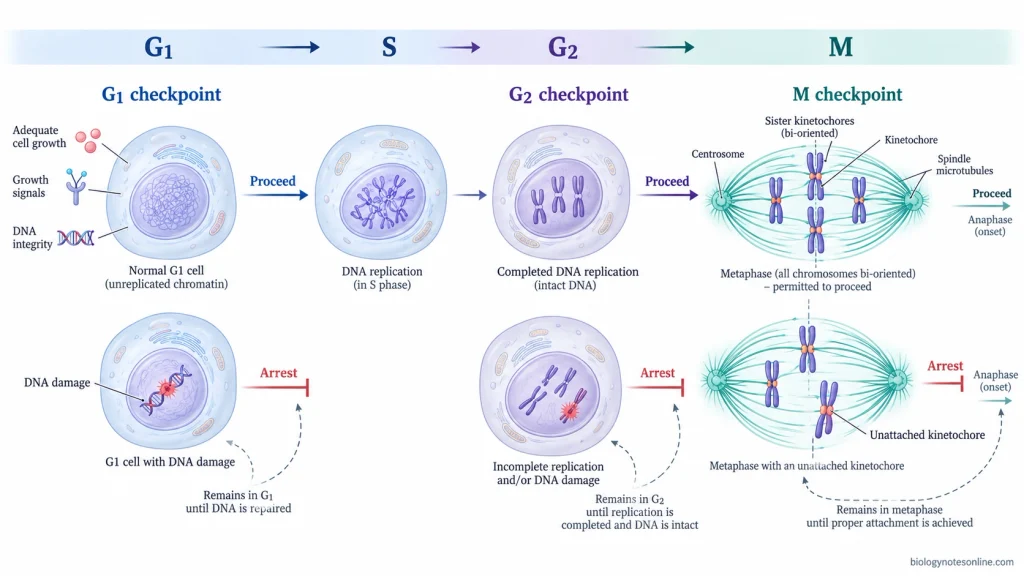
There are three major checkpoints in the cell cycle, which are as follows-
G1 Checkpoint (G1/S Checkpoint)
The G1 checkpoint is a control point present near the end of G1 phase, before the beginning of DNA replication. It controls the progression of cell from G1 to S phase.
- The cell size, availability of nutrients and growth signals are checked at this point. It also detects whether any damage is present in the DNA.
- When the cell has reached a suitable size and sufficient nutrients and growth signals are available, it can proceed into S phase. In this phase, DNA replication takes place.
- In absence of suitable conditions, the cell may remain in G1 without entering S phase. The cell cycle can also be arrested when DNA damage is detected. During this period, the damaged DNA may undergo repair before the cell continues its division.
- Some cells may leave the active cell cycle and enter into the G0 phase instead of proceeding towards DNA replication. They can remain in this non-dividing state until suitable signals become available.
G2 Checkpoint (G2/M Checkpoint)
The G2 checkpoint is present at the end of G2 phase and controls the entry of cell into mitosis. It mainly checks the completion of DNA replication and the presence of any damaged DNA.
- The cell checks whether DNA replication has been completed properly. Incomplete replication can prevent the cell from entering into M phase.
- Any damage present in the replicated DNA is also detected at this checkpoint. When DNA damage or incomplete replication is detected, the progression of the cell cycle is delayed. The cell remains arrested while the required DNA repair or completion of replication takes place.
- After the problems have been resolved, the cell can continue its progression towards mitosis. If important problems remain unresolved, the checkpoint continues to prevent the entry into M phase.
M Checkpoint (Spindle Assembly Checkpoint)
The M checkpoint, also known as the spindle assembly checkpoint, occurs during mitosis before the beginning of anaphase. It controls the separation of sister chromatids by checking their attachment to the mitotic spindle.
- During metaphase, the chromosomes become attached to the spindle microtubules through their kinetochores. The checkpoint detects whether the chromosomes have formed the required attachments with spindle microtubules coming from opposite poles.
- In absence of proper attachment, the cell does not enter into anaphase. Even a single unattached kinetochore can produce a signal that delays the separation of sister chromatids.
- When the required spindle attachments are formed, the checkpoint inhibition is removed and the sister chromatids can separate. They are then moved towards the opposite poles of the cell.
- Incorrect chromosome attachment or failure of the spindle checkpoint may cause unequal distribution of chromosomes between the daughter cells. Some daughter cells may receive extra or missing chromosomes, which is referred to as aneuploidy.
Biological Importance of the Cell Cycle
Cell cycle is the series of events through which a cell grows, replicates its genetic material and undergoes division. It is required for the formation of new cells in both unicellular and multicellular organisms.
The important functions and biological significance of the cell cycle are as follows-
- Growth- The growth of multicellular organisms takes place by an increase in the number of cells. During this process, repeated cell divisions produce new cells which contribute to the growth of different parts of the body.
- Embryonic Development- The development of a multicellular organism from a single fertilized egg involves numerous cycles of cell division. These newly formed cells later contribute to the formation of different tissues and organs.
- Cell Replacement- Many cells of the body are continuously lost during normal cellular activities. These cells are replaced by the formation of new cells. The cells of the intestinal lining and skin are examples where cell replacement takes place regularly.
- Tissue Repair- When a tissue is damaged or injured, new cells are required to replace the lost cells. Some surviving cells start dividing and help in the repair of damaged tissues. The cell cycle is also involved in wound healing. In liver, the cells can re-enter the cell cycle and divide after injury.
- Asexual Reproduction- In unicellular eukaryotic organisms, cell division may produce new individuals. Budding yeast is an example of an organism which reproduces by cell division.
- Genetic Continuity- During the cell cycle, the deoxyribonucleic acid (DNA) is replicated before nuclear division. The replicated chromosomes are separated and distributed between the two daughter nuclei. Each daughter cell normally receives a complete set of genetic information. In mitotic division, the chromosome number remains same as that of the parent cell.
- Cell Growth and Division- The cell increases in size and synthesizes different cellular components before division. These cellular materials are distributed between the daughter cells during cell division.
- Maintenance of Cell Number- The rate of cell division is regulated according to the requirements of different tissues. Some cells undergo division regularly. Other cells may remain outside the active cell cycle for a prolonged period.
- Prevention of Abnormal Cell Division- The cell cycle checkpoints can stop the progression of cells having damaged DNA or incomplete DNA replication. During this period, the damaged DNA may undergo repair. In absence of proper cell cycle regulation, abnormal cell proliferation can take place, which may contribute to the development of cancer.
Cell Cycle at a Glance – Quick Summary Table for Exam
| Features | Key Points |
|---|---|
| Definition | The cell cycle is a series of events in which a cell grows, replicates its DNA and divides into daughter cells. |
| Main Phases | Interphase and M phase (mitotic phase). |
| Interphase | Consists of G1, S and G2 phases. It occupies most of the cell cycle. |
| G1 Phase (First Gap) | Cell growth, synthesis of RNA and proteins, increase in cellular components and preparation for DNA replication. |
| S Phase (Synthesis) | DNA replication takes place. Each chromosome forms two sister chromatids. DNA content doubles, but chromosome number remains unchanged. |
| G2 Phase (Second Gap) | Further cell growth, synthesis of proteins required for mitosis and checking of DNA replication and damage. |
| M Phase (Mitotic Phase) | Includes nuclear division (mitosis) and cytoplasmic division (cytokinesis). |
| Stages of Mitosis | Prophase → Prometaphase → Metaphase → Anaphase → Telophase. |
| Cytokinesis | Division of cytoplasm. Cleavage furrow forms in animal cells, whereas a cell plate forms in plant cells. |
| G0 Phase (Quiescent Phase) | Non-dividing state. Cells remain metabolically active. Some cells can re-enter the cell cycle. |
| Cell Cycle Regulation | Mainly controlled by cyclins, cyclin-dependent kinases (CDKs), CDK inhibitors and other regulatory proteins. |
| G1 Checkpoint | Checks cell size, nutrients, growth signals and DNA damage before S phase. |
| G2 Checkpoint | Checks completion of DNA replication and DNA damage before mitosis. |
| M Checkpoint | Checks chromosome attachment to spindle microtubules before anaphase. |
| Cell Cycle Duration | Approximately 24 hours in certain cultured human cells. Duration varies among cell types and organisms. |
| Longest Phase | Interphase generally occupies the longest period, often represented as about 90% in textbook examples. G1 is often the longest individual phase in many somatic cells. |
| Shortest Phase | M phase generally occupies a much shorter period than interphase. |
| Daughter Cells | Normal mitotic cell division produces two daughter cells with the same chromosome number as the parent cell. |
| Biological Importance | Growth, embryonic development, tissue repair, cell replacement, asexual reproduction and maintenance of genetic continuity. |
References
- Alberts, B., Johnson, A., Lewis, J., Raff, M., Roberts, K., & Walter, P. (2002). Molecular biology of the cell (4th ed.). Garland Science. https://www.ncbi.nlm.nih.gov/books/NBK21054/
- Barr, F. A., & Gruneberg, U. (2007). Cytokinesis: Placing and making the final cut. Cell, 131(5), 847–860. https://doi.org/10.1016/j.cell.2007.11.011
- Bertoli, C., Skotheim, J. M., & de Bruin, R. A. M. (2013). Control of cell cycle transcription during G1 and S phases. Nature Reviews Molecular Cell Biology, 14(8), 518–528. https://doi.org/10.1038/nrm3629
- Clark, M. A., Douglas, M., & Choi, J. (2018). Biology 2e. OpenStax. https://openstax.org/books/biology-2e/pages/1-introduction
- Coller, H. A., Sang, L., & Roberts, J. M. (2006). A new description of cellular quiescence. PLoS Biology, 4(3), Article e83. https://doi.org/10.1371/journal.pbio.0040083
- Cooper, G. M. (2000). The cell: A molecular approach (2nd ed.). Sinauer Associates. https://www.ncbi.nlm.nih.gov/books/NBK9839/
- Glotzer, M. (2005). The molecular requirements for cytokinesis. Science, 307(5716), 1735–1739. https://doi.org/10.1126/science.1096896
- Hartwell, L. H., & Weinert, T. A. (1989). Checkpoints: Controls that ensure the order of cell cycle events. Science, 246(4930), 629–634. https://doi.org/10.1126/science.2683079
- Jürgens, G. (2005). Plant cytokinesis: Fission by fusion. Trends in Cell Biology, 15(5), 277–283. https://doi.org/10.1016/j.tcb.2005.03.005
- Kastan, M. B., & Bartek, J. (2004). Cell-cycle checkpoints and cancer. Nature, 432(7015), 316–323. https://doi.org/10.1038/nature03097
- King, R. W., Deshaies, R. J., Peters, J.-M., & Kirschner, M. W. (1996). How proteolysis drives the cell cycle. Science, 274(5293), 1652–1659. https://doi.org/10.1126/science.274.5293.1652
- Malumbres, M., & Barbacid, M. (2009). Cell cycle, CDKs and cancer: A changing paradigm. Nature Reviews Cancer, 9(3), 153–166. https://doi.org/10.1038/nrc2602
- Morgan, D. O. (1997). Cyclin-dependent kinases: Engines, clocks, and microprocessors. Annual Review of Cell and Developmental Biology, 13, 261–291. https://doi.org/10.1146/annurev.cellbio.13.1.261
- Morgan, D. O. (2007). The cell cycle: Principles of control. New Science Press. https://books.google.com/books?id=_7ygQAOK1DUC
- Musacchio, A. (2015). The molecular biology of spindle assembly checkpoint signaling dynamics. Current Biology, 25(20), R1002–R1018. https://doi.org/10.1016/j.cub.2015.08.051