

Golgi apparatus is a membrane-bound cell organelle present in eukaryotic cells. It forms an important part of the endomembrane system and cellular secretory pathway. Proteins and lipids coming through the endoplasmic reticulum (ER) pathway reach the Golgi, where these undergo further processing and different modifications. The materials are then sorted. They are directed towards their particular cellular locations or for secretion from the cell. Structurally, Golgi apparatus is formed of flattened membrane-bound sacs called cisternae, which are arranged in stacks and have a distinct polarity from the cis to the trans region.
What is the Golgi Apparatus?
Golgi apparatus is a complex membrane-bound cell organelle found in the cytoplasm of eukaryotic cells, which is also known as the Golgi complex or Golgi body. It is made up of flattened stacked pouches known as cisternae. Each cisterna is surrounded by a single membrane.
The Golgi apparatus is found in the middle of the cells’ secretory pathway. As proteins and lipids are transported from the endoplasmic reticulum (ER) into the Golgi, they pass through its different compartments where the materials are processed and modified in various ways. The products are then sorted. Some are packed into transport vesicles and carried to their particular cellular locations, while others are transported towards the cell surface for secretion.
Thus, its major role is in processing, sorting and distribution of proteins and lipids received through the ER pathway. The different membranous compartments of the Golgi allow these cellular products to undergo their required modifications before reaching the final destination.
General Characteristics of Golgi Apparatus
- Golgi apparatus is a membrane-bound cell organelle of eukaryotic cells. It is absent in prokaryotic cells.
- It forms a part of the endomembrane system and is closely associated with the secretory pathway of cell.
- The organelle is formed of a number of flattened membrane sacs called cisternae. These cisternae remain arranged one above another forming stacks and each cisterna is bounded by a single membrane.
- A Golgi stack shows definite polarity. It has a cis face towards the receiving region and a trans face towards the exit region, with medial cisternae occurring between them.
- The different Golgi cisternae are not exactly similar in their composition. They contain different resident enzymes, which carry out processing of proteins and lipids as these materials pass through Golgi.
- Proteins and lipids coming from the endoplasmic reticulum (ER) are transported towards the cis region. Many of these undergo modification inside the Golgi before being sent further.
- Glycosylation and modification of already formed carbohydrate chains are among its important activities. Some proteins are also sulfated and proteolytically processed in Golgi compartments.
- At the trans side is present a membranous region called trans-Golgi network (TGN). This is an important sorting region from where cellular products are directed to their different destinations.
- Golgi apparatus is associated with many small transport vesicles and membrane carriers. These carry materials between Golgi and other cellular compartments.
- The Golgi is not a fixed or completely static structure. Its membranes continuously receive and lose materials during intracellular transport.
- Its arrangement varies in different types of cells. In many mammalian cells the Golgi stacks are interconnected forming a Golgi ribbon, whereas plant cells generally contain many separate Golgi stacks distributed in the cytoplasm.
- Golgi cisternae do not have ribosomes attached on their membrane. The organelle also has no genetic material of its own.
Discovery and History of Golgi Apparatus
- Golgi apparatus was first discovered by the Italian scientist Camillo Golgi in 1898, while studying nerve cells of the cerebellum of an owl.
- Golgi observed a darkly stained network present inside the nerve cells. For its observation he used the chromo-silver impregnation method, related to his famous “black reaction” (reazione nera) used for nervous tissues.
- He initially named this newly observed structure “apparato reticolare interno”, meaning internal reticular apparatus.
- After its discovery, similar structures were reported in many other types of eukaryotic cells. But its actual existence remained doubtful for several years.
- During the early decades of the 20th century, many scientists considered the Golgi apparatus as an artifact produced during fixation or metallic staining. Thus, whether it was a true cellular structure or simply a staining product remained under controversy for a long period.
- The development of electron microscopy finally provided much clearer evidence for its existence. In 1954, Albert J. Dalton and Marie D. Felix produced important high-resolution electron microscopic observations showing the characteristic membranous structure of the Golgi apparatus.
- With electron microscopic studies during the 1950s, the Golgi apparatus was accepted as a true cell organelle. Its flattened cisternae and associated vesicles could now be identified more clearly.
- Further studies during the following decades showed that Golgi is associated with the processing and movement of secretory products. Studies of intracellular secretion and membrane traffic later established its major position in the secretory pathway of eukaryotic cells.
- The organelle is now named the Golgi apparatus or Golgi complex in honour of Camillo Golgi, after the scientist who first reported it.
Location and Occurrence of Golgi Apparatus
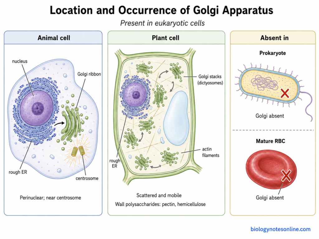
- Golgi apparatus is found in the cytoplasm of eukaryotic cells. Its position and arrangement, however, are not the same in all organisms and different cell types.
- In many mammalian cells, the Golgi is present close to the nucleus. It generally occupies a perinuclear region and is often concentrated around the centrosome.
- The Golgi is closely connected with the endoplasmic reticulum (ER) through the secretory pathway. Materials leaving the ER are transported towards the cis region of Golgi for further processing.
- Its intracellular position can also change according to the type and condition of the cell. In polarized epithelial cells, for example, the Golgi may be placed between the nucleus and the apical region of the cell.
- Golgi organization shows considerable differences among eukaryotes. Some cells contain separate stacks, whereas in many vertebrate cells these stacks are connected together into a larger ribbon-like structure.
- A classical Golgi apparatus is absent from prokaryotic cells. Prokaryotes do not possess the usual membrane-bound endomembrane organelles found in eukaryotic cells.
- It is also not present in every mature eukaryotic cell. Mature mammalian erythrocytes (RBCs) lose the Golgi apparatus along with nucleus, ER, mitochondria and other organelles during their development.
Golgi in Animal Cells
- In many animal cells, especially mammalian cells, several Golgi stacks are interconnected with one another forming a continuous Golgi ribbon. It is generally present in the region around the nucleus.
- The mammalian Golgi ribbon is commonly situated close to the centrosome, which is the major microtubule-organizing region of the cell. Microtubules also help in maintaining this characteristic position.
- Its arrangement is not fixed in all animal cells. The Golgi can change its position and organization during cell division, cell migration and other cellular conditions.
- Golgi apparatus is particularly prominent in cells actively involved in secretion. Goblet cells, pancreatic secretory cells and several other glandular cells have a highly developed Golgi associated with their large secretory activity.
Golgi in Plant Cells
- Plant cells generally contain many separate Golgi stacks distributed throughout the cytoplasm rather than one compact perinuclear Golgi ribbon.
- These individual Golgi stacks are also commonly called dictyosomes.
- Plant Golgi stacks are highly dynamic. They move through the cytoplasm, and their movement is mainly associated with the actin cytoskeleton and myosin motors.
- The stacks are often seen closely associated with the ER network during their movement. They are therefore sometimes described as mobile secretory units of the plant cell.
- Plant Golgi performs the normal processing, sorting and secretion of proteins and lipids. It has another major function also.
- Many non-cellulosic polysaccharides of the plant cell wall, particularly pectins and hemicelluloses, are synthesized in the Golgi. These products are then carried towards the cell surface for secretion and wall formation.
Golgi Apparatus in Plant and Animal Cells
| Feature | Animal Cells | Plant Cells |
|---|---|---|
| General Organization | In many mammalian cells, Golgi stacks are concentrated near the nucleus and commonly interconnected to form a Golgi ribbon. | Usually many separate Golgi stacks are distributed throughout the cytoplasm. |
| Common Term | Commonly called Golgi apparatus or Golgi complex. | Individual Golgi stacks are also commonly referred to as dictyosomes. |
| Basic Structure | Formed of polarized cis, medial and trans cisternae. | The same basic polarized cisternal organization is present. |
| Protein Processing | Proteins coming through the ER pathway undergo modification and processing in the Golgi. | Protein processing and modification also take place in the same basic manner. |
| Lipid Processing | Modification and synthesis of different lipids takes place. | Lipid processing is also present. |
| Cargo Sorting | Proteins and lipids are sorted for transport towards plasma membrane, endosomes, lysosomal pathway and secretion. | Cargo is also sorted towards plasma membrane, vacuolar compartments and other cellular destinations. |
| Secretion | Constitutive and, in specialized cells, regulated secretion are present. | Golgi-mediated secretion is present and has a major role in delivery of proteins and cell wall materials. |
| Cell-wall Polysaccharides | Not applicable because animal cells do not have a cell wall. | A major additional function. Pectins and hemicelluloses are mainly synthesized in Golgi and transported towards the cell surface. |
| Golgi Movement | The perinuclear Golgi ribbon of many mammalian cells is relatively concentrated, although its organization can change with cellular condition. | Individual Golgi stacks are highly dynamic and commonly move through the cytoplasm. |
| TGN Organization | The trans-Golgi network (TGN) remains closely associated with the trans side of Golgi and acts in cargo sorting. | Both Golgi-associated TGN (GA-TGN) and detached or Golgi-independent/free TGN states may occur. |
Structure of the Golgi Apparatus
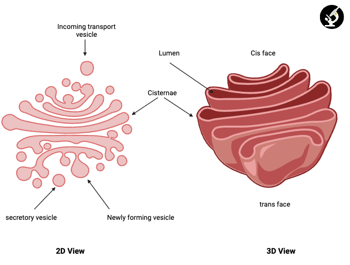
- Golgi apparatus is made up of a group of flattened membrane-bound sacs known as cisternae. These cisternae are arranged one above another to form a stack.
- Each cisterna is surrounded by a single membrane. An enclosed space or lumen is present inside it.
- The membrane of Golgi cisternae is smooth. Ribosomes are not attached on its outer surface.
- The cisternae are generally flattened in the middle and may become wider towards their margins. Small tubules and vesicles are also commonly present around these margins.
- A Golgi stack has two different sides, the cis face and trans face. The medial cisternae are present between these two regions.
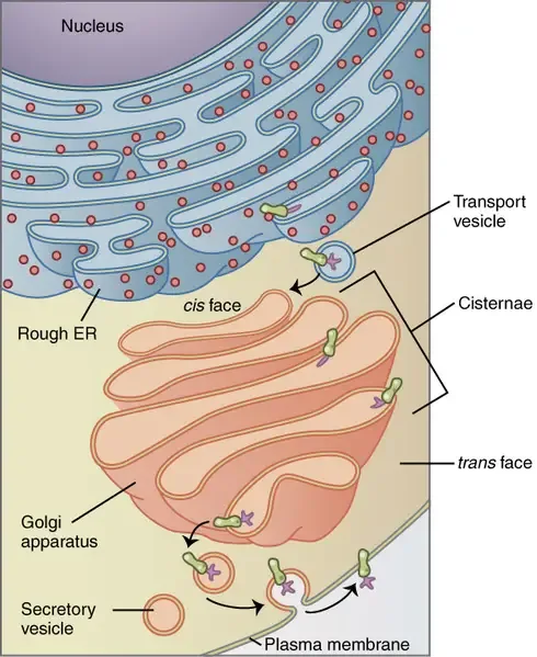
- The cis face is the receiving side of the Golgi apparatus. It is usually located towards the endoplasmic reticulum (ER) and receives transport materials coming from it.
- The middle portion of Golgi consists of medial cisternae. Different enzymes are present in these cisternae which take part in modification of proteins and lipids.
- The trans face is present on the opposite side of cis face. Materials which have passed through the Golgi reach this side before being transported further.
- At the trans side, a network of membrane tubules and vesicles is formed. This region is called the trans-Golgi network (TGN).
- The cis, medial and trans cisternae are not exactly similar. Different enzymes and membrane proteins are found in these regions.
- Many small transport vesicles are associated with the Golgi apparatus. Some vesicles bring materials towards the Golgi, while other vesicles carry materials away from it.
- The number of cisternae in a Golgi stack is variable. It differs in different organisms and also between different cell types.
- In many mammalian cells, separate Golgi stacks are connected with each other and form a larger Golgi ribbon. In plant cells, many individual Golgi stacks are usually scattered throughout the cytoplasm.
- Openings or fenestrae can also be present in some Golgi cisternae, especially towards their peripheral regions.
Golgi Apparatus Diagram
How Does the Golgi Apparatus Work?
The working of Golgi apparatus involves transport of proteins and lipids from the endoplasmic reticulum (ER), their movement through different Golgi compartments and finally sorting from the trans side. The process is not one-way. Materials also move backward for recycling of Golgi and ER components.
1. Transport from the ER to the Golgi
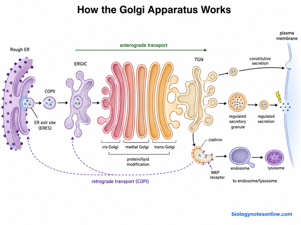
- Proteins entering the secretory pathway are first synthesized through the rough endoplasmic reticulum (RER) pathway. They enter the ER during their synthesis, where folding and some early modifications take place.
- Properly folded proteins that are ready for transport collect at specialized regions of ER called ER exit sites (ERES).
- In this step, COPII coat proteins take part in selection and export of many ER cargo molecules. COPII-associated transport carriers are then formed from the ER membrane.
- After leaving the ER, the coat is removed. In mammalian cells, much of this cargo reaches a tubulovesicular compartment present between ER and Golgi, called the ER-Golgi intermediate compartment (ERGIC).
- The transport can be written in a simple form as follows-
Rough ER → ER exit sites → COPII-associated transport → ERGIC → cis-Golgi
- The ER and Golgi apparatus are not directly continuous membranes. Materials are moved between these compartments by transport carriers and intermediate membrane compartments.
- Not all proteins leaving the ER should remain in the forward pathway. Some resident ER proteins escape during transport, these are recognized and returned back towards the ER.
- COPI-associated retrograde transport has an important role in this retrieval. It also helps to recover different proteins and transport machinery from early Golgi and pre-Golgi compartments.
2. Movement of Cargo Through the Golgi
- After entering the cis side, cargo moves in the general cis to trans direction. It passes through cis, medial and later Golgi compartments.
- The different Golgi compartments contain different processing enzymes. During this movement proteins and lipids undergo modification in a stepwise manner.
- Golgi cisternae are not simply considered as fixed sacs through which every cargo is moved forward by separate vesicles. A major model for this movement is the cisternal maturation model.
- In this model, a newly formed cis cisterna gradually matures. It changes into a medial and then later Golgi cisterna, carrying much of its cargo along during the process.
- But the Golgi enzymes belonging to an earlier compartment need to remain in their proper region. These resident proteins are moved backward from later cisternae to earlier ones.
- COPI-associated carriers have an important role in this backward recycling of Golgi resident proteins and other machinery.
- Cisternal maturation is an important mechanism of intra-Golgi transport, although transport tubules, vesicles and other carrier mechanisms can also participate. Golgi transport is therefore not represented by only one type of carrier movement.
Anterograde Transport
- Anterograde transport refers to movement in the forward direction of the secretory pathway.
- The general direction is from ER → Golgi → trans-Golgi network → later cellular destinations.
- Within Golgi, cargo progresses towards later cisternae and the trans region. This forward movement can occur with maturation of the cisternae as well as other membrane transport mechanisms.
Retrograde Transport
- Retrograde transport is movement in the reverse direction, towards an earlier compartment.
- It takes place from later Golgi regions towards earlier Golgi cisternae and also from Golgi or pre-Golgi compartments back towards the ER.
- This transport recovers resident enzymes, escaped ER proteins, receptors and different transport machinery. They can then be reused.
- COPI is strongly associated with this type of transport in the early secretory pathway.
3. Sorting at the Trans-Golgi Network
- After passing through the Golgi stack, processed cargo reaches the trans-Golgi network (TGN). This is the major sorting region towards the exit side of Golgi.
- Different proteins are not sent through one common route. Cargo signals, sorting receptors and cytosolic adaptor proteins help in selection of materials for different transport carriers.
- Some materials are continuously transported from the TGN towards the cell surface. This is known as constitutive secretion and does not require a special external stimulus for every release.
- In certain specialized secretory cells, proteins are packed into regulated secretory granules. These materials remain stored and are released after receiving an appropriate cellular signal.
- Membrane proteins and lipids are also transported towards the plasma membrane. Fusion of these carriers with plasma membrane adds their membrane components to the cell surface.
- Another group of cargo is directed towards endosomes. From endosomal compartments, some materials can continue further into the lysosomal pathway.
- Many soluble lysosomal enzymes receive a mannose-6-phosphate (M6P) sorting tag. M6P receptors recognize these enzymes at the TGN and help in their transport towards endosomes.
- Clathrin is involved in formation of some TGN-derived carriers, particularly those associated with transport towards the endosomal-lysosomal system. Adaptors such as GGA proteins and AP-1 connect selected cargo or cargo receptors with the coat machinery.
- All carriers leaving the Golgi are not clathrin-coated. Different secretory vesicles, tubules and other membrane carriers are formed depending on the cargo and where it has to be transported.
Functions of the Golgi Apparatus
- Protein Modification– Golgi apparatus modifies and processes proteins coming from the endoplasmic reticulum (ER). Different Golgi compartments contain different enzymes for these reactions.
- N-linked Glycosylation– The N-linked oligosaccharides already added to proteins in the ER are further processed in Golgi. Some sugar residues are removed, while other sugars are added.
- O-linked Glycosylation– It also carries out O-linked glycosylation of many proteins. Here, sugar residues are mainly added to serine or threonine residues.
- Sulfation– Golgi takes part in sulfation of different cellular molecules. Sulfate groups can be added to carbohydrates and some proteins during their processing.
- Protein Processing– Some inactive protein precursors are processed in the late Golgi, trans-Golgi network (TGN) or associated secretory compartments. This processing can produce their active forms.
- Lipid Processing– The Golgi is involved in modification and formation of different lipids. Glycolipids and sphingomyelin are formed from lipid precursors coming through the ER pathway.
- Cargo Sorting– Processed proteins and lipids are sorted mainly at the trans-Golgi network (TGN). They are directed towards plasma membrane, endosomes, lysosomal pathway or different secretory carriers.
- Secretion– Golgi apparatus has an important role in secretion of cellular products. Some materials are released continuously by constitutive secretion, while others are stored in secretory granules and released after a proper signal.
- Membrane Supply– Proteins and lipids transported from Golgi become part of the plasma membrane after fusion of transport carriers with cell surface. In this way new membrane components are also supplied.
- Lysosomal Targeting– Many lysosomal enzymes in animal cells receive a mannose-6-phosphate (M6P) marker during Golgi processing. These enzymes are then recognized by M6P receptors and directed towards the endosomal-lysosomal pathway.
- Protein Retrieval– Golgi also takes part in return transport of different cellular proteins. Escaped ER proteins and some resident transport components can be carried back towards earlier Golgi regions or the ER.
- Wall Polysaccharides– In plant cells, Golgi apparatus synthesizes many non-cellulosic cell wall polysaccharides. Pectins and hemicelluloses are major examples and are transported towards the cell surface after their formation.
- Cellulose Formation– Cellulose is not synthesized inside the Golgi. It is mainly formed by cellulose synthase complexes located in the plasma membrane.
- Cell Plate Formation– During plant cell division, Golgi-derived and TGN-associated vesicles transport membrane and cell wall materials towards the developing cell plate. These vesicles take part in formation of the new partition between the daughter cells.
What Happens When Golgi Function Is Disrupted?
- Protein Processing– Normal processing of proteins becomes affected when Golgi function is disturbed. Proteins coming from the endoplasmic reticulum (ER) may not receive their proper modifications before being transported further.
- Glycosylation Defects– Many glycoproteins and glycolipids are modified inside Golgi. Defects in Golgi enzymes, transporters or organization can therefore produce abnormal carbohydrate chains, and severe inherited defects are found in different congenital disorders of glycosylation (CDGs).
- Cargo Sorting– Golgi is a major sorting region of the secretory pathway. When this sorting is disturbed, proteins and lipids may reach an incorrect cellular location or their normal transport becomes reduced.
- Secretion– Release of secretory proteins can become abnormal. This is particularly important in cells which produce and secrete large quantities of hormones, enzymes, mucus or other secretory materials.
- Lysosomal Targeting– Lysosomal enzymes require proper Golgi processing and sorting. Failure in the mannose-6-phosphate (M6P) targeting system can prevent these enzymes from reaching lysosomes, and some enzymes are instead secreted outside the cell. I-cell disease is a classical example of this type of defect.
- Membrane Transport– Movement of membrane proteins and lipids towards plasma membrane, endosomes and other cellular compartments can be disturbed. The normal balance of membrane traffic is then affected.
- Golgi Stress– When the processing capacity of Golgi becomes insufficient compared with the cellular demand, a Golgi stress response can be activated. Different stress pathways increase Golgi-related functions, while prolonged or severe stress can also become associated with cell damage and apoptotic signaling.
- Golgi Fragmentation– Abnormal disruption can cause the organized Golgi ribbon or stacks to become dispersed into smaller fragments. Golgi fragmentation also occurs normally during some cellular processes such as mitosis, but persistent fragmentation is found in several pathological conditions.
- Cell Polarity– Golgi-dependent membrane trafficking helps to maintain cell polarity by delivering proteins and lipids to particular regions of the cell. Disturbance of this system can therefore interfere with polarized transport and cell organization.
- Cellular Disease– Defects involving Golgi structure, glycosylation or membrane trafficking are associated with different human diseases. These include some congenital glycosylation disorders, neurodegenerative diseases and other inherited trafficking disorders. Golgi abnormalities are also reported in several cancers and infectious conditions, although the Golgi change is not always the primary cause of disease.
Golgi Apparatus vs Endoplasmic Reticulum
| Feature | Golgi Apparatus | Endoplasmic Reticulum (ER) |
|---|---|---|
| Basic Structure | Made up of flattened membrane-bound sacs called cisternae, arranged in stacks. | Formed of an interconnected network of membrane tubules and flattened sacs called cisternae. |
| Membrane | It is a single membrane-bound organelle. | It is also bounded by a single membrane. |
| Occurrence | Present in eukaryotic cells. | Present in eukaryotic cells. |
| Location | In many animal cells it is found near the nucleus and ER. Plant cells generally contain many dispersed Golgi stacks. | Spread through the cytoplasm and is continuous with the outer membrane of the nuclear envelope. |
| Organization | Has distinct cis, medial and trans regions. | Mainly divided into rough ER (RER) and smooth ER (SER). |
| Ribosomes | Ribosomes are not attached on the Golgi membrane. | Ribosomes are present on the cytosolic surface of rough ER, while smooth ER lacks ribosomes. |
| Protein Role | Proteins received through the ER pathway are further modified, processed and sorted here. | Rough ER is involved in synthesis, folding and early modification of many secretory and membrane proteins. |
| Lipid Role | Takes part in modification and synthesis of several lipids and glycolipids. | Smooth ER is a major site for synthesis of many cellular lipids. |
| Glycosylation | Further processing of N-linked oligosaccharides and much O-linked glycosylation take place here. | Initial N-linked glycosylation of many proteins begins in the ER. |
| Cargo Sorting | Major sorting takes place mainly at the trans-Golgi network (TGN). | ER mainly selects proteins for export and sends them towards the Golgi through ER exit sites. |
| Transport | Materials move through Golgi and are finally transported towards plasma membrane, endosomes, lysosomal/vacuolar pathway or secretion. | ER-derived cargo is exported mainly by COPII-associated transport towards the Golgi pathway. |
| Secretory Pathway | It is the major processing and sorting region of the secretory pathway. | It forms the early entry region of the secretory pathway. |
| Calcium Storage | Not a major intracellular calcium-storage compartment. | Smooth ER is an important site for intracellular Ca²⁺ storage and release. |
| Detoxification | It has no major general role in cellular detoxification. | Smooth ER takes part in detoxification of drugs and other compounds, especially in liver cells. |
| Plant Cell Role | In plant cells, it also synthesizes many pectins and hemicelluloses for the cell wall. | ER supplies proteins and lipids and is involved in formation and maintenance of the cellular endomembrane system. |
Cis Face vs Trans Face of Golgi Apparatus
| Feature | Cis Face | Trans Face |
|---|---|---|
| Position | Present on the receiving side of the Golgi apparatus. | Present on the opposite or exit side of the Golgi stack. |
| Other Name | Also called the forming face or entry face. | Also called the maturing face or exit face. |
| Relation with ER | Usually located towards the endoplasmic reticulum (ER) and receives ER-derived cargo. | Located away from the ER side and towards later secretory compartments. |
| Cargo Movement | Proteins and lipids entering Golgi first reach this region. | Processed materials reach this region before their final sorting and transport. |
| Golgi Region | Associated with the cis-Golgi network (CGN) and early Golgi cisternae. | Associated with late Golgi cisternae and the trans-Golgi network (TGN). |
| Main Role | Receives newly arrived cargo and begins Golgi processing. | Takes part mainly in final processing, sorting and distribution of cargo. |
| Protein Modification | Early Golgi modifications take place here, including initial processing of some carbohydrate chains. | Later modifications are completed in trans cisternae and TGN-associated regions. |
| Sorting | Some sorting and retrieval of proteins towards earlier compartments can occur. | Major sorting of processed cargo towards different cellular destinations takes place here. |
| Transport Direction | Cargo generally enters from the ER side and progresses towards medial cisternae. | Cargo leaves towards the plasma membrane, secretory vesicles, endosomes or lysosomal/vacuolar pathways. |
| Shape | Often described as the more convex side of a Golgi stack. | Commonly forms the more concave side of the stack. |
| Associated Vesicles | Transport carriers arriving from ER/ERGIC and retrograde carriers are commonly associated around this region. | Different secretory and sorting carriers are formed from the trans region and TGN. |
| Enzymes | Contains enzymes characteristic of the early Golgi compartments. | Contains a different group of enzymes associated with later Golgi processing. |
References
- Alberts, B., Johnson, A., Lewis, J., Raff, M., Roberts, K., & Walter, P. (2002). Molecular biology of the cell (4th ed.). Garland Science. https://www.ncbi.nlm.nih.gov/books/NBK21054/
- Appenzeller-Herzog, C., & Hauri, H.-P. (2006). The ER-Golgi intermediate compartment (ERGIC): In search of its identity and function. Journal of Cell Science, 119(11), 2173–2183. https://doi.org/10.1242/jcs.03019
- Bentivoglio, M. (1998). 1898: The Golgi apparatus emerges from nerve cells. Trends in Neurosciences, 21(5), 195–200. https://doi.org/10.1016/S0166-2236(98)01229-6
- Cooper, G. M. (2000). The cell: A molecular approach (2nd ed.). Sinauer Associates. https://www.ncbi.nlm.nih.gov/books/NBK9839/
- Coutinho, M. F., Prata, M. J., & Alves, S. (2012). Mannose-6-phosphate pathway: A review on its role in lysosomal function and dysfunction. Molecular Genetics and Metabolism, 105(4), 542–550. https://doi.org/10.1016/j.ymgme.2011.12.012
- De Matteis, M. A., & Luini, A. (2008). Exiting the Golgi complex. Nature Reviews Molecular Cell Biology, 9(4), 273–284. https://doi.org/10.1038/nrm2378
- Downes, K. W., & Zanetti, G. (2025). Mechanisms of COPII coat assembly and cargo recognition in the secretory pathway. Nature Reviews Molecular Cell Biology, 26(12), 910–925. https://doi.org/10.1038/s41580-025-00839-y
- Dröscher, A. (1998). The history of the Golgi apparatus in neurones from its discovery in 1898 to electron microscopy. Brain Research Bulletin, 47(3), 199–203. https://doi.org/10.1016/S0361-9230(98)00080-X
- Dröscher, A. (2011). Cellular dimensions and cell dynamics, or the difficulty over capturing time and space in the era of electron microscopy. Studies in History and Philosophy of Biological and Biomedical Sciences, 42(4), 395–402. https://doi.org/10.1016/j.shpsc.2011.07.005
- Francisco, R., Brasil, S., Poejo, J., Jaeken, J., Pascoal, C., Videira, P. A., & dos Reis Ferreira, V. (2023). Congenital disorders of glycosylation (CDG): State of the art in 2022. Orphanet Journal of Rare Diseases, 18, 329. https://doi.org/10.1186/s13023-023-02879-z
- Guo, Y., Sirkis, D. W., & Schekman, R. (2014). Protein sorting at the trans-Golgi network. Annual Review of Cell and Developmental Biology, 30, 169–206. https://doi.org/10.1146/annurev-cellbio-100913-013012
- Huang, S., & Wang, Y. (2017). Golgi structure formation, function, and post-translational modifications in mammalian cells. F1000Research, 6, 2050. https://doi.org/10.12688/f1000research.11900.1
- Kang, B.-H., Anderson, C. T., Arimura, S.-I., Bayer, E. M., Bezanilla, M., Botella, M. A., Brandizzi, F., Burch-Smith, T. M., Chapman, K. D., Dünser, K., Gu, Y., Jaillais, Y., Kirchhoff, H., Otegui, M. S., Rosado, A., Tang, Y., Kleine-Vehn, J., Wang, P., & Zolman, B. K. (2022). A glossary of plant cell structures: Current insights and future questions. The Plant Cell, 34(1), 10–52.
- https://doi.org/10.1093/plcell/koab247
- Linders, P. T. A., Peters, E., ter Beest, M., Lefeber, D. J., & van den Bogaart, G. (2020). Sugary logistics gone wrong: Membrane trafficking and congenital disorders of glycosylation. International Journal of Molecular Sciences, 21(13), 4654. https://doi.org/10.3390/ijms21134654
- Moras, M., Lefevre, S. D., & Ostuni, M. A. (2017). From erythroblasts to mature red blood cells: Organelle clearance in mammals. Frontiers in Physiology, 8, 1076. https://doi.org/10.3389/fphys.2017.01076
- Nakano, A. (2022). The Golgi apparatus and its next-door neighbors. Frontiers in Cell and Developmental Biology, 10, 884360. https://doi.org/10.3389/fcell.2022.884360
- Neumann, U., Brandizzi, F., & Hawes, C. (2003). Protein transport in plant cells: In and out of the Golgi. Annals of Botany, 92(2), 167–180. https://doi.org/10.1093/aob/mcg134
- Qian, Y., van Meel, E., Flanagan-Steet, H., Yox, A., Steet, R., & Kornfeld, S. (2015). Analysis of mucolipidosis II/III GNPTAB missense mutations identifies domains of UDP-GlcNAc enzyme GlcNAc-1-phosphotransferase involved in catalytic function and lysosomal enzyme recognition. Journal of Biological Chemistry, 290(5), 3045–3056. https://doi.org/10.1074/jbc.M114.612507
- Ravichandran, Y., Goud, B., & Manneville, J.-B. (2020). The Golgi apparatus and cell polarity: Roles of the cytoskeleton, the Golgi matrix, and Golgi membranes. Current Opinion in Cell Biology, 62, 104–113. https://doi.org/10.1016/j.ceb.2019.10.003
- Reily, C., Stewart, T. J., Renfrow, M. B., & Novak, J. (2019). Glycosylation in health and disease. Nature Reviews Nephrology, 15(6), 346–366. https://doi.org/10.1038/s41581-019-0129-4
- Sasaki, K., & Yoshida, H. (2019). Golgi stress response and organelle zones. FEBS Letters, 593(17), 2330–2340. https://doi.org/10.1002/1873-3468.13554
- Sinclair, R., Ruiz Rosquete, M., & Drakakaki, G. (2018). Post-Golgi trafficking and transport of cell wall components. Frontiers in Plant Science, 9, 1784. https://doi.org/10.3389/fpls.2018.01784
- Strzyz, P. (2019). Sorting it out at the Golgi. Nature Reviews Molecular Cell Biology, 20, 2–3. https://doi.org/10.1038/s41580-018-0087-x
- Watson, E. T., Wegeng, W. R., Aravani, S., Ernst, A. M., & von Blume, J. (2025). Mechanistic insights into cargo sorting and export from the Golgi apparatus. Nature Reviews Molecular Cell Biology, 26(12), 940–956. https://doi.org/10.1038/s41580-025-00907-3
- Wei, J.-H., & Seemann, J. (2010). Unraveling the Golgi ribbon. Traffic, 11(11), 1391–1400. https://doi.org/10.1111/j.1600-0854.2010.01114.x
- Zobaroğlu-Özer, P., & Bora-Akoğlu, G. (2024). Split but merge: Golgi fragmentation in physiological and pathological conditions. Molecular Biology Reports, 51(1), 214. https://doi.org/10.1007/s11033-023-09153-2
- Morré, D. J., & Mollenhauer, H. H. (Eds.). (2009). The Golgi apparatus: The first 100 years. Springer. https://doi.org/10.1007/978-0-387-74347-9
- Mironov, A. A., & Pavelka, M. (Eds.). (2008). The Golgi apparatus: State of the art 110 years after Camillo Golgi’s discovery. Springer. https://doi.org/10.1007/978-3-211-76310-0