

What Are Secretory Vesicles?
Secretory vesicles are small membrane-bound intracellular carriers that transport substances which are to be delivered to the cell surface or released outside the cell. They are a part of the secretory pathway of eukaryotic cells. The vesicular membrane surrounds the transported materials and separates them from the cytoplasm.
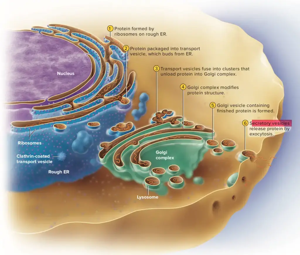
The secretory pathway generally begins in the endoplasmic reticulum (ER), from where newly formed proteins are transported to the Golgi apparatus. After processing and sorting, many secretory proteins and membrane components leave the trans-Golgi network (TGN) in membrane-bound carriers. Their major post-Golgi destination is the plasma membrane.
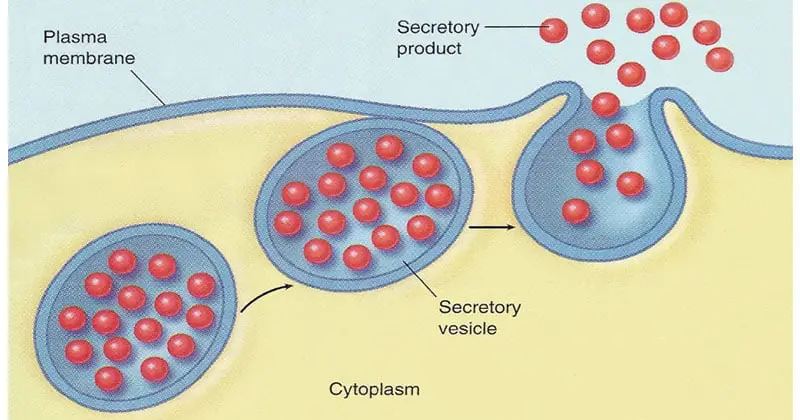
Secretory vesicles reaching the plasma membrane can fuse with it and discharge their soluble contents outside the cell. This process is called exocytosis. At the same time, membrane proteins and lipids present in the vesicle membrane can become a part of the plasma membrane.
In specialized secretory cells, some secretory products are not released immediately. They are concentrated and stored inside vesicles until a particular signal is received. Such storage is common in endocrine, neuroendocrine and exocrine cells. Hormones and digestive enzymes are some examples of substances stored in these vesicles before their release.
Thus, the term secretory vesicle can be used broadly for vesicular carriers involved in secretion. However, all of these vesicles do not necessarily behave in the same manner. Some post-Golgi carriers continuously move towards the cell surface, whereas specialized vesicles of the regulated pathway can remain stored in the cytoplasm before exocytosis.
Terminology Note
Secretory vesicle ≠ always exactly the same thing as secretory granule.
The term secretory vesicle is a broader term and can refer to membrane-bound carriers taking secretory materials towards the plasma membrane. Secretory granule is especially used for the specialized storage organelles of the regulated secretory pathway.
These granules contain concentrated secretory products and commonly appear electron-dense under electron microscopy. For this reason, they are also frequently called dense-core vesicles or dense-core granules. They remain stored in the cell and release their contents after receiving an appropriate stimulus.
Where Are Secretory Vesicles Found?
Secretory vesicles are found in the cytoplasm of eukaryotic cells. They are more common in cells involved in active secretion. In specialized secretory cells, large number of these vesicles may be present.
Newly formed secretory vesicles are initially present around the trans-Golgi network (TGN). From here, they are transported through the cytoplasm towards the cell cortex and plasma membrane. Some vesicles remain stored within the cytoplasm.
In endocrine cells, secretory vesicles are present for storage of hormones. Pancreatic β-cells contain insulin-containing secretory granules in the cytoplasm. Some of these granules are found close to the plasma membrane from where insulin is released.
Pancreatic acinar cells contain large number of zymogen granules. They are mainly present towards the apical region of the cell. Digestive enzymes are stored inside these granules and released from the apical plasma membrane.
In neurons, dense-core secretory vesicles are formed in the cell body and transported to different regions of the neuron. They contain mainly neuropeptides and other secretory substances. Neuroendocrine cells also possess large number of dense-core vesicles for storage and release of hormones or neuropeptides.
Different immune cells also contain specialized secretory granules. Cytotoxic T cells and natural killer (NK) cells contain lytic granules, while mast cells and neutrophils also possess different storage granules. These are released when the cells are activated.
Thus, the distribution of secretory vesicles depends on the type and secretory activity of cell. Cells specialized for secretion generally contain more vesicles than cells with less secretory activity.
Characteristics of Secretory Vesicles
- Membrane bound– Secretory vesicles are small intracellular structures enclosed by a membrane. The membrane is composed of lipid bilayer.
- Occurrence– They are found in eukaryotic cells, particularly well developed in cells which actively release proteins, hormones or enzymes.
- Formation– Most of the post-Golgi secretory vesicles arise from the trans-Golgi network (TGN). Here, the secretory materials are sorted and packed before their transport.
- Contents– Proteins, peptide hormones, enzymes and other secretory products can be present inside them. The content varies with the cell.
- Transport– Their major movement is towards the plasma membrane. Secretory materials formed within the cell are thus carried to the cell surface.
- Different pathways– Secretion by these vesicles may be constitutive or regulated. In constitutive pathway, vesicles move continuously towards the plasma membrane without being stored for a long period.
- Storage– In regulated secretory cells, the situation is different. Vesicles containing secretory products are stored within the cytoplasm until a signal for secretion is received.
- Specialized cells– Such storage vesicles are particularly common in endocrine, neuroendocrine and exocrine cells, where the products need to be kept before their release.
- Signal response– Release of regulated vesicles depends on a stimulus. Increase in intracellular Ca²⁺ is a common signal in many secretory cells.
- Exocytosis– On reaching the cell membrane, the vesicle membrane fuses with it. The contents are then discharged outside the cell by exocytosis.
- Fusion proteins– Several proteins take part during transport, docking and fusion. Rab proteins and SNARE proteins are some of the important proteins associated with these events.
- Variable size– Secretory vesicles do not have a single fixed size or composition. It depends on the type of vesicle, cell and material carried.
- Concentrated products– Regulated storage organelles may contain secretory products in highly concentrated form. Many of them therefore show a dense core under electron microscope.
- Secretory granules– These dense storage organelles are commonly called secretory granules or dense-core vesicles (DCVs). However, the general term secretory vesicle is broader and does not always mean a secretory granule.
- Membrane recovery– During exocytosis the vesicle membrane becomes incorporated with the plasma membrane. Its membrane components can later be taken back into the cell through endocytic processes.
Structure and Composition of a Secretory Vesicle
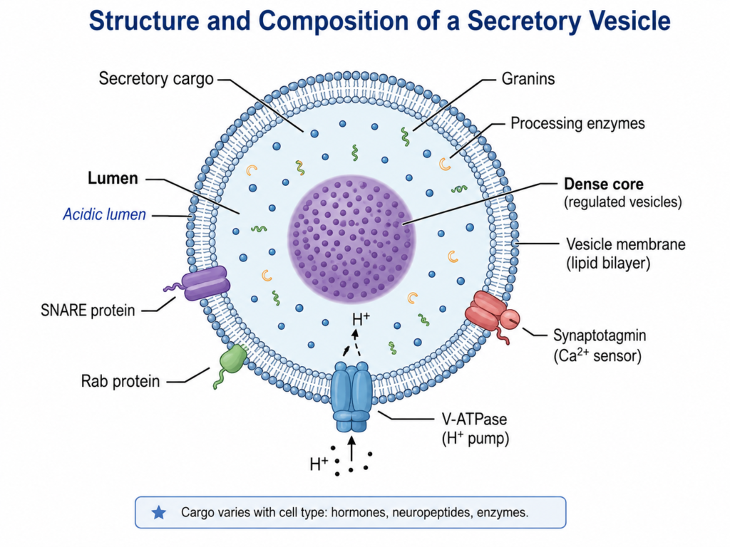
- Outer membrane– A secretory vesicle is bounded by a single membrane. It is a lipid bilayer containing different membrane proteins. The membrane separates vesicular contents from the cytoplasm.
- Vesicle lumen– The space enclosed by the membrane forms its lumen. Soluble substances that are to be secreted are present here.
- Secretory cargo– Proteins form an important part of vesicular contents. Depending on the cell, peptide hormones, neuropeptides, digestive enzymes and other secretory products can be packed inside. Small secretory molecules are also present in some types of vesicles.
- Dense core– Regulated secretory vesicles often contain the material in highly concentrated form. Due to this, many show a dense central core when seen under electron microscope and are referred to as dense-core vesicles or secretory granules.
- Granins– Chromogranins and secretogranins are common lumenal proteins of many neuroendocrine secretory granules. They occur together with peptide hormones and neuropeptides. However, these proteins are not the composition of every type of secretory vesicle.
- Membrane proteins– Different proteins are embedded in the vesicular membrane. Some are required in transport and others during docking or fusion with the target membrane.
- Fusion machinery– SNARE proteins are associated with membrane fusion of secretory vesicles. Different Rab proteins also take part in vesicle targeting and docking. Their particular types differ among secretory organelles and cells.
- Calcium sensor– In several regulated secretory vesicles, membrane proteins such as synaptotagmins participate in Ca²⁺-dependent secretion. Synaptotagmin has also been found in peptide-containing large dense-core vesicles.
- Proton pump– The membrane of regulated secretory granules contains V-type H⁺-ATPase (V-ATPase). Proton movement by this pump makes the inside of the granule acidic. Acidification also develops during maturation of secretory vesicles.
- Processing proteins– Many peptide hormones and neuropeptides enter secretory granules initially as precursor proteins. Proteolytic processing can then occur during formation and maturation of the vesicle. Thus, processing enzymes may also form part of the lumenal contents in these granules.
- Lipids– Phospholipids form the basic membrane structure along with other membrane lipids. The lipid composition is not exactly same in every secretory vesicle, and specialized secretory granules can contain particular lipid domains involved in sorting and granule formation.
- Concentrated material– During maturation, contents of regulated secretory vesicles become more concentrated and the membrane around the stored material becomes closely arranged. Mature granules are therefore densely filled with secretory products.
- Variable composition– Composition of a secretory vesicle is not fixed for all cells. It changes with the type of cell and the material being secreted. An insulin granule, neuropeptide-containing dense-core vesicle and an enzyme-containing zymogen granule therefore do not contain exactly the same substances.
Formation and Maturation of Secretory Vesicles
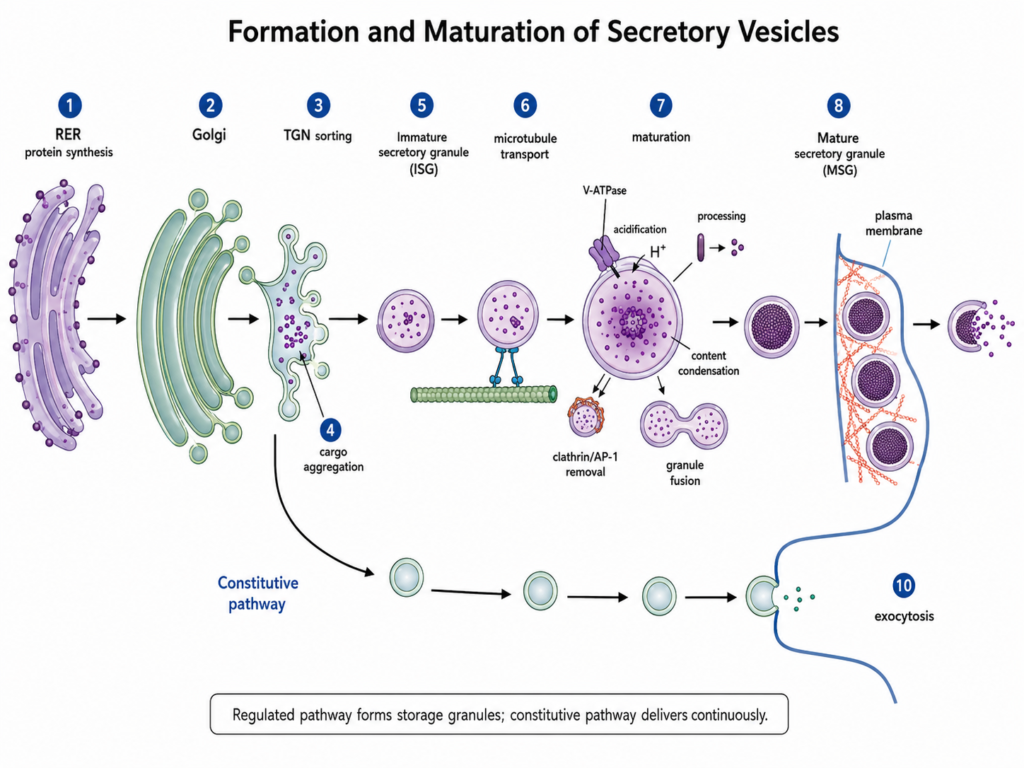
- Protein formation– Secretory proteins are first synthesized in the rough endoplasmic reticulum (RER). They enter the ER lumen where initial folding and processing takes place.
- Golgi transport– From the ER, the proteins are carried towards the Golgi apparatus. Further modification takes place here before reaching the trans side of Golgi.
- TGN sorting– The trans-Golgi network (TGN) is the major sorting region. Proteins for regulated secretion are collected and concentrated in this region.
- Cargo aggregation– Many regulated secretory proteins form aggregates within the TGN lumen. This helps in their packing into developing secretory granules.
- Granule budding– A portion of TGN membrane surrounds the collected secretory material and buds away. The newly formed structure is called an immature secretory granule (ISG). It is not yet a completely mature storage granule.
- Granule movement– Soon after formation, immature granules are transported away from the Golgi region. Microtubules take part in their movement towards the cell periphery and cortical region.
- Acidification– During maturation, lumen of the immature granule becomes more acidic. V-type H⁺-ATPase (V-ATPase) present in the membrane is associated with this acidification.
- Protein processing– In endocrine and neuroendocrine cells, many hormones and neuropeptides are initially present as precursor proteins. These are processed within the developing granule into their active or mature products.
- Content condensation– Secretory material becomes more concentrated as maturation proceeds. Thus, the lumen becomes densely packed and a characteristic dense core is produced in many regulated secretory granules.
- Membrane removal– Excess membrane and some proteins which should not remain in the mature granule are removed from immature granules. Clathrin and adaptor proteins such as AP-1 are involved in this membrane-remodelling process.
- Granule fusion– Immature granules can also undergo fusion with one another during maturation. Such changes occur together with processing, acidification and rearrangement of granule contents.
- Mature granule– After these changes, an immature granule develops into a mature secretory granule (MSG). It contains concentrated secretory products and can remain stored within the cell.
- Cortical storage– Mature granules are commonly present in the actin-rich cell cortex, many close to the plasma membrane. Here, they may remain stored until the cell receives a suitable stimulus.
- Final release– After stimulation, mature granules become involved in regulated membrane fusion. Their contents are released outside the cell by exocytosis.
- Constitutive vesicles– Not all secretory vesicles undergo this type of long maturation. Vesicles of the constitutive secretory pathway generally move from the TGN towards the plasma membrane and continuously deliver their contents. The formation of immature and mature storage granules is mainly a feature of the regulated secretory pathway.
Types of Secretory Pathways
The secretory pathway is mainly of two types. These are constitutive secretory pathway and regulated secretory pathway.
- Constitutive pathway– This pathway occurs in all eukaryotic cells. Vesicles continuously move from the trans-Golgi network (TGN) towards the plasma membrane. No particular external signal is required for their secretion. Soluble proteins present inside the vesicle are released outside, while membrane proteins and lipids become added to the plasma membrane. Thus, it is a continuous secretory process. In polarized cells, the materials can also be carried separately towards apical or basolateral surfaces.
- Regulated pathway– It is mainly present in specialized secretory cells. In this pathway, secretory products are concentrated and stored inside secretory vesicles or secretory granules instead of being released immediately. The vesicles remain inside the cell until a suitable signal is received. After stimulation, they fuse with the plasma membrane and secretion takes place by exocytosis. Increase in intracellular Ca²⁺ commonly acts during this release in many cells. Hormone secretion from endocrine cells, digestive enzyme secretion from pancreatic acinar cells and regulated secretion in neurons are common examples.
Constitutive vs Regulated Secretion
| Features | Constitutive Secretion | Regulated Secretion |
|---|---|---|
| Occurrence | It occurs in almost all eukaryotic cells. | Mainly present in specialized secretory cells. |
| Nature | It is a continuous process. | Secretion occurs only when the cell is stimulated. |
| Storage | Secretory products are generally not stored for a long period. | Products are stored inside secretory vesicles or granules before release. |
| Signal requirement | No specific secretory signal is required. | A suitable cellular signal is required for secretion. |
| Ca²⁺ role | Usually not dependent on a stimulus-induced rise of intracellular Ca²⁺. | Increase in intracellular Ca²⁺ commonly causes release in many regulated secretory cells. |
| Vesicle formation | Transport carriers are formed mainly from the trans-Golgi network (TGN) and move towards plasma membrane. | Immature secretory granules arise from the TGN and undergo maturation. |
| Maturation | Prolonged storage-granule maturation is absent. | Vesicles undergo concentration, processing and maturation before secretion. |
| Movement | Vesicles are continuously carried towards the plasma membrane. | Mature vesicles may remain stored in cytoplasm or close to plasma membrane. |
| Release | Vesicles fuse with plasma membrane continuously and their contents are released by exocytosis. | Fusion with plasma membrane takes place after receiving the required stimulus. |
| Main function | It supplies membrane proteins, lipids and continuously secreted proteins to cell surface or extracellular region. | Mainly used for controlled release of stored hormones, enzymes, neuropeptides and other secretory products. |
| Examples | Secretion of extracellular matrix proteins and continuous delivery of membrane proteins and lipids. | Insulin release from pancreatic β-cells, digestive enzyme secretion from pancreatic acinar cells and neuropeptide secretion. |
How Secretory Vesicles Move Through the Cell
The movement of secretory vesicles from the Golgi region to the cell surface is carried out with the help of cytoskeleton and different motor proteins. The following are the major events involved in their movement-
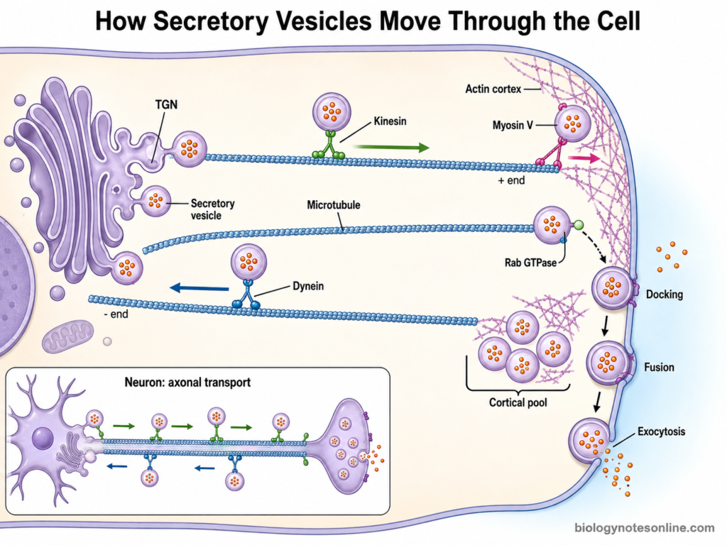
- Golgi release– Secretory vesicles are formed from the trans-Golgi network (TGN). After formation, they move away from the Golgi region towards their site of secretion.
- Cytoskeletal tracks– Vesicles do not simply move freely through cytoplasm. Microtubules and actin filaments provide tracks for their intracellular movement.
- Microtubule transport– Microtubules are mainly involved in movement over longer distance. Secretory vesicles can therefore be carried from the central region of cell towards the cell periphery.
- Kinesin proteins– Kinesins are microtubule motor proteins. Many kinesins move vesicles towards the plus end of microtubules, which in many cells is directed towards the cell periphery. Energy for this movement is obtained from ATP.
- Dynein proteins– Cytoplasmic dynein generally moves in the opposite direction, towards the minus end of microtubules. Thus, vesicles and other membrane-bound structures can also show movement back towards the central region.
- Actin movement– Near the cell surface, vesicles encounter the actin-rich cortex. Actin filaments are mainly associated with shorter range movement and positioning of secretory vesicles in this region.
- Myosin motors– Different myosin proteins take part in movement and handling of vesicles on actin. Myosin V is particularly associated with transport of several secretory vesicles through the cortical actin region. Its importance, however, differs with cell and vesicle type.
- Vesicle targeting– Movement must bring the vesicle to its correct region of plasma membrane. Rab GTPases and their effector proteins participate in vesicle targeting and positioning, together with other components of the trafficking machinery.
- Cortical positioning– After long-distance transport, many regulated secretory vesicles are brought near the plasma membrane. Some remain here or within a nearby storage pool until secretion is required. Constitutive secretory carriers generally continue towards fusion.
- Neuronal transport– A long movement is especially seen in neurons. Neuropeptide-containing secretory vesicles are formed in the cell body and carried along axonal microtubules towards distant nerve terminals. Motor proteins are used during this transport.
- Final movement– At the secretion site, vesicles are brought close to the plasma membrane followed by docking and preparation for fusion. In regulated secretion they can wait until proper signal is received. The vesicular membrane then fuses with plasma membrane and its contents are released by exocytosis.
The exact movement is not same for every secretory vesicle. Use of microtubules, actin and particular motor proteins varies with cell type, distance travelled and the type of secretion.
Mechanism of Secretory Vesicle Exocytosis
Exocytosis involves the fusion of secretory vesicle membrane with the plasma membrane followed by release of its contents outside the cell. The mechanism includes vesicle recognition, docking and membrane fusion. In regulated secretory cells, a signal is also required before final fusion.

The following are the major steps involved in secretory vesicle exocytosis-
- Vesicle arrival– Secretory vesicle is first brought close to its particular region of the plasma membrane. Vesicles arriving here are now prepared for the further fusion process.
- Tethering– In this step, the vesicle is loosely captured near the target membrane. Rab GTPases and their effector proteins have important role in this process. They help in placing the correct vesicle at its proper membrane region.
- Docking– The tethered vesicle now comes very close to the plasma membrane. This closer attachment is referred to as docking. Proteins present on both membranes begin to interact.
- Priming– In regulated secretion, some docked vesicles are converted into a fusion-ready state. This process is called priming. Proteins such as Munc13 and Munc18 have major roles in priming of neuronal and several neuroendocrine secretory vesicles. The exact proteins are not same in every secretory cell.
- SNARE assembly– SNARE proteins present on vesicle and target membrane now associate with one another. A vesicular R-SNARE binds with complementary Q-SNAREs present on target membrane. In neurons, VAMP2 (synaptobrevin-2) is present on vesicle whereas syntaxin-1 and SNAP-25 are present mainly on plasma membrane. Other secretory cells can use different SNARE members.
- SNARE zippering– The SNARE proteins form a tight four-helix bundle. Assembly proceeds in a zipper-like manner and pulls the two membranes towards each other. Thus, the distance between vesicle membrane and plasma membrane becomes very small.
- Calcium signal– In many forms of regulated exocytosis, intracellular Ca²⁺ level increases after stimulation. In neurons and several neuroendocrine cells, Ca²⁺ binds with synaptotagmin proteins associated with secretory vesicles. This helps to trigger rapid final fusion. However, every exocytic pathway does not use the same Ca²⁺ trigger or same synaptotagmin.
- Membrane fusion– After the fusion machinery is activated, lipid bilayers of both membranes start to merge. A hemifusion-like intermediate can occur during this process, followed by complete joining of the membranes. SNARE complex provides an important driving force for this fusion.
- Fusion pore– A small opening is then produced between vesicle lumen and the extracellular region. This opening is called the fusion pore. Secretory contents can now pass through it.
- Cargo release– The fusion pore may enlarge and the stored substances are released outside of cell. Hormones, enzymes, neuropeptides or other secretory products are released depending upon the type of vesicle.
- Fusion mode– In full-collapse fusion, the vesicle membrane becomes completely merged with plasma membrane. In some secretory systems the pore can remain open for a short period and close again without complete collapse. This is referred to as kiss-and-run exocytosis.
- Membrane addition– After complete fusion, vesicular lipids and membrane proteins become a part of the plasma membrane. Thus, exocytosis releases soluble contents as well as adds membrane components to cell surface.
- SNARE disassembly– Following fusion, SNARE proteins are present together in the same membrane as a cis-SNARE complex. NSF and α-SNAP then separate this complex using energy from ATP, allowing the SNARE proteins to be used again.
- Membrane recovery– Vesicular membrane added during exocytosis can later be recovered by endocytosis. The recovered components may then be recycled for another round of vesicle transport and secretion.
Functions of Secretory Vesicles
The major functions of secretory vesicles are as follows-
- Cargo transport– Secretory vesicles carry proteins and other substances from the trans-Golgi network (TGN) towards the plasma membrane. It is one of their major functions.
- Protein secretion– Soluble proteins present inside the vesicle are released to the outside of cell. Many extracellular proteins are secreted by this process.
- Storage– In specialized secretory cells, vesicles also act as storage structures. Hormones, neuropeptides, digestive enzymes and other products can remain packed inside them until secretion is required.
- Regulated release– Some secretory vesicles do not release their contents continuously. They remain stored and fuse with plasma membrane only after receiving a particular stimulus. This is referred to as regulated secretion.
- Hormone secretion– Secretory granules of endocrine cells store hormones and release them when required. Insulin secretion from pancreatic β-cells is a common example.
- Neuropeptide release– In neurons and neuroendocrine cells, dense-core vesicles carry and store neuropeptides. These substances are released during cell signalling.
- Enzyme secretion– Digestive enzymes are stored in secretory granules of many exocrine cells. Pancreatic acinar cells, for example, release their digestive enzymes from zymogen granules.
- Membrane supply– During exocytosis, membrane of the secretory vesicle becomes incorporated into the plasma membrane. Thus, vesicular transport also supplies lipids and membrane proteins to the cell surface.
- Surface proteins– Membrane proteins carried in constitutive secretory vesicles become inserted into the plasma membrane after fusion. They may function there as receptors, channels or other surface proteins.
- Extracellular matrix– Cells use the constitutive secretory pathway for secretion of many extracellular matrix components. Proteoglycans and glycoproteins are some of the important substances delivered outside the cell.
- Cell communication– Release of hormones, neuropeptides and other signalling substances allows communication between cells. Secretory vesicles therefore have major role in endocrine and neuronal signalling.
- Polarized transport– In polarized cells, secretory materials are not always delivered to the same surface. Different proteins and lipids can be carried towards particular apical or basolateral regions of plasma membrane.
- Cargo concentration– Regulated secretory vesicles concentrate selected secretory products inside them. During maturation, the contents may become highly condensed, forming the dense material of secretory granules or dense-core vesicles.
- Rapid secretion– Storage of secretory products makes their rapid release possible when a cell is stimulated. This is particularly important in cells secreting hormones, neuropeptides and digestive enzymes.
- Membrane balance– Vesicle membrane added to plasma membrane during exocytosis does not remain permanently in every case. Membrane components can be removed again by endocytosis, helping in maintaining the cell surface membrane.
Examples of Secretory Vesicles in Different Cells
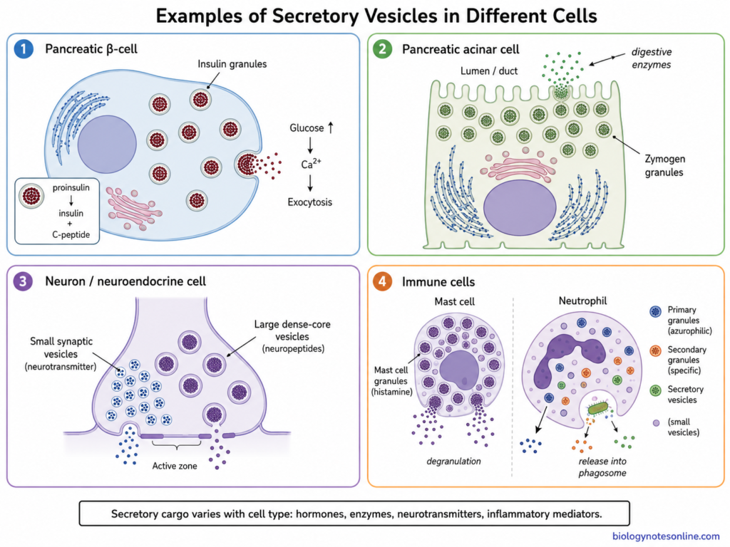
Secretory vesicles are present in different types of cells and their contents are also different. Some cells store hormones while others contain enzymes, neurotransmitters or inflammatory substances. Some of the important examples are-
Insulin Secretory Granules
- Insulin secretory granules are present in pancreatic β-cells. These are dense-core secretory granules containing insulin and related processing products.
- Newly formed granules initially contain proinsulin. During granule maturation, proinsulin is processed into insulin and C-peptide and the contents become concentrated.
- Insulin is not continuously released from these granules. The granules remain stored in the β-cell and secretion takes place by the regulated secretory pathway.
- Increased glucose stimulates insulin secretion. Glucose metabolism increases the ATP level, ATP-sensitive K⁺ channels close and the cell membrane becomes depolarized. Voltage-gated Ca²⁺ channels are then opened.
- Entry of Ca²⁺ into the β-cell provides an important signal for exocytosis. Insulin granules now fuse with the plasma membrane and insulin is released outside the cell.
- Insulin granules are one of the well-studied examples of regulated secretory vesicles. Their formation, maturation, transport, docking, priming and final fusion with plasma membrane have been extensively studied.
Zymogen Granules
- Zymogen granules are large secretory granules found abundantly in pancreatic acinar cells. They are mainly collected towards the apical region of these cells.
- Pancreatic acinar cells are specialized for synthesis, storage and secretion of digestive enzymes. Many digestive enzymes are stored as inactive enzyme precursors or zymogens, although some pancreatic digestive enzymes are stored in active forms.
- Zymogen granules are formed through the trans-Golgi network (TGN). Their contents become concentrated during maturation and mature granules are moved towards the apical side of cell.
- On stimulation, these granules fuse with the apical plasma membrane. Digestive contents are then released into the pancreatic duct system by regulated exocytosis.
- The term zymogen granule has an established relationship with secretory vesicles. NLM MeSH lists Zymogen Granules as an entry term under Secretory Vesicles.
Secretory Vesicles in Neurons and Neuroendocrine Cells
- Neurons contain different types of secretory vesicles. They should not be considered as a single identical group. Small synaptic vesicles and large dense-core vesicles (LDCVs) are two important types.
- Small synaptic vesicles are concentrated particularly at presynaptic terminals. Classical small-molecule neurotransmitters such as glutamate, GABA and acetylcholine are generally stored in these vesicles.
- Large dense-core vesicles are larger and possess electron-dense contents. Neuropeptides are commonly stored within them. Depending upon neuron type, some other signalling substances or neurotransmitters can also occur in dense-core vesicles.
- Neuropeptide-containing dense-core vesicles are formed through the Golgi secretory pathway. They can be transported from neuronal cell body through the axon towards their sites of secretion.
- Neuroendocrine cells also contain dense-core secretory granules. Hormones and neuropeptides are stored here and released after stimulation.
- Both small synaptic vesicles and dense-core vesicles undergo regulated exocytosis, but their formation, localization and release are not exactly same. Dense-core vesicles therefore should not simply be treated as enlarged synaptic vesicles.
Secretory Granules in Immune Cells
- Different immune cells contain specialized granules used during immune responses. Mast cells and granulocytes are common examples, but their granules are not all of the same type.
- Mast cells contain numerous secretory granules. Histamine, proteases, proteoglycans and some other preformed inflammatory mediators are stored inside them.
- When a mast cell is activated, granule contents can be released rapidly by regulated exocytosis. This release of stored granules is commonly referred to as degranulation. Histamine released during this process has an important role in inflammatory and allergic responses.
- Granulocytes such as neutrophils also possess several granule populations. Neutrophils contain primary (azurophilic), secondary (specific) and tertiary granules, together with a separate population called secretory vesicles. Thus, all neutrophil granules should not be called secretory vesicles as if they are identical.
- Different antimicrobial substances are stored in these neutrophil granules. Myeloperoxidase, defensins and elastase occur mainly in primary granules, while substances such as lactoferrin are associated with secondary granules.
- After activation, neutrophil granules can fuse with the plasma membrane or with a phagosome. Their substances are therefore released outside the cell or directly around an engulfed microorganism. This regulated granule release forms an important part of antimicrobial defence.
Secretory Vesicles in Plant Cells
Secretory vesicles are also present in plant cells and form an important part of plant secretory pathway. They carry different proteins, membrane materials and cell wall substances to their required sites.
- In plant cells, proteins formed in the endoplasmic reticulum (ER) are transported through the Golgi apparatus. The trans-Golgi network (TGN) acts as an important sorting region for further transport.
- From the Golgi/TGN, secretory vesicles move towards the plasma membrane. The vesicle membrane fuses with it and the contents are released into the extracellular region.
- Plant secretory pathway has an important role in formation of the cell wall. Many wall matrix polysaccharides are synthesized in Golgi and then carried outside by secretory vesicles.
- Pectins and many hemicelluloses are important extracellular glycans transported through this pathway. These substances are released into the apoplast and become components of the cell wall. Cellulose, however, is mainly synthesized directly at the plasma membrane and not transported as finished cellulose inside secretory vesicles.
- During plant cell growth, secretion supplies wall materials and different membrane components to the expanding cell surface. Thus, secretory transport is continuously required when the cell is enlarging.
- Secretory vesicles have a major role during plant cytokinesis also. Golgi/TGN-derived vesicles are transported towards the division plane and begin to fuse with each other.
- The fused vesicles gradually form the developing cell plate. More membrane and polysaccharide materials are added during this process, and finally the cell plate becomes connected with the parental cell wall.
What Happens When Secretory Vesicle Function Is Disrupted?
Secretory vesicles are required for proper transport, storage and release of different cellular products. Any defect in their formation, movement or exocytosis can therefore affect the normal function of cell. The major effects are as follows-
- Transport defect– Secretory products may not reach the plasma membrane properly. Some proteins remain inside the cell or are transported to an incorrect region.
- Formation defect– Abnormal vesicle formation can reduce the number of properly formed secretory vesicles. Packing of secretory materials may also become affected.
- Maturation defect– Regulated secretory granules need maturation before release. Defect in this process can affect concentration and processing of their stored substances.
- Movement defect– Vesicles can be formed normally but their movement through cytoplasm becomes disturbed. They may not reach the required region of plasma membrane.
- Docking defect– Some vesicles reach the cell surface but fail to dock properly. Secretory material therefore remains inside the cell.
- Fusion defect– Abnormality in SNARE proteins, Rab proteins or other fusion-associated proteins can affect membrane fusion. Exocytosis is then reduced or may fail.
- Insulin secretion– In pancreatic β-cells, defects in insulin granule formation, docking or exocytosis can decrease insulin release. Impaired glucose-stimulated insulin secretion is an important feature associated with type 2 diabetes.
- Neuronal secretion– Defect in synaptic or dense-core vesicle function can alter release of neurotransmitters and neuropeptides. Normal communication between neurons is therefore affected.
- Enzyme release– Pancreatic acinar cells depend on regulated granule exocytosis for release of digestive enzymes. Disturbance of this process can reduce proper enzyme secretion.
- Immune response– Cytotoxic T cells and natural killer (NK) cells use specialized lytic granules for killing target cells. Defects in granule exocytosis can greatly reduce this function.
- Degranulation defect– Mast cells and granulocytes also depend on regulated granule release. Abnormal degranulation can change inflammatory and antimicrobial responses.
- Cargo accumulation– When secretion is blocked, some secretory products may accumulate inside the cell. Excess intracellular accumulation can disturb normal cellular functions.
- Membrane delivery– Constitutive secretory vesicles also supply proteins and lipids to cell surface. Defects in this pathway can therefore change composition and function of the plasma membrane.
- Variable effects– The final effect is not same in every cell. It depends on the secretory product and which step of vesicle formation, transport or release is affected.
How Are Secretory Vesicles Studied?
Different microscopic, biochemical and genetic methods are used to study secretory vesicles. These methods can show their structure, movement, contents and release from the cell.
- Transmission electron microscopy– Transmission electron microscopy (TEM) is used to study the fine structure of secretory vesicles. Dense-core granules, their membrane and association with Golgi can be observed. Since the cells are fixed, their actual movement cannot be followed by this method.
- Fluorescence microscopy– It is used to locate secretory vesicles and their proteins inside the cell. Fluorescent dyes or fluorescent proteins are attached with the particular vesicle components. The labelled vesicles can then be seen as fluorescent structures.
- Live-cell imaging– Living cells can be observed continuously for vesicle movement. In this method, movement from the Golgi region, transport through cytoplasm and final exocytosis can be followed with time.
- Fluorescent proteins– Vesicle proteins or secretory cargo can be tagged with GFP, mCherry and other fluorescent proteins. pHluorin, a pH-sensitive GFP, is also widely used. It becomes brighter when the acidic vesicle lumen is exposed to the extracellular medium during exocytosis.
- TIRF microscopy– Total internal reflection fluorescence microscopy (TIRF) is especially useful for vesicles present very close to the plasma membrane. Only a thin region near the glass-cell surface is illuminated. Thus, docking and individual fusion events can be detected with less background fluorescence.
- Biochemical methods– Secretory granules can also be isolated from disrupted cells. Differential centrifugation and density-gradient centrifugation are commonly used for this purpose. Their proteins and other components can then be studied by immunoblotting, electrophoresis, proteomics or other biochemical methods.
- Secretion assays– The amount of material released from a cell can be measured after stimulation. Hormones, enzymes or other secretory products present in extracellular medium are measured depending upon the cell studied. Membrane capacitance and amperometry are also used for studying exocytosis, particularly in neuroendocrine and excitable cells.
- Genetic manipulation– Genes associated with vesicle formation, transport or fusion can be removed or altered. Knockout, knockdown and CRISPR-Cas9 methods are used, and the effect on secretion is then observed. This method has helped to identify proteins required for vesicle trafficking and exocytosis.
- Plant-cell imaging– Secretory transport in plant cells has also been studied by electron microscopy and fluorescence imaging. Electron tomography gives detailed three-dimensional structure of Golgi-associated vesicles, while fluorescent markers allow their movement in living plant cells to be followed.
Secretory Vesicles vs Other Cellular Vesicles
Secretory Vesicle vs Transport Vesicle
| Features | Secretory Vesicle | Transport Vesicle |
|---|---|---|
| Meaning | Secretory vesicles carry substances mainly towards the cell surface for secretion. | Transport vesicles is a broader group of vesicles that carry materials between different cell compartments. |
| Main pathway | Mainly associated with the secretory pathway and post-Golgi transport. | They occur throughout intracellular membrane trafficking. |
| Origin | Many secretory vesicles arise from the trans-Golgi network (TGN). | They may arise from ER, Golgi, endosomes or other membrane compartments. |
| Destination | Their important destination is the plasma membrane. | Destination depends on the type of transport, such as ER to Golgi, Golgi to endosome or other compartments. |
| Contents | Secretory proteins, hormones, enzymes and membrane components may be present. | Contents depend on the organelles between which transport takes place. |
| Storage | Regulated secretory vesicles may remain stored before release. | Most transport vesicles are temporary carriers and normally do not act as long-term storage structures. |
| Final process | Contents can be released by exocytosis. | Usually fuse with another intracellular membrane. |
| Relation | A secretory vesicle can also be considered a specialized transport carrier. | Transport vesicle is the more general term. |
Secretory Vesicle vs Extracellular Vesicle/Exosome
| Features | Secretory Vesicle | Extracellular Vesicle / Exosome |
| Location | Present inside the cell before secretion. | Present outside the cell after their release. |
| Major role | Carries secretory cargo towards the plasma membrane. | Carries proteins, lipids, nucleic acids and other molecules between cells. |
| Formation | Many are formed from the TGN during secretory transport. | Extracellular vesicles have different origins. Exosomes are formed through the endosomal multivesicular body pathway. |
| Membrane fusion | Vesicle membrane generally fuses with plasma membrane during exocytosis. | The whole vesicle itself is released into extracellular space. |
| Cargo release | Soluble lumenal contents are discharged outside the cell. | Cargo remains enclosed within the released vesicle until interaction with another cell or extracellular breakdown. |
| Examples | Insulin granules, zymogen granules and dense-core secretory vesicles. | Exosomes and plasma-membrane-derived microvesicles. |
| Important difference | It is primarily an intracellular secretory carrier. | It is an extracellular membrane-bound particle after release from a cell. |
Secretory Vesicle vs Lysosome
| Features | Secretory Vesicle | Lysosome |
| Main function | Mainly involved in transport, storage and secretion of cellular products. | Mainly involved in intracellular digestion and degradation. |
| Contents | May contain hormones, enzymes, neuropeptides or other secretory substances. | Contains different acid hydrolases used for degradation of macromolecules. |
| Internal condition | Regulated secretory granules can have an acidic lumen. | Lysosomes have a strongly acidic lumen required for lysosomal enzymes. |
| Destination | Usually transported towards the plasma membrane for secretion. | Mainly receives substances from endosomes, autophagosomes and other degradative pathways. |
| Storage | Some secretory vesicles store products before their release. | It is not mainly a storage organelle for products to be secreted. |
| Exocytosis | Exocytosis is a major function of secretory vesicles. | Lysosomes can also undergo lysosomal exocytosis in particular cells and conditions, but degradation is their major function. |
| Cargo fate | Cargo is commonly released outside the cell. | Cargo is mostly broken down and recycled within the cell. |
Secretory Granules vs Synaptic Vesicles
| Features | Secretory Granules | Synaptic Vesicles |
| Structure | Generally larger vesicles with concentrated contents, often showing a dense core. | Small, usually clear vesicles present mainly at presynaptic nerve terminals. |
| Other name | Often called dense-core vesicles or dense-core granules. | Commonly referred to as small synaptic vesicles. |
| Contents | Mainly peptide hormones, neuropeptides, enzymes and other regulated secretory products. | Mainly classical small-molecule neurotransmitters such as glutamate, GABA and acetylcholine. |
| Formation | Formed through the Golgi/TGN secretory pathway and undergo maturation. | Their membrane components are supplied through biosynthetic trafficking, but mature synaptic vesicles are extensively regenerated locally by endocytic recycling at nerve terminals. |
| Maturation | Contents become concentrated and precursor proteins may be processed during maturation. | They do not undergo the same dense-core granule maturation process. |
| Distribution | Present in endocrine, neuroendocrine and several other secretory cells. Large dense-core vesicles are also present in neurons. | Mainly concentrated at presynaptic terminals of neurons. |
| Release | Released by regulated, commonly Ca²⁺-dependent exocytosis. | Neurotransmitter release is also mainly Ca²⁺-dependent, usually at specialized active zones. |
| Recycling | After secretion, membrane components can be recovered, but newly formed dense-core granules are generally supplied from the cell body/Golgi pathway. | Synaptic vesicle membrane is rapidly recovered and reused locally through endocytosis. |
| Important difference | Mainly stores larger secretory products such as peptides and hormones. | Mainly stores and rapidly releases small-molecule neurotransmitters. |
Frequently Asked Questions (FAQs)
1. What are secretory vesicles?
Secretory vesicles are membrane-bound structures that carry substances from inside the cell towards the plasma membrane. Some vesicles also store secretory products before their release.
2. What is the main function of secretory vesicles?
The main function is transport and release of cellular products. Proteins, hormones, enzymes and other substances can be carried in these vesicles and released by exocytosis.
3. Where are secretory vesicles formed?
Most post-Golgi secretory vesicles are formed from the trans-Golgi network (TGN). Secretory proteins reach this region after synthesis in the ER and processing through the Golgi apparatus.
4. Are secretory vesicles formed by the Golgi apparatus?
Yes. Many secretory vesicles bud from the trans-Golgi network. In regulated secretory cells, newly formed immature granules can undergo further maturation before storage and secretion.
5. What do secretory vesicles contain?
Their contents depend on the type of cell. They may contain proteins, peptide hormones, neuropeptides, digestive enzymes and other secretory substances.
6. Where are secretory vesicles found in a cell?
They are found in the cytoplasm of eukaryotic cells. Newly formed vesicles occur around the Golgi region, while mature vesicles may be present near the cell cortex and plasma membrane.
7. How do secretory vesicles move through the cytoplasm?
Their movement takes place with the help of the cytoskeleton. Microtubules, actin filaments and motor proteins such as kinesins, dynein and myosins participate in vesicle transport.
8. How do secretory vesicles release their contents?
The vesicle membrane fuses with the plasma membrane and the stored material is released outside the cell. This process is called exocytosis.
9. What is the relationship between secretory vesicles and exocytosis?
Secretory vesicles are the intracellular carriers involved in secretion, whereas exocytosis is the process by which their membrane fuses with plasma membrane and releases the contents.
10. What is the difference between secretory vesicles and transport vesicles?
Transport vesicle is a broader term for vesicles carrying materials between cellular compartments. Secretory vesicles mainly carry materials towards the plasma membrane for secretion.
11. What is the difference between constitutive and regulated secretion?
In constitutive secretion, vesicles continuously move towards the plasma membrane and release their contents. In regulated secretion, products remain stored in secretory vesicles or granules until a suitable signal is received.
12. Are secretory vesicles and secretory granules the same?
Not always. Secretory vesicle is a broader term. Secretory granule is commonly used for specialized storage organelles of the regulated secretory pathway, especially those containing concentrated secretory products.
13. What are dense-core vesicles?
Dense-core vesicles (DCVs) are regulated secretory organelles containing concentrated material that appears electron-dense under electron microscope. They commonly store peptide hormones and neuropeptides.
14. Are secretory vesicles present in plant cells?
Yes. Plant cells use secretory vesicles for transport of proteins, membrane components and many cell-wall materials from the Golgi/TGN towards the plasma membrane. They also have an important role during formation of the cell plate.
15. What happens to the secretory-vesicle membrane after exocytosis?
After fusion, the vesicle membrane becomes part of the plasma membrane. Some membrane proteins and lipids can later be recovered through endocytosis and reused by the cell.
References
- lberts, B., Johnson, A., Lewis, J., Raff, M., Roberts, K., & Walter, P. (2002). Molecular biology of the cell (4th ed.). Garland Science. https://www.ncbi.nlm.nih.gov/books/NBK21054/
- Arvan, P., & Castle, D. (1998). Sorting and storage during secretory granule biogenesis: Looking backward and looking forward. Biochemical Journal, 332(3), 593–610. https://doi.org/10.1042/bj3320593
- Augustine, G. J., Burns, M. E., DeBello, W. M., Pettit, D. L., & Schweizer, F. E. (1996). Exocytosis: Proteins and perturbations. Annual Review of Pharmacology and Toxicology, 36, 659–701. https://doi.org/10.1146/annurev.pa.36.040196.003303
- Bartolomucci, A., Possenti, R., Mahata, S. K., Fischer-Colbrie, R., Loh, Y. P., & Salton, S. R. J. (2011). The extended granin family: Structure, function, and biomedical implications. Endocrine Reviews, 32(6), 755–797. https://doi.org/10.1210/er.2010-0027
- Bond, L. M., Brandstaetter, H., Sellers, J. R., Kendrick-Jones, J., & Buss, F. (2011). Myosin motor proteins are involved in the final stages of the secretory pathways. Biochemical Society Transactions, 39(5), 1115–1119. https://doi.org/10.1042/BST0391115
- Bonnemaison, M. L., Eipper, B. A., & Mains, R. E. (2013). Role of adaptor proteins in secretory granule biogenesis and maturation. Frontiers in Endocrinology, 4, 101. https://doi.org/10.3389/fendo.2013.00101
- Burgoyne, R. D., & Morgan, A. (2003). Secretory granule exocytosis. Physiological Reviews, 83(2), 581–632. https://doi.org/10.1152/physrev.00031.2002
- Cooper, G. M. (2000). The cell: A molecular approach (2nd ed.). Sinauer Associates. https://www.ncbi.nlm.nih.gov/books/NBK9839/
- Dernick, G., Alvarez de Toledo, G., & Lindau, M. (2007). The patch amperometry technique: Design of a method to study exocytosis of single vesicles. In A. C. Michael & L. M. Borland (Eds.), Electrochemical methods for neuroscience (Chap. 15). CRC Press/Taylor & Francis. https://doi.org/10.1201/9781420005868.ch15
- Fukuda, M. (2007). The role of synaptotagmin and synaptotagmin-like protein (Slp) in regulated exocytosis. In R. Regazzi (Ed.), Molecular mechanisms of exocytosis (pp. 42–61). Springer. https://doi.org/10.1007/978-0-387-39961-4_4
- Gandasi, N. R., Yin, P., Riz, M., Chibalina, M. V., Cortese, G., Lund, P.-E., Matveev, V., Rorsman, P., Sherman, A., Pedersen, M. G., & Barg, S. (2017). Ca²⁺ channel clustering with insulin-containing granules is disturbed in type 2 diabetes. The Journal of Clinical Investigation, 127(6), 2353–2364. https://doi.org/10.1172/JCI88491
- Gorr, S.-U., Venkatesh, S. G., & Darling, D. S. (2005). Parotid secretory granules: Crossroads of secretory pathways and protein storage. Journal of Dental Research, 84(6), 500–509. https://doi.org/10.1177/154405910508400604
- Howell, S. L., Fink, C. J., & Lacy, P. E. (1969). Isolation and properties of secretory granules from rat islets of Langerhans. I. Isolation of a secretory granule fraction. The Journal of Cell Biology, 41(1), 154–161. https://doi.org/10.1083/jcb.41.1.154
- Husain, S., & Thrower, E. (2009). Molecular and cellular regulation of pancreatic acinar cell function. Current Opinion in Gastroenterology, 25(5), 466–471. https://doi.org/10.1097/MOG.0b013e32832ebfac
- Jahn, R., Cafiso, D. C., & Tamm, L. K. (2024). Mechanisms of SNARE proteins in membrane fusion. Nature Reviews Molecular Cell Biology, 25(2), 101–118. https://doi.org/10.1038/s41580-023-00668-x
- Jahn, R., & Fasshauer, D. (2012). Molecular machines governing exocytosis of synaptic vesicles. Nature, 490(7419), 201–207. https://doi.org/10.1038/nature11320
- Jahn, R., & Scheller, R. H. (2006). SNAREs—engines for membrane fusion. Nature Reviews Molecular Cell Biology, 7(9), 631–643. https://doi.org/10.1038/nrm2002
- Jena, B. P., Gumkowski, F. D., Konieczko, E. M., von Mollard, G. F., Jahn, R., & Jamieson, J. D. (1994). Redistribution of a Rab3-like GTP-binding protein from secretory granules to the Golgi complex in pancreatic acinar cells during regulated exocytosis. The Journal of Cell Biology, 124(1–2), 43–53. https://doi.org/10.1083/jcb.124.1.43
- Jeon, J.-H., Hong, C.-W., Kim, E. Y., & Lee, J. M. (2020). Current understanding on the metabolism of neutrophils. Immune Network, 20(6), e46. https://doi.org/10.4110/in.2020.20.e46
- Jiang, K., Yang, L.-T., & Xue, M. (2025). Breaking the synaptic vesicle cycle: Mechanistic insights into presynaptic dysfunctions in epilepsy. Epilepsy Currents, 25(2), 119–124. https://doi.org/10.1177/15357597251317898
- Karatekin, E. (2018). Toward a unified picture of the exocytotic fusion pore. FEBS Letters, 592(21), 3563–3585. https://doi.org/10.1002/1873-3468.13270
- Kim, S.-J., & Brandizzi, F. (2014). The plant secretory pathway: An essential factory for building the plant cell wall. Plant and Cell Physiology, 55(4), 687–693. https://doi.org/10.1093/pcp/pct197
- Kim, T., Gondré-Lewis, M. C., Arnaoutova, I., & Loh, Y. P. (2006). Dense-core secretory granule biogenesis. Physiology, 21(2), 124–133. https://doi.org/10.1152/physiol.00043.2005
- Kögel, T., & Gerdes, H.-H. (2010). Maturation of secretory granules. Results and Problems in Cell Differentiation, 50, 1–20. https://doi.org/10.1007/400_2009_31
- Lacy, P. (2006). Mechanisms of degranulation in neutrophils. Allergy, Asthma & Clinical Immunology, 2(3), 98–108. https://doi.org/10.1186/1710-1492-2-3-98
- Laguerre, F., Anouar, Y., & Montero-Hadjadje, M. (2020). Chromogranin A in the early steps of the neurosecretory pathway. IUBMB Life, 72(4), 524–532. https://doi.org/10.1002/iub.2218
- Lai, Y., Choi, U. B., Leitz, J., Rhee, H. J., Lee, C., Altas, B., Zhao, M., Pfuetzner, R. A., Wang, A. L., Brose, N., Rhee, J.-S., & Brunger, A. T. (2017). Molecular mechanisms of synaptic vesicle priming by Munc13 and Munc18. Neuron, 95(3), 591–607.e10. https://doi.org/10.1016/j.neuron.2017.07.004
- Li, C., & Kim, K. (2008). Neuropeptides. WormBook, 1–36. https://doi.org/10.1895/wormbook.1.142.1
- Li, W.-H., & Li, D. (2013). Fluorescent probes for monitoring regulated secretion. Current Opinion in Chemical Biology, 17(4), 672–681. https://doi.org/10.1016/j.cbpa.2013.04.026
- Lippincott-Schwartz, J., Roberts, T. H., & Hirschberg, K. (2000). Secretory protein trafficking and organelle dynamics in living cells. Annual Review of Cell and Developmental Biology, 16, 557–589. https://doi.org/10.1146/annurev.cellbio.16.1.557
- Loubéry, S., & Coudrier, E. (2008). Myosins in the secretory pathway: Tethers or transporters? Cellular and Molecular Life Sciences, 65(18), 2790–2800. https://doi.org/10.1007/s00018-008-8350-5
- Manna, P. T., Chiang, S. C. C., Bryceson, Y. T., Orange, J. S., & Ammann, S. (2021). Editorial: Membrane trafficking in immunology—How membrane transport and exocytosis defects underlie immunodeficiencies. Frontiers in Immunology, 12, 769815. https://doi.org/10.3389/fimmu.2021.769815
- Moon, T. C., Befus, A. D., & Kulka, M. (2014). Mast cell mediators: Their differential release and the secretory pathways involved. Frontiers in Immunology, 5, 569. https://doi.org/10.3389/fimmu.2014.00569
- Morgan, A. (1995). Exocytosis. Essays in Biochemistry, 30, 77–95. https://pubmed.ncbi.nlm.nih.gov/8822150/
- Nagao, M., Lagerstedt, J. O., & Eliasson, L. (2022). Secretory granule exocytosis and its amplification by cAMP in pancreatic β-cells. Diabetology International, 13(3), 471–479. https://doi.org/10.1007/s13340-022-00580-3
- Omar-Hmeadi, M., & Idevall-Hagren, O. (2021). Insulin granule biogenesis and exocytosis. Cellular and Molecular Life Sciences, 78(5), 1957–1970. https://doi.org/10.1007/s00018-020-03688-4
- Othman, A., Sekheri, M., & Filep, J. G. (2022). Roles of neutrophil granule proteins in orchestrating inflammation and immunity. The FEBS Journal, 289(14), 3932–3953. https://doi.org/10.1111/febs.15803
- Pain, C., & Farley, E. (2026). From EM to AI: Multiscale imaging of the secretory pathway with insights from plant cells. Journal of Microscopy, 303(1), 3–20. https://doi.org/10.1111/jmi.70115
- Pang, Z. P., & Südhof, T. C. (2010). Cell biology of Ca²⁺-triggered exocytosis. Current Opinion in Cell Biology, 22(4), 496–505. https://doi.org/10.1016/j.ceb.2010.05.001
- Park, J. J., & Loh, Y. P. (2008). How peptide hormone vesicles are transported to the secretion site for exocytosis. Molecular Endocrinology, 22(12), 2583–2595. https://doi.org/10.1210/me.2008-0209
- Purves, D., Augustine, G. J., Fitzpatrick, D., Katz, L. C., LaMantia, A.-S., McNamara, J. O., & Williams, S. M. (Eds.). (2001). Neuroscience (2nd ed.). Sinauer Associates. https://www.ncbi.nlm.nih.gov/books/NBK10799/
- Rohli, K. E., Boyer, C. K., Blom, S. E., & Stephens, S. B. (2022). Nutrient regulation of pancreatic islet β-cell secretory capacity and insulin production. Biomolecules, 12(2), 335. https://doi.org/10.3390/biom12020335
- Rudolf, R., Salm, T., Rustom, A., & Gerdes, H.-H. (2001). Dynamics of immature secretory granules: Role of cytoskeletal elements during transport, cortical restriction, and F-actin-dependent tethering. Molecular Biology of the Cell, 12(5), 1353–1365. https://doi.org/10.1091/mbc.12.5.1353
- Ruiz Rosquete, M., Davis, D. J., & Drakakaki, G. (2018). The plant trans-Golgi network: Not just a matter of distinction. Plant Physiology, 176(1), 187–198. https://doi.org/10.1104/pp.17.01239
- Shupliakov, O., & Haucke, V. (2006). Synaptic endosomes. In I. Dikic (Ed.), Endosomes (pp. 36–49). Springer. https://doi.org/10.1007/978-0-387-39951-5_4
- Skendros, P., Mitroulis, I., & Ritis, K. (2018). Autophagy in neutrophils: From granulopoiesis to neutrophil extracellular traps. Frontiers in Cell and Developmental Biology, 6, 109. https://doi.org/10.3389/fcell.2018.00109
- Stinchcombe, J. C., & Griffiths, G. M. (2001). Normal and abnormal secretion by haemopoietic cells. Immunology, 103(1), 10–16. https://doi.org/10.1046/j.1365-2567.2001.01225.x
- Thurmond, D. C. (2007). Regulation of insulin action and insulin secretion by SNARE-mediated vesicle exocytosis. In A. R. Saltiel & J. E. Pessin (Eds.), Mechanisms of insulin action (pp. 52–70). Springer. https://doi.org/10.1007/978-0-387-72204-7_3
- Tooze, S. A., Flatmark, T., Tooze, J., & Huttner, W. B. (1991). Characterization of the immature secretory granule, an intermediate in granule biogenesis. The Journal of Cell Biology, 115(6), 1491–1503. https://doi.org/10.1083/jcb.115.6.1491
- Tooze, S. A., Martens, G. J. M., & Huttner, W. B. (2001). Secretory granule biogenesis: Rafting to the SNARE. Trends in Cell Biology, 11(3), 116–122. https://doi.org/10.1016/S0962-8924(00)01907-3
- Trexler, A. J., & Taraska, J. W. (2017). Two-color total internal reflection fluorescence microscopy of exocytosis in endocrine cells. In Y. Markaki & H. Harz (Eds.), Light microscopy: Methods and protocols (pp. 151–165). Humana Press. https://doi.org/10.1007/978-1-4939-6810-7_11
- Walch-Solimena, C., Takei, K., Marek, K. L., Midyett, K., Südhof, T. C., De Camilli, P., & Jahn, R. (1993). Synaptotagmin: A membrane constituent of neuropeptide-containing large dense-core vesicles. The Journal of Neuroscience, 13(9), 3895–3903. https://doi.org/10.1523/JNEUROSCI.13-09-03895.1993
- Zhao, B., Wang, H.-B., Lu, Y.-J., Hu, J.-W., Bao, L., & Zhang, X. (2011). Transport of receptors, receptor signaling complexes and ion channels via neuropeptide-secretory vesicles. Cell Research, 21(5), 741–753. https://doi.org/10.1038/cr.2011.29