

Cellular vesicles are small membrane-bound structures that are found within the cell. They are surrounded by a lipid bilayer, similar to the membrane of different cell organelles and plasma membrane. The membrane separates the material present inside the vesicle from the cytoplasm. The substances present or transported inside these vesicles are generally called cargo.
The cargo of vesicles can contain proteins, lipids, enzymes and different molecules required by the cell. Some vesicles carry these materials from one part of cell to another. This movement is an important part of intracellular transport. Vesicles are mainly involved in transport between the endoplasmic reticulum (ER), Golgi apparatus, endosomes, lysosomes and plasma membrane.
Vesicles are also used for secretion of substances from the cell. In this process, the vesicle containing cargo moves towards plasma membrane and fuses with it. The material present inside is then released outside the cell. This process is referred to as exocytosis.
They also take part in the uptake of substances into the cell. During this process, a portion of plasma membrane surrounds the material and forms a vesicle towards inside of the cell. This is referred to as endocytosis. Different dissolved substances, macromolecules and receptor-bound materials can be taken into the cell by this process.
The vesicles described here are cellular vesicles which are involved in transport and other cellular processes. These are not the skin vesicles or blisters, which are fluid-filled lesions formed within or below the layers of skin.
What Are Vesicles?
Vesicles are small membrane-bound compartments present in the cell. They are surrounded by a lipid bilayer. This forms an internal space inside the vesicle, called the lumen. In comparison to larger membrane organelles, vesicles are usually much smaller in size. The lumen remains separated from the surrounding cytosol by the vesicle membrane.
Vesicles are found in different forms. Intracellular vesicles are present inside the cell and take part in storage and movement of different cellular substances. Some vesicles are released outside the cell. These are called extracellular vesicles (EVs), which may contain proteins, lipids, RNA and other cellular materials.
Many intracellular vesicles are associated with the endomembrane system. They are formed from different cellular membranes and carry materials between the endoplasmic reticulum (ER), Golgi apparatus, endosomes, lysosomes and plasma membrane. A vesicle can bud from one membrane and then fuse with another membrane. During this process, the material carried within vesicle is transferred to another cellular compartment.
Some vesicles mainly have transport function. Others are used for storage of substances within the cell, until these are required. Proteins, enzymes, lipids, signaling substances and different molecules may be present inside them. Synaptic vesicles, for example, store neurotransmitters before their release from nerve cells.
Characteristics of Cellular Vesicles
The following are some of the important characteristics of cellular vesicles–
- Membrane-bound- Vesicles are small membrane-bound structures of the cell. A lipid bilayer forms their outer boundary and separates the materials present inside from the surrounding cytosol.
- Lumen- An internal aqueous space is present inside the vesicle, which is called the lumen. Proteins and other soluble substances can be present within this region.
- Small size- Vesicles are generally small in comparison to the larger membrane-bound organelles. Their size, however, varies according to the type of vesicle and its function.
- Cargo- Vesicles contain different cellular materials which are referred to as cargo. These may include proteins, lipids, enzymes and other molecules.
- Intracellular transport- Many vesicles are involved in transport of materials within the cell. They move proteins and lipids between the endoplasmic reticulum (ER), Golgi apparatus, endosomes, lysosomes and plasma membrane.
- Budding- Transport vesicles are generally formed by budding from a donor membrane. Different coat proteins such as COPI, COPII and clathrin are involved in formation of specific vesicles.
- Targeting- Vesicles are transported towards particular cellular membranes. Rab GTPases, tethering proteins and SNARE proteins take part in vesicle recognition, docking and membrane fusion.
- Dynamic nature- Vesicles are continuously formed and transported in the cell. They can dock and fuse with other membranes during different membrane trafficking processes.
- Storage- Some vesicles are used for storage of different substances. Secretory vesicles can store their materials for some time before these are released from the cell.
- Secretion- Vesicles can carry substances towards the plasma membrane. After fusion with the membrane, their contents are released outside through exocytosis.
- Endocytosis- Vesicles are also formed during uptake of substances from outside the cell. In this process, the plasma membrane surrounds the material and forms a vesicle towards inside.
- Extracellular vesicles- Some membrane-enclosed vesicles are released outside the cell. These are called extracellular vesicles (EVs) and may contain proteins, lipids, RNA and other cellular materials.
Structure and Composition of Vesicles
The structure and major components of cellular vesicles are as follows-
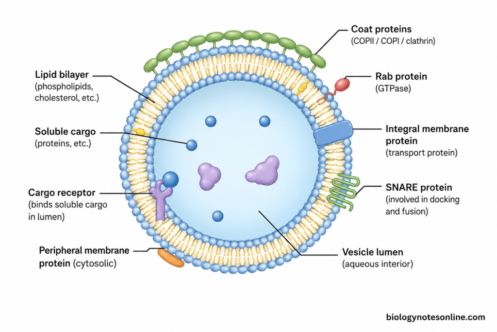
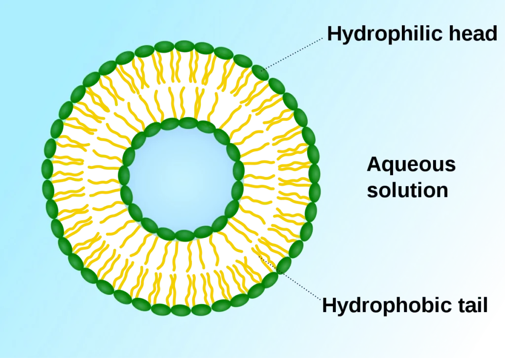
- Lipid bilayer- Vesicles are surrounded by a thin lipid bilayer. It forms the main structural boundary of the vesicle. The membrane is mainly formed of amphipathic lipids with different membrane proteins embedded within it.
- Membrane lipids- Phospholipids form a major part of the vesicular membrane. Other lipids such as cholesterol, sphingolipids and glycolipids can also occur, but their amount is different according to the membrane from which the vesicle is formed and the vesicle type.
- Vesicle lumen- The space surrounded by the vesicle membrane is called the lumen. It contains an aqueous environment which remains separated from cytosol. Soluble proteins and other substances being transported can be present in this region.
- Membrane proteins- Different proteins are present in the vesicle membrane. Some pass through the lipid bilayer, while others remain associated with its surface. Transport proteins, cargo receptors and proteins required for membrane targeting can be present depending upon the vesicle.
- Soluble cargo- Proteins and other water-soluble materials can be enclosed within the lumen of transport vesicles. During ER export, for example, soluble secretory proteins can be selected through transmembrane cargo receptors and packed into the developing carrier.
- Membrane cargo- Vesicles also transport membrane proteins and membrane lipids. These remain as a part of vesicle membrane during transport rather than being free within its lumen.
- Coat proteins- Some newly forming transport vesicles have a protein coat on their cytosolic surface. COPII, COPI and clathrin are important coat systems used in different trafficking pathways. The coat helps in cargo selection and membrane deformation during formation of the carrier.
- Cargo receptors- These are transmembrane proteins which help in selection of particular soluble cargo. The receptor binds cargo on the lumenal side and can interact with coat machinery through its cytosolic region.
- Rab proteins- Different vesicles and membrane compartments contain specific Rab GTPases on their cytosolic surface. These proteins are associated with vesicle trafficking and recruitment of different tethering and motor proteins.
- SNARE proteins- SNAREs are membrane proteins involved during docking and fusion of a vesicle with its particular target membrane. Different SNARE combinations are present in different trafficking pathways.
- Variable composition- All vesicles do not contain the same lipids, proteins or lumenal materials. Their composition depends on the site of formation and the function performed by the vesicle. Secretory vesicles, transport vesicles and endocytic vesicles therefore carry different sets of materials.
- Extracellular vesicles- Vesicles released outside the cell also have a lipid membrane and contain selected cellular components. Proteins, lipids and different forms of RNA have been detected in purified extracellular vesicles (EVs), although their composition differs between EV populations and cells of origin.
How Are Vesicles Formed?
Vesicles are generally formed by budding from a pre-existing cellular membrane. The process is not same for all vesicles. Different coat proteins and accessory proteins take part according to the site of formation and type of transport vesicle.
The major steps involved in formation of vesicles are as follows-
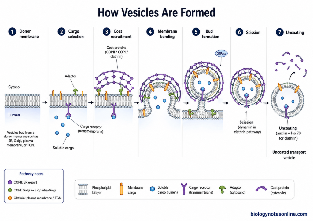
- Donor membrane- Vesicle formation begins at a particular region of the donor membrane. This may be the endoplasmic reticulum, Golgi membrane, plasma membrane or another membrane compartment.
- Cargo selection- The molecules that have to be transported are selected and concentrated at the site of vesicle formation. Membrane cargo can interact with coat machinery directly or through adaptor proteins. Soluble cargo may use transmembrane cargo receptors for its selection.
- Coat recruitment- Different coat proteins are now recruited on the cytosolic side of membrane. COPII is mainly involved in export from the ER, COPI in retrograde Golgi-ER and intra-Golgi transport, while clathrin takes part in different pathways from plasma membrane and trans-Golgi network.
- Membrane bending- Assembly of coat proteins causes deformation of the membrane. The flat membrane starts to curve out and forms a developing bud containing the selected cargo. Coat proteins have an important role both in membrane deformation and cargo collection.
- Bud formation- As more coat components are assembled, the membrane becomes increasingly curved. A vesicular bud is formed which remains connected with the donor membrane by a narrow neck. In COPII vesicle formation, recruitment of Sar1-GTP, Sec23/Sec24 and Sec13/Sec31 occurs during this process.
- Membrane scission- The narrow neck is finally separated from the donor membrane, releasing the newly formed vesicle. Proteins involved in this step are different for different pathways. During clathrin-mediated endocytosis at plasma membrane, dynamin has a major role in membrane scission.
- Uncoating- After vesicle formation, many coated vesicles lose their coat proteins. In clathrin-coated vesicles, auxilin and Hsc70 take part in removal of clathrin coat after membrane scission. The released vesicle can then proceed further in the trafficking pathway.
Types of Vesicles
Different types of cellular vesicles are present according to their origin, cargo and function. The important types are described below-
1. Transport Vesicles
Transport vesicles are membrane carriers used for movement of proteins and lipids between different cell compartments. These bud from a donor membrane and carry selected cargo towards another membrane. COPII, COPI and clathrin are the major coat systems associated with different transport pathways.
a. COPII-Coated Carriers
- COPII-coated carriers are mainly associated with export of proteins and lipids from the endoplasmic reticulum (ER). They are formed at ER exit sites.
- The COPII machinery contains Sar1, Sec23/Sec24 and Sec13/Sec31. Cargo selection and membrane deformation takes place during their assembly.
- From ER, the exported cargo is moved towards the ERGIC/cis-Golgi region. Small vesicles are formed in some cases, while larger or tubular COPII-associated carriers are also present for particular cargo.
b. COPI-Coated Carriers
- COPI-coated carriers are mainly associated with Golgi membranes. The coat is formed by the coatomer complex, with ARF1 taking part in coat recruitment.
- A major function is the return of escaped ER proteins and transport components from Golgi towards the ER. This is a retrograde pathway.
- COPI also has roles within the Golgi itself. Resident Golgi proteins and enzymes can be recycled between Golgi cisternae during their maturation.
c. Clathrin-Coated Vesicles
- Clathrin-coated vesicles have a characteristic clathrin coat on their cytosolic surface during formation.
- At the plasma membrane, they have an important role in endocytosis. Receptors together with their bound cargo can be concentrated into clathrin-coated pits and taken inside the cell.
- Clathrin-coated carriers are also formed from the trans-Golgi network (TGN). From here, selected proteins are transported towards endosomes.
- Clathrin does not select most cargo alone. Adaptor and accessory proteins connect the coat with membrane and particular cargo proteins.
2. Secretory Vesicles
Secretory vesicles are formed from the later Golgi or trans-Golgi network (TGN) and transport substances towards the plasma membrane. Proteins are first processed and sorted in Golgi before their movement into the secretory pathway.
- In constitutive secretion, carriers continuously move towards and fuse with plasma membrane. New membrane proteins and lipids are also supplied by this pathway.
- Some cells have regulated secretory vesicles. Their contents remain stored until a particular signal causes secretion.
- Hormones are released in this manner from many endocrine cells. Digestive enzymes from pancreatic acinar cells are another example of regulated secretion.
- During secretion, the vesicle membrane fuses with plasma membrane and the soluble contents are released outside. This process is called exocytosis.
3. Synaptic Vesicles
Synaptic vesicles are small secretory vesicles found in the presynaptic terminals of neurons. Neurotransmitters are stored within them before their release.
- They are concentrated near specialized regions of presynaptic membrane called active zones.
- When an action potential reaches the nerve terminal, voltage-gated calcium channels open. Ca²⁺ enters the terminal.
- Increase in Ca²⁺ causes rapid fusion of primed synaptic vesicles with the presynaptic membrane. The neurotransmitter is then released into the synaptic cleft.
- Synaptic vesicle membrane is not simply lost after exocytosis. It is recovered by endocytosis and vesicle components are recycled for further rounds of neurotransmitter release.
4. Endocytic Vesicles
Endocytic vesicles are formed from the plasma membrane during uptake of substances into the cell. A region of membrane bends towards inside and separates to form an intracellular carrier.
- In receptor-mediated endocytosis, particular extracellular molecules bind to their cell-surface receptors. These receptor-cargo complexes are then concentrated for internalization.
- Clathrin-mediated endocytosis is an important form of receptor-mediated uptake. Clathrin-coated pits develop at plasma membrane and later separate as coated vesicles.
- After formation, endocytic cargo is delivered towards the endosomal system. Sorting can then occur for recycling towards cell surface or further transport towards late endosomes and lysosomes.
5. Extracellular Vesicles
Extracellular vesicles (EVs) are membrane-enclosed particles released from cells. They are surrounded by a lipid bilayer and cannot replicate on their own, according to the definition retained in MISEV2023.
- EVs can contain different cellular materials. Proteins, lipids and nucleic acids are among the cargo found in different EV populations, and the composition varies with their cell of origin and formation pathway.
- These vesicles can interact with other cells and have roles in intercellular communication. Their cargo may influence functions of recipient cells.
- MISEV2023 recommends use of the general term extracellular vesicle when the exact biogenesis is not demonstrated. Terms such as exosome and ectosome/microvesicle describe proposed routes of formation and should not be applied only from particle size.
- Exosome specifically indicates an EV arising through the endosomal or multivesicular body pathway. Small EV and exosome are therefore not the same term.
COPI vs COPII vs Clathrin-Coated Vesicles
| Feature | COPI Vesicles | COPII Vesicles | Clathrin-Coated Vesicles |
|---|---|---|---|
| Main coat | Coat is formed by COPI coatomer complex. | Coat is formed mainly by Sec23/Sec24 and Sec13/Sec31. | Coat is mainly formed by clathrin together with adaptor proteins. |
| Small GTPase | ARF1 is mainly involved in coat formation. | Sar1 starts assembly of the COPII coat. | ARF1 or other adaptor-associated mechanisms are involved depending on the membrane site. |
| Main origin | Mostly formed from Golgi membranes. | Mainly formed from endoplasmic reticulum (ER). | Formed from plasma membrane and trans-Golgi network (TGN). |
| Main direction | Mainly Golgi-to-ER and intra-Golgi transport. | Mainly ER-to-Golgi transport. | Plasma membrane-to-endosome and TGN-to-endosomal transport. |
| Transport type | Mostly retrograde transport. | Mainly anterograde transport. | Mainly involved in endocytosis and selected post-Golgi transport. |
| Cargo | Returns ER proteins, transport machinery and other membrane components. | Carries newly synthesized proteins and lipids from ER. | Carries receptors, ligands and selected proteins from plasma membrane or TGN. |
| Cargo selection | Coatomer and cargo signals take part in selection. | Sec24 has an important role in cargo selection. | Adaptor proteins help in binding particular cargo with clathrin coat. |
| Budding site | Golgi and related membranes. | ER exit sites (ERES). | Coated pits of plasma membrane and regions of TGN. |
| Scission | Vesicle or carrier separates after coat assembly and membrane deformation. | Carrier is formed after assembly of COPII machinery. | At plasma membrane, dynamin commonly helps in final membrane scission. |
| Uncoating | COPI coat is removed after carrier formation. | COPII components are released during further transport. | Clathrin coat is removed after vesicle formation. |
| Major function | Retrieval and recycling towards ER. | Export of secretory and membrane proteins from ER. | Uptake of extracellular materials and sorting from TGN. |
How Does Vesicular Transport Work?
Vesicular transport is the process of movement of proteins, lipids and other materials between membrane compartments of the cell. It takes place by formation of transport vesicles from one membrane and their delivery towards another membrane. The major steps are as follows-
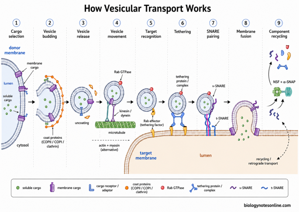
- Cargo selection- In the first step, the proteins and lipids which have to be transported are selected at the donor membrane. Some cargo proteins bind directly with the coat machinery, while others require cargo receptors or adaptor proteins.
- Vesicle budding- Coat proteins are assembled on the cytosolic surface of membrane. The membrane now starts to bend and forms a bud. COPII, COPI and clathrin are some important coat systems used in different transport pathways.
- Vesicle release- The developing bud is separated from the donor membrane and a free transport vesicle is formed. This separation of the membrane is referred to as membrane scission. Many coated vesicles then lose their coat before the later stages of transport.
- Vesicle movement- The vesicle is transported towards its required cellular region. Many vesicles and membrane organelles move along microtubules with the help of motor proteins such as kinesin and dynein. Actin filaments and myosin motors are also used for some vesicular movements.
- Target recognition- A vesicle does not normally fuse with any membrane present near it. Rab GTPases and their effector proteins take part in recognizing the appropriate target membrane. Rab proteins also recruit different tethering factors required during this process.
- Tethering- The vesicle is first captured near the target membrane by tethering proteins or tethering complexes. This brings the two membranes close to each other. Rab proteins have an important role in this step.
- SNARE pairing- SNARE proteins present on the vesicle and target membrane now interact with each other. Their pairing pulls the vesicle membrane and target membrane into close contact. Different trafficking pathways use particular sets of SNARE proteins.
- Membrane fusion- Finally, the lipid bilayers of vesicle and target membrane fuse. The lumen of vesicle becomes continuous with the lumen of the target compartment, or with extracellular region when fusion occurs with plasma membrane. The vesicle cargo is then delivered to its destination.
- Component recycling- After fusion, many proteins used during vesicular transport are recycled for further use. NSF and α-SNAP, for example, disassemble the formed SNARE complex so that SNARE proteins can be reused. Membrane and resident proteins can also undergo retrograde transport back towards earlier compartments.
Major Pathways of Vesicle-Mediated Transport
The major pathways of vesicle-mediated transport are as follows-
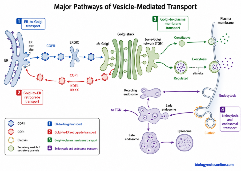
ER-to-Golgi Transport
- Newly synthesized proteins which have passed the quality control of endoplasmic reticulum (ER) are moved towards specific regions called ER exit sites (ERES). Proteins and lipids that have to be transported are collected in these regions.
- COPII coat proteins are mainly involved during transport from ER towards Golgi. Sar1 is recruited first, followed by Sec23/Sec24 and Sec13/Sec31. These proteins take part in cargo selection and formation of ER-derived transport carriers.
- The membrane starts to curve during assembly of COPII proteins. Cargo is now packed within the developing carrier, which separates from the ER membrane.
- The ER-derived cargo then moves towards the cis-Golgi. In many animal cells, these materials first enter the ER-Golgi intermediate compartment (ERGIC) before reaching Golgi.
- Transport does not always occur through small spherical vesicles. Tubular and larger transport carriers can also be formed, especially for transport of large cargo molecules.
Golgi-to-ER Retrograde Transport
- Some proteins and membrane components that reach Golgi have to be transported again towards the ER. This backward movement is called retrograde transport.
- COPI-coated carriers are mainly involved in transport from Golgi towards ER. COPI also takes part in transport between different regions of Golgi.
- Some soluble proteins normally present in ER may escape and enter Golgi. Many of these proteins contain a KDEL sequence. KDEL receptors recognize these proteins in Golgi and help in their transport back to ER.
- Resident ER membrane proteins can also contain retrieval signals, such as KKXX sequences. These signals help their interaction with COPI transport machinery.
- Transport receptors, SNARE proteins and some other components are also returned towards ER. In this way, components used during forward transport can be used again.
Golgi-to-Plasma Membrane Transport
- Proteins and lipids reaching the later part of Golgi are sorted at the trans-Golgi network (TGN). From this region, different transport carriers are formed.
- Some transport carriers continuously move towards the plasma membrane. This is referred to as the constitutive secretory pathway. It supplies proteins and lipids to plasma membrane and also releases soluble proteins outside the cell.
- In some specialized cells, secretory products are first stored within secretory vesicles or granules. They are not immediately released.
- Release of these stored materials occurs after the cell receives a particular signal. This type is called the regulated secretory pathway.
- The transport vesicle finally fuses with the plasma membrane. Vesicle membrane becomes a part of plasma membrane while soluble contents present inside are released outside the cell by exocytosis.
Endocytosis and Endosomal Transport
- During endocytosis, materials are taken from the cell surface towards inside of the cell. A portion of plasma membrane bends inward and forms an endocytic vesicle.
- Clathrin-mediated endocytosis is one of the important pathways of this process. Cells also contain several clathrin-independent endocytic pathways.
- Newly internalized materials are generally transported towards the early endosome. It is an important sorting compartment of the endocytic pathway.
- Some receptors and membrane proteins are returned back to plasma membrane. This may occur directly from early endosomes or through recycling endosomes.
- Other materials are transported further towards late endosomes. Cargo which has to be degraded is finally delivered to lysosomal compartments, where it is broken down by lysosomal enzymes.
- Some proteins present in endosomes can also be transported towards the trans-Golgi network instead of plasma membrane or lysosome. This forms another retrograde transport pathway within the cell.
Are Vesicles Present in All Cells?
Vesicles are not present in same form in all cells. They are commonly found in eukaryotic cells. Animal, plant and fungal cells contain different membrane-bound vesicles, which take part in transport and storage of cellular materials. The number and type of them are also different.
In eukaryotic cells, vesicles are mostly associated with the endomembrane system. These move between endoplasmic reticulum, Golgi apparatus, endosomes, lysosomes or vacuoles and plasma membrane. Some cells contain many vesicles. Secretory cells are one of them.
However, every specialized cell does not retain the complete vesicular system. Mature mammalian red blood cells (RBCs) lose nucleus and most membrane-bound organelles during maturation. The typical ER-Golgi transport system is therefore absent. Still, small membrane vesicles can be released from surface of these cells.
Prokaryotic cells are different. They do not contain the typical ER-Golgi endomembrane system, hence classical intracellular transport vesicles are absent in them. Bacteria and archaea, however, can form membrane vesicles and release them outside the cell. These are extracellular membrane vesicles.
Functions of Vesicles
The following are some of the important functions of cellular vesicles–
- Intracellular transport- Proteins, lipids and other materials are carried from one cell compartment to another.
- Secretion- Secretory vesicles carry hormones, enzymes and proteins. These fuse with plasma membrane and release their content outside the cell.
- Endocytosis- In this process, substances from outside are taken into the cell by formation of vesicles.
- Storage- Some vesicles act as storage sacs. Different substances remain stored inside until they are required by the cell.
- Neurotransmitter release- Synaptic vesicles contain neurotransmitters. On stimulation, these are released at the synapse.
- Membrane supply- New lipids and membrane proteins are carried to the plasma membrane. This helps in maintenance of cell membrane.
- Recycling- Internalized receptors and some membrane proteins are returned again to the cell surface.
- Degradation- Unwanted materials can be carried towards lysosomes. Here, these materials are broken down.
- Cargo sorting- Different cellular materials are sorted and sent towards their required location.
- Retrograde transport- Some proteins and membrane components move back towards the earlier cell compartments.
- Cell communication- Extracellular vesicles (EVs) carry proteins, lipids and RNA between cells. They have a role in intercellular signaling.
Examples of Vesicles in Different Cells
Some examples of vesicles found in different cells are as follows-
- Neurons- In neurons, synaptic vesicles are present at the presynaptic terminal. Neurotransmitters are stored in these vesicles and released during nerve stimulation.
- Pancreatic β-cells- Insulin is stored in secretory granules of pancreatic β-cells. When blood glucose increases, the granules fuse with cell membrane. Insulin is released.
- Pancreatic acinar cells- These cells possess a large number of zymogen granules, mainly towards the apical side. They contain digestive enzymes and inactive enzyme precursors before secretion into pancreatic ducts.
- Mast cells- Numerous granules are present in the cytoplasm of mast cells. Histamine, heparin, proteases and other inflammatory substances are stored in them. On activation of mast cell, these contents are released.
- Platelets- Two major granules are found in platelets, α-granules and dense granules. Different proteins are present in α-granules. Dense granules mainly contain ADP, ATP, calcium and serotonin.
- Macrophages- During phagocytosis, a microorganism or large particle is surrounded by the cell membrane and enclosed inside a phagosome. It later joins with lysosomal compartments where the material is degraded.
- Goblet cells- Secretory granules filled with mucins are found in goblet cells. After their release, mucins become hydrated and form mucus over epithelial surface.
- Red blood cells- Mature red blood cells also release membrane-derived extracellular vesicles (EVs). Selected membrane proteins, lipids and some cytoplasmic materials can be present in these vesicles.
Biological Importance of Vesicles
Some of the important biological roles of vesicles are-
- Cell organization- Vesicular transport keeps different membrane compartments connected. Proteins and lipids can reach their particular cellular location by this system.
- Protein transport- Newly formed secretory and membrane proteins are carried from ER to Golgi and then towards other regions of cell. Defect in this transport can cause improper localization and loss of proteins.
- Secretion- Secretory vesicles move cellular products towards plasma membrane. Fusion releases their contents outside the cell. This process is important for normal exocytosis.
- Cellular uptake- By endocytic vesicles, receptors and other materials are taken from cell surface. Some receptors are again recycled back instead of degradation.
- Neural activity- Synaptic vesicles are necessary for neurotransmitter release at synapses. Release occurs from these vesicles in response to calcium during nerve signaling.
- Membrane recycling- Cell-surface receptors do not always remain permanently internalized after endocytosis. Endosomal transport returns many of them again to plasma membrane and also affects their signaling.
- Cellular degradation- Endosomes and autophagic vesicles carry materials towards lysosomes. Damaged or unwanted cellular materials can be degraded through these pathways.
- Cell communication- Extracellular vesicles (EVs) can transfer cellular materials between cells. Transfer of mRNA and microRNA through extracellular vesicles has also been demonstrated.
Intracellular vs Extracellular Vesicles
| Feature | Intracellular Vesicles | Extracellular Vesicles |
|---|---|---|
| Definition | Membrane-bound vesicles present within the cell. | Membrane-enclosed vesicles released outside the cell. |
| Location | Present in the cytoplasm and associated with different membrane compartments. | Found in extracellular fluid, blood and other body fluids. |
| Formation | Commonly formed by budding from ER, Golgi, endosomes or plasma membrane. | Formed by outward budding of plasma membrane or released from multivesicular bodies. |
| Main types | Transport vesicles, secretory vesicles, synaptic vesicles and endocytic vesicles. | Exosomes, microvesicles and other extracellular vesicle populations. |
| Membrane | Surrounded by a lipid bilayer. | Also enclosed by a lipid bilayer. |
| Cargo | Proteins, lipids, enzymes and other cellular materials are carried. | Proteins, lipids, RNA and other selected cellular components may be present. |
| Major role | Mainly involved in transport, storage, secretion and uptake inside the cell. | Mainly involved in transfer of materials between cells and cell communication. |
| Transport pathway | Commonly works as a part of the endomembrane system. | Released from one cell and may reach nearby or distant cells. |
| Examples | COPI vesicles, COPII vesicles, clathrin-coated vesicles, synaptic vesicles. | Exosomes and microvesicles are common examples. |
| Occurrence | Common in eukaryotic cells with active membrane trafficking. | Released by many different cell types, including animal cells and some microorganisms. |
Vesicle vs Vacuole vs Lysosome vs Endosome
| Feature | Vesicle | Vacuole | Lysosome | Endosome |
|---|---|---|---|---|
| Definition | Small membrane-bound sac used mainly for transport or storage of cellular materials. | Large membrane-bound compartment mainly used for storage, ion balance and degradation. | Membrane-bound organelle containing hydrolytic enzymes for digestion of cellular materials. | Membrane compartment involved in sorting of materials taken up by endocytosis. |
| Size | Usually small. | Generally larger than common transport vesicles. | Small to medium-sized organelle. | Variable in size and shape. |
| Membrane | Surrounded by a lipid bilayer. | Surrounded by a membrane called tonoplast in plant cells. | Single lipid bilayer membrane. | Single lipid bilayer membrane. |
| Main contents | Proteins, lipids, enzymes, neurotransmitters or other cargo. | Water, ions, metabolites, pigments and waste materials. | Acid hydrolases and materials undergoing degradation. | Internalized receptors, membrane proteins and extracellular cargo. |
| Main function | Transport, secretion, uptake and temporary storage. | Storage, maintenance of turgor and degradation of materials. | Intracellular digestion and recycling. | Sorting, recycling and delivery of endocytosed materials. |
| Formation | Commonly formed by budding from cellular membranes. | Formed through fusion and maturation of membrane compartments. | Develops through the endosomal-lysosomal pathway and receives lysosomal enzymes from Golgi. | Formed mainly from endocytic vesicles after uptake from plasma membrane. |
| pH | Depends on vesicle type. | Often acidic, particularly plant vacuoles. | Strongly acidic, usually around pH 4.5–5.0. | Becomes progressively acidic from early to late endosome. |
| Occurrence | Present in many eukaryotic cells. | Especially prominent in plant and fungal cells. | Characteristic of animal cells, with similar lytic compartments present in other eukaryotes. | Present in eukaryotic cells carrying out endocytosis. |
| Examples | COPI, COPII, clathrin-coated vesicles, synaptic vesicles. | Central vacuole, food vacuole, contractile vacuole. | Primary and degradative lysosomal compartments. | Early endosome, recycling endosome, late endosome. |
Frequently Asked Questions (FAQs)
1. What are vesicles in a cell?
Vesicles are small membrane-bound sacs present inside the cell. They contain an internal space or lumen and are mainly used for transport, storage and secretion of cellular materials.
2. What is the main function of vesicles?
The major function is transport of proteins, lipids and other materials. Some vesicles also store substances, release them outside the cell or take materials into the cell.
3. What are vesicles made of?
A vesicle is surrounded by a lipid bilayer. Different membrane proteins are present within it, while proteins, enzymes and other cargo may occur inside the lumen.
4. Are vesicles membrane-bound organelles?
Vesicles are membrane-bound cellular compartments. They are generally much smaller and more temporary than large membrane organelles such as the Golgi apparatus or lysosome.
5. Where are vesicles found in cells?
They are found throughout the cytoplasm of eukaryotic cells. Large number of vesicles can be present around the ER, Golgi apparatus, endosomes and plasma membrane.
6. What do vesicles contain?
The contents are different according to vesicle type. Proteins, enzymes, neurotransmitters, hormones and other soluble substances can occur inside, while lipids and membrane proteins are present in the membrane.
7. How are vesicles formed?
Most intracellular transport vesicles are formed by budding from a pre-existing membrane. Cargo is selected, membrane bends and the developing carrier finally separates from donor membrane.
8. What are the major types of vesicles?
Some major types are transport vesicles, secretory vesicles, synaptic vesicles, endocytic vesicles and extracellular vesicles. COPI, COPII and clathrin-coated carriers are important transport types.
9. What are transport vesicles?
These are membrane carriers that move proteins and lipids between different cell compartments. They are an important part of the endomembrane system.
10. What is vesicular transport?
Vesicular transport is the movement of cellular cargo from one membrane compartment to another through membrane-bound carriers. It includes budding, movement, docking and membrane fusion.
11. How do vesicles move through the cell?
Some short movements can occur by diffusion. For directed movement, vesicles can move along microtubules with kinesin or dynein motors. Actin and myosin are also used in some cells.
12. How do vesicles know which membrane to fuse with?
Targeting is controlled by several proteins. Rab GTPases, tethering factors and particular SNARE proteins help a vesicle recognize and fuse with the required membrane.
13. What are SNARE proteins?
SNAREs are membrane proteins involved in vesicle fusion. SNAREs from opposing membranes pair together and bring both membranes close enough for fusion.
14. What is the difference between COPI and COPII vesicles?
COPII mainly carries cargo from ER towards Golgi. COPI, on the other hand, is mainly involved in Golgi-to-ER retrieval and recycling within the Golgi system.
15. What is the difference between a vesicle and a vacuole?
A vesicle is generally small and commonly involved in transport or temporary storage. Vacuoles are usually larger compartments, especially in plant and fungal cells, where they perform storage, ion balance and degradative functions.
16. Are vesicles present in prokaryotic cells?
Prokaryotes do not possess the classical ER-Golgi vesicular transport system of eukaryotic cells. However, bacteria and archaea can form and release membrane vesicles, mainly as extracellular membrane vesicles.
References
- Alberts, B., Johnson, A., Lewis, J., Raff, M., Roberts, K., & Walter, P. (2002). Molecular biology of the cell (4th ed.). Garland Science. https://www.ncbi.nlm.nih.gov/books/NBK21054/
- Barr, F. A. (2013). Rab GTPases and membrane identity: Causal or inconsequential? Journal of Cell Biology, 202(2), 191–199. https://doi.org/10.1083/jcb.201306010
- Bas, L., Papinski, D., Licheva, M., Torggler, R., Rohringer, S., Schuschnig, M., & Kraft, C. (2018). Reconstitution reveals Ykt6 as the autophagosomal SNARE in autophagosome–vacuole fusion. Journal of Cell Biology, 217(10), 3656–3669. https://doi.org/10.1083/jcb.201804028
- Borchers, A.-C., Langemeyer, L., & Ungermann, C. (2021). Who’s in control? Principles of Rab GTPase activation in endolysosomal membrane trafficking and beyond. Journal of Cell Biology, 220(9), e202105120. https://doi.org/10.1083/jcb.202105120
- Bowman, S. L., Shiwarski, D. J., & Puthenveedu, M. A. (2016). Distinct G protein-coupled receptor recycling pathways allow spatial control of downstream G protein signaling. Journal of Cell Biology, 214(7), 797–806. https://doi.org/10.1083/jcb.201512068
- Chen, Z., & Schmid, S. L. (2020). Evolving models for assembling and shaping clathrin-coated pits. Journal of Cell Biology, 219(9), e202005126. https://doi.org/10.1083/jcb.202005126
- Cooper, G. M. (2000). The cell: A molecular approach (2nd ed.). Sinauer Associates. https://www.ncbi.nlm.nih.gov/books/NBK9839/
- Dao, D.-P. D., & Le, P. H. (2023). Histology, goblet cells. In StatPearls. StatPearls Publishing. https://www.ncbi.nlm.nih.gov/books/NBK553208/
- Deshpande, O. A., & Wadhwa, R. (2023). Phagocytosis. In StatPearls. StatPearls Publishing. https://www.ncbi.nlm.nih.gov/books/NBK556043/
- Donovan, K. W., & Bretscher, A. (2015). Tracking individual secretory vesicles during exocytosis reveals an ordered and regulated process. Journal of Cell Biology, 210(2), 181–189. https://doi.org/10.1083/jcb.201501118
- Ganley, I. G., Espinosa, E., & Pfeffer, S. R. (2008). A syntaxin 10-SNARE complex distinguishes two distinct transport routes from endosomes to the trans-Golgi in human cells. Journal of Cell Biology, 180(1), 159–172. https://doi.org/10.1083/jcb.200707136
- Gill, S., Catchpole, R., & Forterre, P. (2019). Extracellular membrane vesicles in the three domains of life and beyond. FEMS Microbiology Reviews, 43(3), 273–303. https://doi.org/10.1093/femsre/fuy042
- Gomez-Navarro, N., & Miller, E. A. (2016). Protein sorting at the ER–Golgi interface. Journal of Cell Biology, 215(6), 769–778. https://doi.org/10.1083/jcb.201610031
- He, K., Song, E., Upadhyayula, S., Dang, S., Gaudin, R., Skillern, W., Bu, K., Capraro, B. R., Rapoport, I., Kusters, I., Ma, M., & Kirchhausen, T. (2020). Dynamics of Auxilin 1 and GAK in clathrin-mediated traffic. Journal of Cell Biology, 219(3), e201908142. https://doi.org/10.1083/jcb.201908142
- Heidarzadeh-Asl, S., Maurer, M., Kiani, A., Atiakshin, D., Stahl Skov, P., & Elieh-Ali-Komi, D. (2025). Novel insights on the biology and immunologic effects of histamine: A road map for allergists and mast cell biologists. Journal of Allergy and Clinical Immunology, 155(4), 1095–1114. https://doi.org/10.1016/j.jaci.2024.12.1081
- Hua, Z., & Graham, T. R. (2009). The Golgi apparatus. In N. Segev (Ed.), Trafficking inside cells: Pathways, mechanisms and regulation (pp. 42–66). Landes Bioscience/Springer Science+Business Media. https://www.ncbi.nlm.nih.gov/books/NBK6268/
- Ibrahim, S. A., & Khan, Y. S. (2023). Histology, extracellular vesicles. In StatPearls. StatPearls Publishing. https://www.ncbi.nlm.nih.gov/books/NBK562256/
- Jeppesen, D. K., Fenix, A. M., Franklin, J. L., Higginbotham, J. N., Zhang, Q., Zimmerman, L. J., Liebler, D. C., Ping, J., Liu, Q., Evans, R., Fissell, W. H., Patton, J. G., Rome, L. H., Burnette, D. T., & Coffey, R. J. (2019). Reassessment of exosome composition. Cell, 177(2), 428–445.e18. https://doi.org/10.1016/j.cell.2019.02.029
- Kasprowicz, J., Kuenen, S., Swerts, J., Miskiewicz, K., & Verstreken, P. (2014). Dynamin photoinactivation blocks clathrin and α-adaptin recruitment and induces bulk membrane retrieval. Journal of Cell Biology, 204(7), 1141–1156. https://doi.org/10.1083/jcb.201310090
- Krahn, A. H., Pantazopoulou, A., Austin, J., II, Johnson, N., Lee-Smith, C. F., & Glick, B. S. (2026). COPI-dependent intra-Golgi recycling at an intermediate stage of cisternal maturation. Journal of Cell Biology, 225(6), e202509174. https://doi.org/10.1083/jcb.202509174
- Lin, L., Zhou, Y., & Hu, K. (2023). Cell–cell communication and extracellular vesicles in cancer. Cancers, 15(9), 2419. https://doi.org/10.3390/cancers15092419
- Liu, S., Wei, J., Zhong, L., Hai, S., Song, S., Xie, C., Huang, Z., Cheng, Z., Zhang, J., Du, A., Zhang, P., Yan, Y., & Shi, A. (2025). RAB-10 cooperates with EHBP-1 to capture vesicular carriers during post-Golgi exocytic trafficking. Journal of Cell Biology, 224(4), e202410003. https://doi.org/10.1083/jcb.202410003
- Lőrincz, P., Tóth, S., Benkő, P., Lakatos, Z., Boda, A., Glatz, G., Zobel, M., Bisi, S., Hegedűs, K., Takáts, S., Scita, G., & Juhász, G. (2017). Rab2 promotes autophagic and endocytic lysosomal degradation. Journal of Cell Biology, 216(7), 1937–1947. https://doi.org/10.1083/jcb.201611027
- Lund, V. K., Chirco, A., Caliari, M., Larsen, A. H., Tang, K. T., Gether, U., Madsen, K. L., Wierer, M., & Kjaerulff, O. (2026). A Syd and RUFY dynein adaptor complex mediates axonal circulation of dense core vesicles. Journal of Cell Biology, 225(3), e202507071. https://doi.org/10.1083/jcb.202507071
- Ma, S.-R., Xia, H.-F., Gong, P., & Yu, Z.-L. (2023). Red blood cell-derived extracellular vesicles: An overview of current research progress, challenges, and opportunities. Biomedicines, 11(10), 2798. https://doi.org/10.3390/biomedicines11102798
- Maldutyte, J., Li, X.-H., Gomez-Navarro, N., Robertson, E. G., & Miller, E. A. (2025). ER export via SURF4 uses diverse mechanisms of both client and coat engagement. Journal of Cell Biology, 224(1), e202406103. https://doi.org/10.1083/jcb.202406103
- Malhotra, V. (2024). Tailored assemblies of COPII proteins in secretion. Journal of Cell Biology, 223(8), e202404013. https://doi.org/10.1083/jcb.202404013
- Meléndez, A., & Levine, B. (2009). Autophagy in C. elegans. WormBook. https://www.ncbi.nlm.nih.gov/books/NBK116074/
- Norman, C. A., Krishnakumar, S. S., Timofeeva, Y., & Volynski, K. E. (2023). The release of inhibition model reproduces kinetics and plasticity of neurotransmitter release in central synapses. Communications Biology, 6, 1091. https://doi.org/10.1038/s42003-023-05445-2
- Pereira, C., Stalder, D., Anderson, G. S. F., Shun-Shion, A. S., Houghton, J., Antrobus, R., Chapman, M. A., Fazakerley, D. J., & Gershlick, D. C. (2023). The exocyst complex is an essential component of the mammalian constitutive secretory pathway. Journal of Cell Biology, 222(5), e202205137. https://doi.org/10.1083/jcb.202205137
- Purves, D., Augustine, G. J., Fitzpatrick, D., Katz, L. C., LaMantia, A.-S., McNamara, J. O., & Williams, S. M. (Eds.). (2001). Neuroscience (2nd ed.). Sinauer Associates. https://www.ncbi.nlm.nih.gov/books/NBK10799/
- Risinger, M., & Kalfa, T. A. (2020). Red cell membrane disorders: Structure meets function. Blood, 136(11), 1250–1261. https://doi.org/10.1182/blood.2019000946
- Sharda, A., & Flaumenhaft, R. (2018). The life cycle of platelet granules. F1000Research, 7, 236. https://doi.org/10.12688/f1000research.13283.1
- Short, B. (2015). Sorting out endosome form and function. Journal of Cell Biology, 210(6), 870. https://doi.org/10.1083/jcb.2106fta
- Shomron, O., Nevo-Yassaf, I., Aviad, T., Yaffe, Y., Zahavi, E. E., Dukhovny, A., Perlson, E., Brodsky, I., Yeheskel, A., Pasmanik-Chor, M., Mironov, A., Beznoussenko, G. V., Mironov, A. A., Sklan, E. H., Patterson, G. H., Yonemura, Y., Sannai, M., Kaether, C., & Hirschberg, K. (2021). COPII collar defines the boundary between ER and ER exit site and does not coat cargo containers. Journal of Cell Biology, 220(6), e201907224. https://doi.org/10.1083/jcb.201907224
- Sittewelle, M., & Royle, S. J. (2024). Passive diffusion accounts for the majority of intracellular nanovesicle transport. Life Science Alliance, 7(1), e202302406. https://doi.org/10.26508/lsa.202302406
- Stancheva, V. G., Li, X.-H., Hutchings, J., Gomez-Navarro, N., Santhanam, B., Babu, M. M., Zanetti, G., & Miller, E. A. (2020). Combinatorial multivalent interactions drive cooperative assembly of the COPII coat. Journal of Cell Biology, 219(11), e202007135. https://doi.org/10.1083/jcb.202007135
- Südhof, T. C. (1999). Characteristics of synaptic vesicle exocytosis. In G. J. Siegel, B. W. Agranoff, R. W. Albers, S. K. Fisher, & M. D. Uhler (Eds.), Basic neurochemistry: Molecular, cellular and medical aspects (6th ed.). Lippincott-Raven. https://www.ncbi.nlm.nih.gov/books/NBK27986/
- Thomas, V., Mase, H., Michon, L., Picco, A., Kaksonen, M., & Martin, S. G. (2026). High-resolution mapping of the actin fusion focus reveals myosin V-dependent formin transport for aster formation. Journal of Cell Biology, 225(2), e202510018. https://doi.org/10.1083/jcb.202510018
- Tokarev, A. A., Alfonso, A., & Segev, N. (2009). Overview of intracellular compartments and trafficking pathways. In N. Segev (Ed.), Trafficking inside cells: Pathways, mechanisms and regulation. Landes Bioscience/Springer Science+Business Media. https://www.ncbi.nlm.nih.gov/books/NBK7286/
- Valadi, H., Ekström, K., Bossios, A., Sjöstrand, M., Lee, J. J., & Lötvall, J. O. (2007). Exosome-mediated transfer of mRNAs and microRNAs is a novel mechanism of genetic exchange between cells. Nature Cell Biology, 9, 654–659. https://doi.org/10.1038/ncb1596
- Weimer, C., Beck, R., Eckert, P., Reckmann, I., Moelleken, J., Brügger, B., & Wieland, F. T. (2008). Differential roles of ArfGAP1, ArfGAP2, and ArfGAP3 in COPI trafficking. Journal of Cell Biology, 183(4), 725–735. https://doi.org/10.1083/jcb.200806140
- Weiss, M. A., Steiner, D. F., & Philipson, L. H. (2014). Insulin biosynthesis, secretion, structure, and structure-activity relationships. In Endotext. MDText.com, Inc. https://www.ncbi.nlm.nih.gov/books/NBK279029/
- Welsh, J. A., Goberdhan, D. C. I., O’Driscoll, L., Buzas, E. I., Blenkiron, C., Bussolati, B., Cai, H., Di Vizio, D., Driedonks, T. A. P., Erdbrügger, U., Falcon-Perez, J. M., Fu, Q.-L., Hill, A. F., Lenassi, M., Lim, S. K., Lotvall, J., Nieuwland, R., Ochiya, T., Pandey, A., … Witwer, K. W. (2024). Minimal information for studies of extracellular vesicles (MISEV2023): From basic to advanced approaches. Journal of Extracellular Vesicles, 13(2), e12404. https://doi.org/10.1002/jev2.12404