

What Is Epithelial Tissue?
Epithelial tissue is one of the four basic types of animal tissues which is made up of closely packed cells, arranged as sheets over the external surface of body and lining different internal parts. The other tissues are connective, muscular and nervous tissue.
It is also called epithelium. The term epithelial is generally used for the cells, tissues or structures which belong to the epithelium. Thus, epithelium and epithelial tissue refer to the same type of tissue.
The main role of this tissue is covering and lining. It covers the exposed surfaces and lines the cavities, hollow organs and different passageways present inside the body. The epidermis of skin is formed by epithelium. In the gastrointestinal tract, epithelial cells form the lining through which protection, secretion and absorption takes place, while different types of epithelium are also present throughout much of the respiratory tract.
Epithelial cells also give rise to much of the glandular tissue of the body. During formation of many glands, cells of the epithelium grow down into the underlying connective tissue. These cells become specialized for secretion. In exocrine glands, the connection with surface is generally retained as a duct, while endocrine glands lose this connection during development.
Where Is Epithelial Tissue Found?
Epithelial tissue is widely distributed throughout the body. It covers the external body surface and forms the lining of many internal organs, cavities, tubes and passageways. It is also present as the cellular part of many glands. Thus, epithelial tissue forms an important boundary between different body compartments and between the body and external environment.
Some of the major locations of epithelial tissue are-
- Skin – In the skin, epithelial tissue forms the epidermis, which is the outer covering of the body. It is made up mainly of keratinized stratified squamous epithelium. The dermis below it is connective tissue, so the complete skin is not epithelial in nature.
- Digestive tract – Epithelium lines almost the entire digestive passage. The oral cavity and esophagus contain mainly stratified squamous epithelium for protection, whereas the stomach and intestines are lined mainly by columnar epithelial cells. In the intestine, these cells are especially involved in absorption and secretion.
- Respiratory tract – Different epithelial tissues occur along the respiratory passages. Much of the conducting respiratory tract, including the trachea and larger airways, contains ciliated epithelium with mucus-secreting cells. The alveoli are different. Their thin epithelial lining provides the surface where gaseous exchange takes place.
- Cardiovascular system – The inner surface of blood vessels and capillaries is lined by a thin simple squamous epithelium called endothelium. It forms a continuous interface between circulating blood and the tissues around the vessels.
- Urinary system – Epithelial tissue occurs throughout different parts of the urinary system, but the same type is not present everywhere. Kidney tubules contain mainly cuboidal epithelium and thin epithelial cells are associated with the renal corpuscle. The ureters and urinary bladder are lined by specialized transitional epithelium or urothelium, which can change its appearance during stretching.
- Reproductive system – Different regions of reproductive organs also possess specialized epithelial linings. In females, the uterine tubes contain ciliated columnar epithelium, the uterine cavity is lined by columnar epithelium and the vagina has nonkeratinized stratified squamous epithelium. Thus, epithelial structure changes considerably from one reproductive organ to another.
- Glands – Much of the glandular tissue of the body is epithelial. Epithelial cells form secretory portions of many glands and also form the ducts of exocrine glands. Sweat glands, salivary glands and different glands associated with the digestive system are examples.
- Serous membranes – A special simple squamous epithelium called mesothelium covers serous surfaces. It is present in the pleura, pericardium and peritoneum, where it lines body cavities and covers many organs present within them. The smooth mesothelial surface helps the organs move with less friction.
Characteristics of Epithelial Tissue
The following are some of the important characteristics of epithelial tissue–
- Closely packed cells – Epithelial tissue is made up of cells that remain very closely arranged with one another. Only a very small amount of extracellular material is present between the cells. Thus, the cells form continuous sheets or layers.
- Cell polarity – A characteristic feature of epithelial cells is the presence of polarity. The cell has an apical surface, lateral surface and basal surface, which are structurally and functionally different. Apical surface faces the outside of body or a lumen, while the basal region is directed towards the underlying tissue.
- Presence of cell junctions – The neighbouring epithelial cells are joined with each other by specialized cell junctions. These include tight junctions, adherens junctions, desmosomes and gap junctions. They maintain attachment between the cells and are also involved in barrier and communication functions.
- Basement membrane – At the basal surface, epithelium is attached to a thin extracellular layer called the basement membrane. It separates and anchors the epithelial tissue to the underlying connective tissue. Basement membranes can also take part in support, filtration and regulation of cell behaviour depending upon the tissue.
- Generally avascular – Blood vessels are generally absent within the epithelial layer. Oxygen and nutrients required by epithelial cells are supplied mainly by diffusion from blood vessels present in the underlying connective tissue. For example, blood vessels of the dermis supply the overlying epidermis.
- High cellular renewal – Many epithelial tissues have a considerable capacity for cell division and replacement. Cells lost by normal wear or injury can be replaced by dividing cells present within the epithelium. The rate of this renewal is not same in every epithelium and differs according to the location and function.
- Specialized free surface – The apical surface of some epithelial cells contains special structures. Microvilli, cilia or stereocilia may be present according to their function. Microvilli mainly increase surface area, whereas motile cilia help in movement of materials over the epithelial surface.
- Origin from different germ layers – Epithelial tissues can develop from all the three embryonic germ layers, ectoderm, mesoderm and endoderm. Because of this, epithelial tissues are found in different organs and regions throughout the body.
Structure of Epithelial Tissue
Epithelial tissue is formed by closely arranged cells which remain connected with each other and form a continuous sheet. Very little extracellular material is present between these cells. The structure is organized into different surfaces and cell layers.
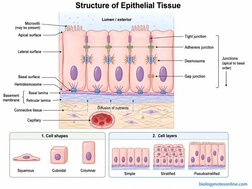
The structure of epithelial tissue can be described as follows-
- Epithelial cells – These are the main structural units of epithelial tissue. The cells are packed very close to one another. Depending upon the tissue, they may be flat, cube-shaped or tall cells and are referred to as squamous, cuboidal and columnar cells.
- Apical surface – It is the free surface of epithelial cell that faces towards the exterior of body or towards the lumen of an internal organ. This surface can be smooth. In some epithelial cells, special structures such as microvilli, cilia or stereocilia are present over it.
- Lateral surface – The lateral surfaces are present on the sides of the epithelial cells, where one cell remains in contact with the neighbouring cells. Different junctional structures are located in this region. It also helps in maintaining the epithelial sheet as a connected layer.
- Basal surface – This is the lower surface of epithelial cells. It faces the underlying connective tissue and remains attached to the basal lamina. The apical and basal regions of epithelial cells are structurally different, giving the cells their characteristic apical-basal polarity.
- Cell junctions – Epithelial cells are connected by specialized junctions. These mainly include tight junctions, adherens junctions, desmosomes and gap junctions. Their position and number differs according to the type of epithelium.
- Basement membrane – A thin layer of extracellular matrix is present below the epithelial cells, known as the basement membrane. It provides attachment and structural support to epithelium and separates it from the underlying connective tissue. The basal surface of epithelial cells is attached to this region through cell-matrix junctions such as hemidesmosomes.
- Arrangement of cell layers – Epithelial tissue may contain a single layer or several layers of cells. A single cell layer forms simple epithelium, whereas two or more layers form stratified epithelium. Pseudostratified epithelium appears to contain several layers, but all of its cells contact the basement membrane.
- Underlying connective tissue – Epithelium is generally supported by connective tissue present below the basement membrane. Blood vessels do not normally enter the epithelial layer, so nutrients and oxygen reach epithelial cells from the underlying vascular connective tissue by diffusion.
Classification of Epithelial Tissue
Epithelia are mainly classified according to the number of cell layers and the shape of the cells at the surface. Two features are therefore considered. One is arrangement of the cells in layers and another is their shape.
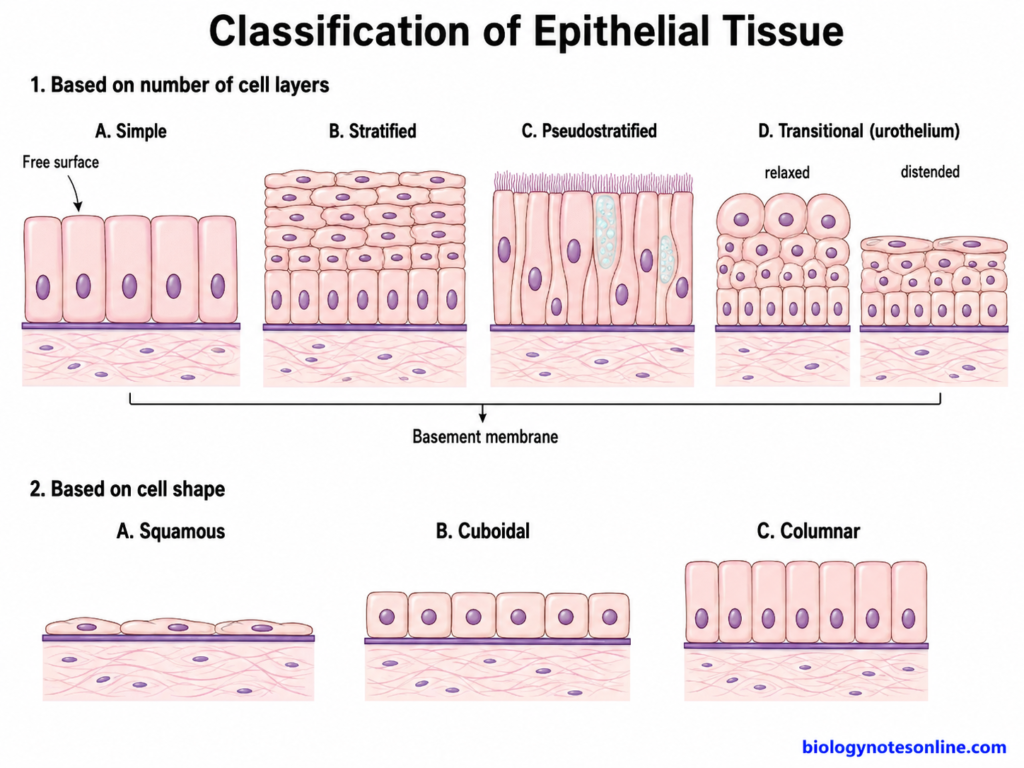
Classification Based on Number of Cell Layers
Based on the number and arrangement of cells, epithelial tissues are mainly of the following types-
- Simple epithelium – It consists of a single layer of epithelial cells. All the cells remain attached to the basement membrane and the tissue is comparatively thin. Based on the shape of cells, it can be simple squamous, simple cuboidal or simple columnar epithelium.
- Stratified epithelium – This type is made up of two or more layers of cells. Only the deepest or basal layer remains directly attached to the basement membrane, while the other layers are arranged above it. It is mainly found where protection against friction and mechanical injury is required.
- Pseudostratified epithelium – It appears like a stratified epithelium, but actually consists of a single layer of cells. The nuclei are located at different levels and due to this the tissue gives an appearance of several layers. All cells contact the basement membrane, although every cell does not reach the free surface. It is commonly formed by columnar cells.
- Transitional epithelium – It is a specialized type of stratified epithelium, also called urothelium. This tissue is mainly associated with urinary passages such as the ureters and urinary bladder. Its superficial cells and the thickness of tissue change with distension. The large surface cells become more flattened when the urinary organ is stretched.
Classification Based on Cell Shape
The epithelial cells are also divided according to their shape. Three main forms are present-
- Squamous epithelium – The cells are thin and flattened in shape. Their width is much greater than their height. These cells form a thin epithelial surface.
- Cuboidal epithelium – These cells are approximately as tall as they are wide, giving them a cube-like appearance in section. The nucleus is generally rounded and located near the central region of cell.
- Columnar epithelium – The cells are taller than their width. They appear elongated or column-like and the nucleus is commonly located towards the basal half of the cell.
How Epithelial Tissues Are Named
Most epithelial tissues are named by combining the number of cell layers with the shape of cells. Thus, the first word usually indicates whether the epithelium is simple or stratified and the second indicates the cell shape.
Some common examples are-
- Simple squamous epithelium – one layer of flattened cells.
- Simple cuboidal epithelium – one layer of cuboidal cells.
- Simple columnar epithelium – one layer of tall columnar cells.
- Stratified squamous epithelium – several layers are present and the cells at the surface are squamous.
In stratified epithelium, the name is determined by the cells present at the apical or superficial surface. It is not named according to the basal cells. Thus, stratified squamous epithelium may have cuboidal or columnar cells in its deeper layer but the flattened cells present at the surface give the tissue its name.
Pseudostratified epithelium and transitional epithelium have their special naming. Pseudostratified tissue only appears multilayered, whereas transitional epithelium has a specialized organization suited for stretching of the urinary tract.
Types of Epithelial Tissue
Epithelial tissues are of different types based on the arrangement and shape of their cells. Some consist of only one cell layer while others have many layers. Their structure also differs according to the function performed at different parts of the body.
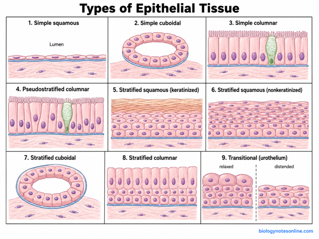
1. Simple Squamous Epithelium
Definition – Simple squamous epithelium is a type of epithelium consisting of a single layer of very thin and flattened cells.
Structure – The cells are wider than their height and have very little cytoplasm. Their nuclei are generally flattened or elliptical and may produce a slight bulge in the cell. Only one layer is present.
Location – It is present in the alveoli of lungs, lining of blood and lymphatic vessels and the parietal layer of Bowman’s capsule. The simple squamous lining of blood vessels is called endothelium, whereas the epithelium covering serous cavities and organs is referred to as mesothelium.
Function – Due to its very small thickness, it allows rapid diffusion and movement of substances. Gas exchange in alveoli and filtration associated with the renal corpuscle are important examples. Mesothelium also provides a smooth lubricated surface.
Microscopic identification – Under the microscope, it appears as a very thin single line of cells. The flattened nuclei are more easily visible than the thin cytoplasm. In blood vessels, these cells can be seen forming the thin lining around the lumen.
2. Simple Cuboidal Epithelium
Definition – Simple cuboidal epithelium is formed by a single layer of approximately cube-shaped cells.
Structure – The height and width of the cells are nearly similar. A round nucleus is generally present near the centre of each cell. These cells are thicker than squamous epithelial cells.
Location – It commonly lines many kidney tubules and small ducts of glands. The follicles of the thyroid gland are also generally lined by simple cuboidal follicular cells, although their height can change with functional activity.
Function – Secretion and absorption are the major functions. In kidney tubules, these epithelial cells take part in absorption and secretion, while glandular cuboidal cells are commonly associated with secretory processes.
Microscopic identification – It appears as a single row of cuboidal cells around a lumen. The round and centrally placed nuclei are usually prominent. In cross section of kidney tubules, they commonly appear like a ring of cells surrounding a clear central space.
3. Simple Columnar Epithelium
Definition – Simple columnar epithelium consists of a single layer of tall cells, in which the cell height is greater than its width.
Structure – The cells are column-like and contain oval or elongated nuclei, generally towards the basal part. Their apical surface can possess microvilli. Goblet cells may also occur between columnar cells at several mucosal surfaces, particularly in intestine.
Location – This epithelium forms the surface lining of the stomach, intestines and gallbladder. The small intestine contains absorptive columnar cells with microvilli and goblet cells. Gallbladder epithelium is also simple columnar with apical microvilli but normally lacks goblet cells. Ciliated simple columnar epithelium is found in places such as the uterine tubes.
Function – Its major functions are absorption and secretion. Microvilli increase the apical surface available for absorption. Secretory columnar cells and goblet cells produce different substances including mucus.
Microscopic identification – Tall cells arranged in one layer are seen. Their nuclei generally form a row towards the basal region. In intestinal epithelium, a brush border and pale goblet cells can also be identified between the absorptive cells.
4. Pseudostratified Columnar Epithelium
Definition – Pseudostratified columnar epithelium appears to have several cell layers, but actually it is a single-layered epithelium. All cells contact the basement membrane. Not all of them reach the free surface.
Structure – Cells are of unequal heights and their nuclei occur at different levels. Because of this arrangement, the nuclei give a false appearance of stratification. Cilia and goblet cells are commonly present in its respiratory form.
Location – Ciliated pseudostratified columnar epithelium forms much of the conducting respiratory epithelium, including the trachea and larger bronchi. Goblet cells occur between the ciliated cells and release mucus.
Another form is found in the epididymis. Its principal columnar cells bear long stereocilia. These are modified microvilli and are not the motile cilia of respiratory epithelium.
Function – In the respiratory tract, mucus traps particles and the beating of motile cilia helps to move this material along the epithelial surface. Epididymal epithelial cells are involved in absorption and secretion associated with sperm maturation.
Microscopic identification – Nuclei are present at several different heights, giving the tissue a multilayered appearance. A basement membrane is present below all of the epithelial cells. In respiratory epithelium, cilia can be seen along the free surface and pale mucus-containing goblet cells occur between the columnar cells.
5. Stratified Squamous Epithelium
Definition – Stratified squamous epithelium is formed of several layers of epithelial cells, with flattened squamous cells at its superficial surface.
Structure – The basal cells may be cuboidal or columnar and become progressively flatter towards the surface. Thus, this tissue is named according to its superficial cells, not the cells present at the base.
Location – It occurs in regions which are regularly subjected to friction and mechanical stress. Depending on the presence or absence of a keratinized surface, it is divided into keratinized and nonkeratinized types.
Function – Its major function is protection against abrasion. Having several cell layers allows superficial cells to be lost without immediately exposing the underlying tissue.
Microscopic identification – Many layers of cells are present. Basal cells appear cuboidal or columnar whereas the cells become progressively flattened towards the free surface.
a. Keratinized Stratified Squamous Epithelium
Definition – Keratinized stratified squamous epithelium is a stratified squamous epithelium having a surface layer of flattened, keratinized cells.
Structure – The superficial region forms the stratum corneum, which contains flattened dead corneocytes with abundant keratin. These cells have lost their nuclei in the fully cornified layer.
Location – The characteristic example is the epidermis of skin.
Function – It forms a strong mechanical barrier. The cornified surface and associated lipids also reduce excessive loss of water from the body and protect against the external environment.
Microscopic identification – Numerous cell layers are present with a distinct keratinized material at the surface. The outermost cells are flattened and lack nuclei. Deeper cells still contain nuclei.
b. Nonkeratinized Stratified Squamous Epithelium
Definition – Nonkeratinized stratified squamous epithelium consists of several cell layers but does not develop the dry cornified surface seen in epidermis.
Structure – Basal cells are more cuboidal while the superficial cells become flattened. The superficial cells remain living and retain their nuclei.
Location – It is found on moist surfaces subjected to abrasion. Important examples are portions of the oral cavity, esophagus and vagina.
Function – It mainly protects the underlying tissues from mechanical abrasion while maintaining a moist epithelial surface.
Microscopic identification – Several cell layers can be seen, but there is no thick anucleate keratin layer over the surface. The flattened superficial cells still have visible nuclei.
| Feature | Keratinized | Nonkeratinized |
|---|---|---|
| Surface | Keratinized, superficial corneocytes | Moist nucleated cell surface |
| Main function | Protection and reduced water loss | Mainly abrasion protection |
| Example | Epidermis | Esophagus |
6. Stratified Cuboidal Epithelium
Definition – Stratified cuboidal epithelium is a relatively uncommon epithelial tissue having two or sometimes more cell layers, with cuboidal cells at the superficial surface.
Structure – It is thicker than simple cuboidal epithelium. In many ducts, two layers of cuboidal cells form the wall around a small lumen.
Location – It is mainly associated with ducts of some larger exocrine glands. A common example is the duct of sweat glands. Stratified cuboidal epithelium can also occur in portions of larger salivary and mammary gland ducts.
Function – It mainly provides protection to the duct and can also have secretory functions depending upon the gland.
Microscopic identification – Two or a few layers are seen around a duct lumen. The superficial cells have a cuboidal appearance with rounded nuclei. It should not be confused with a simple cuboidal duct cut in an irregular plane.
7. Stratified Columnar Epithelium
Definition – Stratified columnar epithelium is a rare type of epithelium in which several cell layers are present and the cells at the free surface are columnar.
Structure – The deeper cells are generally smaller and may be cuboidal. The superficial layer contains the taller columnar cells.
Location – It has a limited distribution in the human body. It occurs in some large glandular ducts and in selected regions of the urethra. Claims that it widely lines different organs should therefore be avoided.
Function – Protection and secretion are its main functions.
Microscopic identification – More than one cell layer is visible, but the cells facing the lumen are tall and column-like. This superficial columnar layer is the important feature used for its identification.
8. Transitional Epithelium (Urothelium)
Definition – Transitional epithelium, also called urothelium, is a specialized stratified epithelium of the urinary tract. It is particularly adapted to repeated stretching and contraction.
Structure – It consists of basal, intermediate and large superficial cells. The characteristic superficial cells are referred to as umbrella cells. Their appearance changes considerably according to whether the urinary organ is relaxed or distended.
Location – Urothelium lines important parts of the urinary tract, particularly the ureters and urinary bladder. It also extends through other urothelial portions of the collecting urinary passages.
Function – It allows the urinary organs to increase in volume without loss of epithelial continuity. At the same time, it forms a highly resistant barrier between urine and the underlying tissues. Tight junctions and specialized apical membranes of umbrella cells contribute to this barrier.
Microscopic identification – In a relaxed bladder, the epithelium appears relatively thick and the superficial umbrella cells are large and rounded or dome-shaped. During distension, the epithelial sheet becomes thinner and the superficial cells become much flatter. The large umbrella cells at the luminal surface are the major identifying feature.
Epithelial Tissue Types, Locations and Functions
Different types of epithelial tissues occur in different regions of the body according to their structure and function. The following table summarizes their major locations and functions-
| Type of epithelial tissue | Major locations | Main functions |
|---|---|---|
| Simple squamous epithelium | Alveoli of lungs, lining of blood and lymphatic vessels (endothelium), parietal layer of Bowman’s capsule, serous membranes (mesothelium) | Rapid diffusion, filtration and exchange of substances. It also provides a smooth surface in serous cavities. |
| Simple cuboidal epithelium | Kidney tubules, thyroid follicles and small glandular ducts | Mainly absorption and secretion. |
| Simple columnar epithelium | Stomach, intestines and gallbladder. Ciliated form occurs in uterine tubes | Absorption and secretion. Microvilli help in absorption, while cilia in ciliated forms help movement of materials. |
| Pseudostratified columnar epithelium | Trachea and much of the conducting respiratory tract. Non-ciliated stereociliated form is found in epididymis | Secretion and movement of mucus in respiratory passages. In epididymis, it is involved in absorption and secretion. |
| Keratinized stratified squamous epithelium | Epidermis of skin | Protection against abrasion and mechanical injury. The keratinized surface also helps to reduce water loss. |
| Nonkeratinized stratified squamous epithelium | Oral cavity, esophagus and vagina | Protects moist surfaces against abrasion and mechanical stress. |
| Stratified cuboidal epithelium | Ducts of sweat glands and portions of some larger glandular ducts | Mainly protection. It may also take part in secretion. |
| Stratified columnar epithelium | Selected large ducts and some parts of urethra | Protection and secretion. It is a relatively uncommon epithelial type. |
| Transitional epithelium (urothelium) | Ureters, urinary bladder and other parts of urinary passages | Allows stretching during filling of urinary organs and forms a strong barrier against urine. |
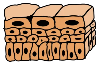
Functions of Epithelial Tissue
Epithelial tissue performs different functions depending upon its structure and location in the body. The epithelium of skin mainly acts as a protective layer, while intestinal epithelium is more involved in absorption and secretion. Some of the important functions of epithelial tissue are-
- Protection – One of the major functions of epithelial tissue is protection of the underlying tissues. Stratified squamous epithelium forms several layers and protects regions exposed to friction and mechanical injury. The epidermis also forms a barrier against water loss and entry of microorganisms.
- Absorption – Epithelial cells are involved in absorption of water, nutrients and other substances. This function is especially important in the small intestine. The presence of microvilli increases the apical surface area and helps in absorption.
- Secretion – Many epithelial cells produce and release different substances. Glandular epithelium is specially modified for this function. Mucus, enzymes and other secretory products can be released by epithelial cells depending upon the organ.
- Diffusion and exchange – Thin epithelial surfaces allow rapid movement of molecules from one side to another. Simple squamous epithelium of the alveoli is suitable for gaseous exchange. Similar thin endothelial surfaces also allow exchange of substances between blood and surrounding tissues.
- Filtration – Some specialized epithelial structures take part in filtration. In the renal corpuscle, the glomerular filtration barrier includes fenestrated capillary endothelium, basement membrane and the visceral epithelial cells called podocytes. These together control movement of fluid and solutes into Bowman’s space.
- Movement of materials – Ciliated epithelial cells can move substances over their free surface. In the respiratory tract, beating of cilia moves mucus containing trapped dust and other particles towards the throat. This forms an important part of mucociliary clearance.
- Selective permeability and transport – Epithelium does not act only as a simple covering. It also controls movement of ions, water and different molecules between two regions. Tight junctions and different membrane transport systems help in maintaining this selective movement.
- Sensory reception – Some specialized epithelia are associated with receiving sensory stimuli. Sensory epithelial structures are involved in functions such as smell, hearing and balance. These contain specialized receptor cells or epithelial-associated sensory cells for receiving particular stimuli.
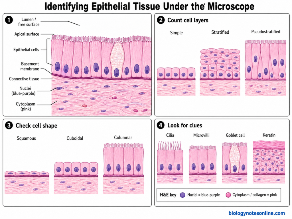
How to Identify Epithelial Tissue Under a Microscope
Epithelial tissue can be identified under a microscope by observing the arrangement of cells, number of cell layers and shape of cells present towards the free surface. The cells are generally closely packed. First locate an epithelial sheet and its relationship with a lumen, cavity or external surface, then other features can be identified.
The following steps can be used to identify epithelial tissue-
- Locate the lumen or free surface – First look for the surface towards which the epithelial cells are facing. This may be a lumen, body cavity or an exposed free surface. The apical side of epithelial cells is directed towards this region. In tubular organs, the lumen generally appears as an empty or lightly stained space surrounded by the lining cells.
- Identify the basement side – The opposite side of the epithelium is its basal region. It is attached to the basement membrane and usually has connective tissue below it. The basement membrane itself may not always be clearly distinguished with routine H&E staining, so the border between closely arranged epithelial cells and underlying connective tissue is useful for finding the basal side.
- Count the apparent cell layers – Observe whether one layer or several layers of cells are present. Simple epithelium has one cell layer, while stratified epithelium consists of two or more layers. Pseudostratified epithelium is different. Its nuclei appear at several levels, but all of the cells contact the basement membrane.
- Examine cells at the apical surface – In stratified epithelium, mainly observe the cells nearest to the free surface. These cells are used for naming the epithelium. Thus, an epithelium with several layers and flattened superficial cells is called stratified squamous epithelium, even when deeper cells have cuboidal or columnar appearance.
- Identify the cell shape – Epithelial cells are mainly recognized as squamous, cuboidal or columnar. Squamous cells are thin and flattened. Cuboidal cells have nearly similar height and width, whereas columnar cells are taller. Looking at the complete tissue arrangement is important because sectioning can sometimes alter the apparent shape of individual cells.
- Examine nuclear shape and position – The nucleus also gives an important clue about the cell type. Squamous epithelial cells commonly have flattened nuclei. Cuboidal cells generally contain rounded nuclei, while columnar cells usually have elongated nuclei. In simple columnar epithelium, nuclei are commonly located towards the basal part. Pseudostratified epithelium shows nuclei at different heights.
- Look for surface and cellular specializations – Some epithelial tissues can be identified by additional structures. Cilia appear as relatively long projections over the apical surface, particularly in respiratory epithelium. Microvilli may form a brush or striated border. Goblet cells usually appear paler than the surrounding columnar cells in routine sections. A thick superficial keratin layer indicates keratinized stratified squamous epithelium.
- Relate the epithelial pattern to the organ – Identification should not depend upon only one cell. The arrangement must also be compared with the tissue and organ present around it. For example, round kidney tubules commonly have cuboidal lining cells, the trachea has pseudostratified ciliated columnar epithelium and urinary passages can contain transitional epithelium (urothelium). The organ pattern therefore gives another useful clue.
Appearance With H&E Staining
Hematoxylin and eosin (H&E) is commonly used for routine examination of tissue structure. Hematoxylin mainly produces a blue to purple colour in nuclei, so the arrangement, shape and position of epithelial nuclei can be easily followed. Eosin stains much of the cytoplasm and extracellular protein material in different shades of pink.
Nuclei – These generally appear dark blue or purple. Nuclear arrangement is especially useful for distinguishing simple columnar from pseudostratified epithelium and for estimating the number of cell layers.
Cytoplasm – The cytoplasm usually appears pink to reddish-pink with eosin. Its amount and appearance differs between different epithelial cells.
Lumen – A lumen commonly appears as a clear or relatively empty space bordered by epithelial cells. This is particularly useful while examining ducts, glands, blood vessels and different hollow organs.
Connective tissue – Underlying connective tissue is present below the epithelial sheet and generally contains more extracellular matrix with scattered cells, unlike the closely packed appearance of epithelium. Its eosinophilic collagen commonly gives the region a pink appearance.
Basement region – It occurs between the epithelial cells and underlying connective tissue. In routine H&E sections this thin basement membrane may not always appear as a distinct line. The basal boundary of the epithelial sheet is therefore commonly easier to identify than the membrane itself.
Simple vs Stratified Epithelium
Simple epithelium and stratified epithelium mainly differ in the number of cell layers present. Simple epithelium contains only one layer, whereas stratified epithelium is formed by two or more layers of cells.
| Feature | Simple Epithelium | Stratified Epithelium |
|---|---|---|
| Number of cell layers | Single layer of cells | Two or more cell layers |
| Contact with basement membrane | All cells contact the basement membrane | Mainly the basal cells contact the basement membrane |
| Main function | Absorption, secretion, diffusion and filtration | Mainly protection against abrasion and mechanical stress |
| Thickness | Generally thin | Comparatively thicker |
| Naming | Named according to shape of the cells in the single layer | Named according to shape of the superficial or apical cells |
| Common types | Simple squamous, simple cuboidal, simple columnar | Stratified squamous, stratified cuboidal, stratified columnar |
| Examples | Alveoli, kidney tubules, intestinal lining | Epidermis, esophagus and some larger ducts |
Pseudostratified epithelium may look like a stratified tissue because nuclei occur at different levels. It is actually a simple epithelium, as all cells remain in contact with the basement membrane.
Epithelial Tissue vs Connective Tissue
Epithelial tissue and connective tissue are two of the four basic animal tissues. They differ mainly in arrangement of cells, amount of extracellular matrix and their major functions.
| Feature | Epithelial Tissue | Connective Tissue |
|---|---|---|
| Cell arrangement | Cells are closely packed together | Cells are generally more widely separated |
| Extracellular matrix | Very little extracellular material is present | Usually contains abundant extracellular matrix |
| Main location | Covers body surfaces and lines organs, cavities and ducts | Present below epithelia and between or around different tissues and organs |
| Basement membrane | Usually rests on a basement membrane | Does not form an epithelial-type free sheet over a basement membrane |
| Blood supply | Epithelium is generally avascular | Most connective tissues are vascular, but some such as cartilage have no blood vessels |
| Major functions | Protection, absorption, secretion, diffusion and filtration | Support, binding, protection, storage and transport |
| Cell polarity | Apical and basal polarity is a common characteristic | Such distinct apical-basal polarity is generally absent |
| Examples | Epidermis, intestinal lining, kidney tubules | Bone, cartilage, adipose tissue, blood and loose connective tissue |
The two tissues are also closely associated in many organs. Epithelial tissue commonly forms the surface or lining, while connective tissue present below it provides structural and nutritional support.
Clinical Significance of Epithelial Tissue
Epithelial tissue has important clinical significance as it forms the covering and lining of many organs and is frequently exposed to injury, infection and different harmful agents. Abnormal growth of epithelial cells is also associated with different precancerous and cancerous conditions.
Some of the important clinical significance of epithelial tissue are-
- Epithelial injury and repair – Epithelial cells can be damaged due to physical injury, inflammation, infection or chemicals. After injury, the cells present near the damaged region start to migrate and divide. In this process, the lost epithelial surface is formed again, which is referred to as re-epithelialization.
- Metaplasia – Metaplasia is a change of one mature cell type into another mature type. It generally takes place during continuous irritation or stress. It is not cancer itself. In some tissues, however, persistent metaplastic changes can be associated with later development of dysplasia and malignancy.
- Dysplasia – It refers to abnormal growth and arrangement of cells in a tissue. The cells show changes in their size, nuclei and normal organization. Epithelial dysplasia can be mild or more severe, and severe dysplastic changes are important during examination of precancerous lesions.
- Carcinoma – A malignant tumor arising from epithelial tissue is called a carcinoma. Since epithelial tissues are widely present in the body, carcinomas can develop in many different organs.
- Adenocarcinoma and squamous cell carcinoma – Adenocarcinoma is a carcinoma showing glandular differentiation. On the other hand, squamous cell carcinoma (SCC) shows differentiation towards squamous epithelial cells. These are two important forms of epithelial cancers.
- Biopsy – Examination of epithelial tissue is commonly used during diagnosis of different lesions. A small tissue sample is collected by biopsy and then examined microscopically. Changes in cell arrangement, nuclei, tissue structure and invasion into the surrounding tissue can be detected.
- Cytokeratin as epithelial marker – Cytokeratins are intermediate filament proteins commonly present in epithelial cells. Antibodies against cytokeratins are used in immunohistochemistry for identification of epithelial origin of many tumors, particularly carcinomas. Different cytokeratin patterns can also help in their further identification. However, these markers are interpreted together with tissue morphology and other diagnostic markers.
Frequently Asked Questions (FAQs)
1. What is epithelial tissue?
Epithelial tissue is one of the four basic types of animal tissues. It is made up of closely arranged cells that cover body surfaces, line different internal organs and also form much of the glandular tissue.
2. What are the main characteristics of epithelial tissue?
Epithelial cells are closely packed and contain very little extracellular material. They show apical-basal polarity, rest on a basement membrane and are generally avascular. Cell junctions are also well developed between neighbouring cells.
3. What are the major functions of epithelial tissue?
The major functions are protection, absorption, secretion, diffusion and filtration. Some epithelial tissues also help in movement of substances and sensory reception.
4. How is epithelial tissue classified?
Epithelial tissue is mainly classified according to the number of cell layers and shape of cells present at the surface. It can be simple, stratified, pseudostratified or specialized transitional epithelium.
5. What are the major types of epithelial tissue?
The major types include simple squamous, simple cuboidal, simple columnar, pseudostratified columnar, stratified squamous, stratified cuboidal, stratified columnar and transitional epithelium.
6. What is simple epithelium?
Simple epithelium consists of only one layer of epithelial cells. All cells remain in contact with the basement membrane. It is commonly associated with absorption, secretion, diffusion or filtration.
7. What is stratified epithelium?
Stratified epithelium is made up of two or more layers of cells. Mainly the basal cells remain attached to the basement membrane. Its major role is protection.
8. What is pseudostratified epithelium?
Pseudostratified epithelium looks like a multilayered tissue because the nuclei occur at different levels. Actually it is a single-layered epithelium. All cells contact the basement membrane, although all of them do not reach the free surface.
9. What is transitional epithelium?
Transitional epithelium, also called urothelium, is a specialized stratified epithelium present in the urinary tract. It can change its appearance during stretching and is especially found in the ureters and urinary bladder.
10. Why is epithelial tissue avascular?
Blood vessels generally do not pass through the epithelial layer. Thus, epithelial tissue is considered avascular. Blood vessels are present in the connective tissue below it.
11. How does epithelial tissue receive nutrients?
Nutrients and oxygen reach epithelial cells mainly by diffusion from blood vessels of the underlying connective tissue. They pass across the basement region before reaching the epithelial cells.
12. What is the basement membrane?
The basement membrane is a thin extracellular layer present below most epithelia. It attaches the epithelium to underlying tissue and provides structural support. It can also have filtration and regulatory functions depending upon its location.
13. Where is epithelial tissue found in the human body?
Epithelial tissue is present in the skin, digestive tract, respiratory tract, blood vessels, urinary system, reproductive organs and glands. It also forms the mesothelium of serous membranes.
14. Which epithelial tissue lines the alveoli of the lungs?
The alveoli are mainly lined by very thin simple squamous epithelial cells, especially type I pneumocytes. Their thin structure allows rapid diffusion of oxygen and carbon dioxide.
15. What is the difference between epithelial and connective tissue?
Epithelial tissue has closely packed cells with very little extracellular matrix. Connective tissue generally contains more extracellular matrix and cells are usually more widely separated. Epithelium mainly covers and lines surfaces, whereas connective tissue gives support, binding and structural connection.
16. How can epithelial tissue be identified under a microscope?
First locate the free surface or lumen. Then observe the number of cell layers and the shape of cells at the surface. Nuclear shape, position, cilia, microvilli, goblet cells and keratin can also help in identification.
References
- Adivitiya, Kaushik, M. S., Chakraborty, S., Veleri, S., & Kateriya, S. (2021). Mucociliary respiratory epithelium integrity in molecular defense and susceptibility to pulmonary viral infections. Biology, 10(2), 95. https://doi.org/10.3390/biology10020095
- Alberts, B., Johnson, A., Lewis, J., Raff, M., Roberts, K., & Walter, P. (2002). Molecular biology of the cell (4th ed.). Garland Science. https://www.ncbi.nlm.nih.gov/books/NBK21054/
- Alonso, L., & Fuchs, E. (2003). Stem cells of the skin epithelium. Proceedings of the National Academy of Sciences of the United States of America, 100(Suppl. 1), 11830–11835. https://doi.org/10.1073/pnas.1734203100
- Bellizzi, A. M. (2020). An algorithmic immunohistochemical approach to define tumor type and assign site of origin. Advances in Anatomic Pathology, 27(3), 114–163. https://doi.org/10.1097/PAP.0000000000000256
- Birder, L. A. (2010). Urothelial signaling. Autonomic Neuroscience, 153(1–2), 33–40. https://doi.org/10.1016/j.autneu.2009.07.005
- Cote, L. E., & Feldman, J. L. (2022). Won’t you be my neighbor: How epithelial cells connect together to build global tissue polarity. Frontiers in Cell and Developmental Biology, 10, 887107. https://doi.org/10.3389/fcell.2022.887107
- Dalghi, M. G., Montalbetti, N., Carattino, M. D., & Apodaca, G. (2020). The urothelium: Life in a liquid environment. Physiological Reviews, 100(4), 1621–1705. https://doi.org/10.1152/physrev.00041.2019
- Emanuel, E., Arifuzzaman, M., & Artis, D. (2024). Epithelial-neuronal-immune cell interactions: Implications for immunity, inflammation, and tissue homeostasis at mucosal sites. Journal of Allergy and Clinical Immunology, 153(5), 1169–1180. https://doi.org/10.1016/j.jaci.2024.02.004
- Falkson, S. R., & Bordoni, B. (2023). Anatomy, abdomen and pelvis: Bowman capsule. In StatPearls. StatPearls Publishing. https://www.ncbi.nlm.nih.gov/books/NBK554474/
- Freeman, S. C., Malik, A., & Basit, H. (2023). Physiology, exocrine gland. In StatPearls. StatPearls Publishing. https://www.ncbi.nlm.nih.gov/books/NBK542322/
- Gurina, T. S., & Simms, L. (2023). Histology, staining. In StatPearls. StatPearls Publishing. https://www.ncbi.nlm.nih.gov/books/NBK557663/
- Ishitsuka, Y., & Roop, D. R. (2022). The epidermis: Redox governor of health and diseases. Antioxidants, 11(1), 47. https://doi.org/10.3390/antiox11010047
- Kurn, H., & Daly, D. T. (2023). Histology, epithelial cell. In StatPearls. StatPearls Publishing. https://www.ncbi.nlm.nih.gov/books/NBK559063/
- Lachaud, C. C., Rodriguez-Campins, B., Hmadcha, A., & Soria, B. (2015). Use of mesothelial cells and biological matrices for tissue engineering of simple epithelium surrogates. Frontiers in Bioengineering and Biotechnology, 3, 117. https://doi.org/10.3389/fbioe.2015.00117
- Larjava, H., Koivisto, L., & Häkkinen, L. (2000–2013). Keratinocyte interactions with fibronectin during wound healing. In Madame Curie Bioscience Database. Landes Bioscience. https://www.ncbi.nlm.nih.gov/books/NBK6391/
- Mescher, A. L. (2024). Epithelial tissue. In Junqueira’s basic histology: Text and atlas (17th ed., Chapter 4). McGraw Hill. https://www.accessscience.com/content/book/9781264930395/chapter/chapter4
- Murphrey, M. B., Miao, J. H., & Zito, P. M. (2022). Histology, stratum corneum. In StatPearls. StatPearls Publishing. https://www.ncbi.nlm.nih.gov/books/NBK513299/
- OpenStax. (2022). Anatomy and physiology 2e. Rice University. https://openstax.org/details/books/anatomy-and-physiology-2e
- Rosner, J., Samardzic, T., & Sarao, M. S. (2024). Physiology, female reproduction. In StatPearls. StatPearls Publishing. https://www.ncbi.nlm.nih.gov/books/NBK537132/