

Transitional epithelium is a specialized epithelial tissue, also called urothelium, which forms a stratified lining in major parts of the urinary tract. It is found in the renal pelvis, ureters, urinary bladder and proximal regions of the urethra. This tissue is specially adapted to accommodate repeated distension as urine is collected and released. At the same time, it forms a strong permeability barrier which separates urine from the underlying tissues.
The urothelium consists of basal cells, intermediate cells and large superficial cells called umbrella cells. During filling of urinary organs, the epithelial layer becomes thinner and the superficial cells become more flattened. When the organ is relaxed, these cells appear larger and dome-shaped. Thus, its appearance changes according to the degree of stretching.
The superficial umbrella cells have specialized apical membrane containing uroplakins and are joined by tight junctions. These features provide very low permeability to water, ions and many urinary solutes, even when the tissue is stretched. This combination of distensibility and barrier function is a characteristic feature of transitional epithelium.
What Is Transitional Epithelium?
Transitional epithelium is a specialized stratified epithelium mainly associated with the urinary system. It is also called urothelium, meaning the epithelial lining of the urinary tract. It lines the renal calyces, renal pelvis, ureters, urinary bladder and parts of the urethra.
The tissue is formed of several layers of cells and is specially adapted for stretching. Its apparent thickness and cell shape can change according to the amount of distension. In a distended urinary bladder, the epithelium becomes thinner and the superficial cells become much flatter. This allows the urinary organs to increase their volume without damaging the epithelial lining.
Why Is It Called Transitional Epithelium?
The name transitional epithelium was given from its changing microscopic appearance. In a relaxed or empty urinary bladder, the epithelium appears thicker. The superficial umbrella cells are large, rounded or dome-shaped in this condition.
During distension, the same epithelial layer becomes stretched and appears thinner. The superficial cells become broad and flattened, giving the tissue a different appearance. This changing appearance historically led to the descriptive term “transitional” epithelium.
The cells, however, do not transform from one epithelial tissue into another. They mainly change their shape and arrangement during stretching and relaxation. Thus, the apparent transition is an adaptation of urothelial cells to repeated distension of the urinary tract.
Where Is Transitional Epithelium Found?
Transitional epithelium is found in the urinary tract. It is also called urothelium. The lining extends from the collecting region of kidney to the urinary bladder and into the proximal part of urethra.
The major locations are-
- Renal calyces- It is found in the calyces of kidney. In the intrarenal region, the urothelium is thinner and regional differences are also present.
- Renal pelvis- The renal pelvis is lined by urothelium. Urine collected from the calyces enters into this region before passing to ureter.
- Ureters- It is present throughout the ureters. The urothelial lining occurs around the lumen, with connective tissue and muscular layers present below it.
- Urinary bladder- Urothelium is well developed in urinary bladder. It forms the inner lining. During filling, this tissue becomes stretched and its apparent thickness becomes less. The bladder urothelium is also the most commonly studied region of this epithelium.
- Proximal urethra- Transitional epithelium is also found in the proximal part of urethra. It does not continue as the same epithelial type throughout the complete urethra.
Characteristics of the Urothelium
The following are the important characteristics of urothelium–
- Urothelium is a specialized type of stratified epithelium present in the urinary tract. It forms the lining between urine and the underlying tissues.
- It is mainly formed of three cell populations, basal cells, intermediate cells and superficial umbrella cells. The basal cells are attached to the basement membrane, while umbrella cells form the luminal surface.
- The thickness and appearance of urothelium are not fixed. It appears thicker when the urinary organ is relaxed and becomes thinner during distension. The superficial cells also become more flattened with stretching.
- Umbrella cells are large and highly differentiated cells. Their luminal surface is covered by specialized membrane plaques made mainly of uroplakin proteins.
- The adjacent umbrella cells are connected by well-developed tight junctions. These junctions limit movement of substances through the spaces between the cells.
- Urothelium forms a strong permeability barrier against water, ions, urea and other substances present in urine. This barrier remains functional even when the urinary bladder undergoes considerable stretching.
- The apical membrane of umbrella cells is highly adaptable. Membrane can be added or removed during filling and emptying, which helps these cells to accommodate changes in surface area.
- Urothelial cells are not only protective cells. They can respond to mechanical and chemical changes in the urinary environment and participate in signalling with underlying nerves and smooth muscle.
- Under normal conditions, the urothelium has a relatively low rate of cell turnover. However, it has considerable regenerative capacity when the epithelial lining is injured.
Structure and Cell Layers
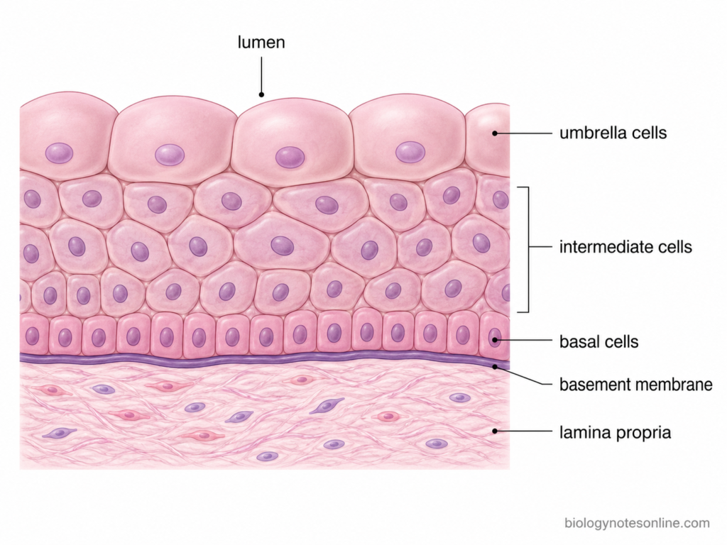
The urothelium is a stratified epithelium made up of different layers of cells. From the basement membrane toward the lumen, these are the basal cells, intermediate cells and superficial umbrella cells. The number of intermediate cell layers can vary and the apparent thickness also changes with distension.
Basal Cells
- Basal cells form the deepest part of the urothelium. They are located immediately above the basement membrane and remain attached to the basal lamina.
- These cells are generally small compared with intermediate and umbrella cells. They form a continuous basal population along the epithelial base.
- Basal cells have an important role in maintenance and renewal of the urothelium. The normal adult urothelium has slow turnover, but basal progenitor populations can become active during tissue injury and repair.
Intermediate Cells
- Intermediate cells are present above the basal layer and below the superficial umbrella cells. Usually several intermediate cells can be present between these two regions.
- They are larger than basal cells and commonly have a polygonal or irregular shape. Their morphology can vary with the degree of stretching of the tissue.
- Some intermediate cells act as progenitor cells and can differentiate toward the superficial cell phenotype. They participate in formation and regeneration of the umbrella cell layer.
- Thus, the intermediate layer forms an important cellular region between the small basal cells and the highly differentiated superficial cells. It also contributes to the multilayered architecture of urothelium.
Umbrella Cells
- Umbrella cells are the outermost cells of the urothelium and directly face the lumen containing urine. They form a single superficial layer over the underlying intermediate cells.
- These are very large cells as compared to cells of the deeper layers. A single umbrella cell can extend across and cover several underlying intermediate cells.
- In relaxed urothelium, the cells have a rounded or characteristic dome-shaped appearance. This is one of the typical microscopic features of transitional epithelium.
- During distension, umbrella cells spread over a larger surface and become considerably flatter. In the filled bladder they may appear almost squamous. The same cells again acquire a thicker appearance after the bladder is emptied.
- Some umbrella cells are binucleated and can also have increased DNA content. Binucleated superficial cells are a recognized feature of differentiated urothelium.
- Umbrella cells are highly differentiated cells. Their apical surface contains specialized membrane plaques and the cells are connected with one another by tight junctions.
- The major function of these cells is formation of the urinary permeability barrier. They prevent uncontrolled passage of water, ions, urea and other urinary solutes into underlying tissues.
- This barrier has to remain functional even when the bladder undergoes repeated filling and emptying. Therefore, umbrella cells have a specially adapted luminal membrane which can tolerate large changes in cell shape and surface area.
Uroplakins, Tight Junctions and Apical Membrane
- The luminal surface of umbrella cells contains specialized plaques formed mainly by uroplakins. These proteins are organized in the apical membrane and are characteristic differentiation components of urothelial superficial cells.
- The uroplakin-rich membrane has very low permeability and helps restrict movement of water and dissolved substances across the superficial cells.
- Adjacent umbrella cells are joined by tight junctions. These junctions limit movement of ions, water and other solutes through the spaces between neighboring cells.
- Both the specialized apical membrane and tight junctions are important during stretching. Even with marked change in umbrella cell shape, the urothelium continues to provide an effective barrier between urine and the underlying tissue.
How Does Transitional Epithelium Stretch?
Transitional epithelium can stretch mainly by changes in the shape and surface area of its cells. This is especially important in the urinary bladder, where the tissue has to accommodate a large increase in volume without losing its barrier function. During distension, the epithelium becomes thinner and the superficial umbrella cells spread over a wider area.
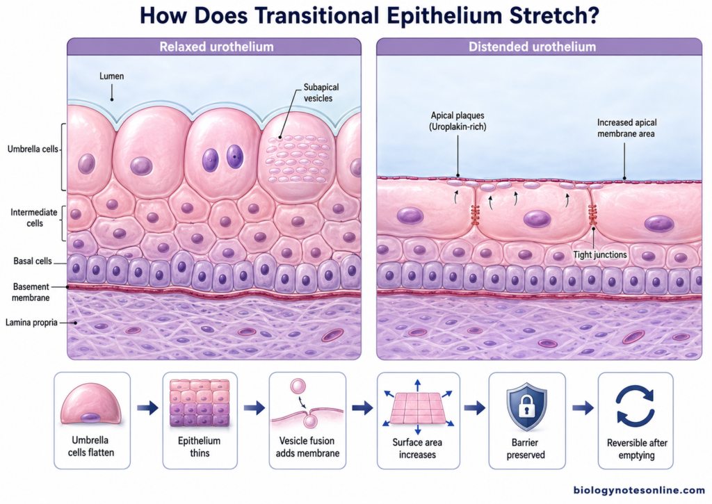
The following changes take place during stretching-
- Flattening of umbrella cells – In the relaxed bladder, umbrella cells are thick and dome-shaped. During filling, these cells become broader and much flatter. Thus, the same superficial cells cover a greater luminal surface.
- Reduction in apparent epithelial thickness – The relaxed urothelium appears comparatively thick. As distension takes place, the cells rearrange and flatten, making the whole epithelial layer appear thinner. This change does not mean that cell layers are converted into another epithelial tissue.
- Increase in apical membrane surface area – Stretching is not produced only by unfolding of the cell surface. Umbrella cells can increase their apical membrane area when mechanical tension develops during bladder filling. Experimental studies show a considerable increase in this surface area during continued stretch.
- Fusion of intracellular vesicles – Umbrella cells contain specialized subapical vesicles. During distension, membrane from these vesicles can be inserted into the apical plasma membrane by exocytosis. This provides additional membrane for expansion of the cell surface.
- Uroplakin-containing membrane adapts to stretching – The luminal surface contains plaques rich in uroplakins. Although these plaques form a strong urinary barrier, the apical membrane is highly compliant and can undergo considerable deformation during bladder filling.
- Cell junctions also expand – The junctional region between neighbouring umbrella cells changes during bladder filling. The apical junctional ring becomes larger as the cells spread, while maintaining contact between adjacent cells. This helps the epithelial sheet remain continuous during distension.
- Tight junctions remain functional – The cells have to stretch without allowing urine to leak between them. Tight junctions between umbrella cells remain an important part of the permeability barrier while the tissue is expanded.
- Changes are reversible after emptying – After urine is removed, umbrella cells return toward their thicker form and the epithelial surface becomes more folded again. Membrane trafficking, including endocytosis, also participates during changes associated with the filling and voiding cycle.
Functions of Transitional Epithelium
The following are some of the important functions of transitional epithelium (urothelium)–
- Formation of urinary barrier – The major function of urothelium is to form a strong permeability barrier between urine and the underlying tissues. It restricts movement of water, ions, urea and other urinary solutes across the epithelial lining.
- Protection of underlying tissues – Urine contains different salts, metabolites and other substances that should remain separated from the tissues of urinary organs. The urothelium provides this protective lining and prevents uncontrolled exposure of deeper tissues to urine.
- Allows distension of urinary organs – Transitional epithelium can accommodate considerable stretching, particularly in the urinary bladder. The superficial umbrella cells become flatter and their apical surface area increases during filling.
- Maintains barrier during stretching – The permeability barrier is not lost when the bladder expands. Uroplakin-rich apical membrane and tight junctions between adjacent umbrella cells help to maintain this function during repeated filling and emptying.
- Sensing mechanical changes – The urothelium can detect mechanical stimuli produced during bladder filling. Stretch can activate receptors and ion channels present in urothelial cells. Thus, these cells also act as a part of the sensory system of urinary bladder.
- Release of signalling molecules – In response to stretch and other stimuli, urothelial cells can release substances such as ATP, nitric oxide, acetylcholine and prostaglandins. These mediators can influence nearby nerves, smooth muscle and other cells of the bladder wall.
- Contribution to bladder sensation – Signals released from urothelial cells can interact with underlying sensory nerves. This is involved in communication about bladder filling and contributes to normal storage and voiding functions.
- Adjustment of luminal surface area – Umbrella cells regulate their apical membrane during filling and emptying. Membrane addition and removal help the urothelium to adapt to repeated changes in bladder volume without disrupting its epithelial surface.
- Tissue renewal and repair – The normal urothelium has relatively slow cell turnover. After injury, however, cells of the urothelium can increase proliferation and restore the damaged epithelial barrier.
Transitional Epithelium Under the Microscope
Microscopic Appearance
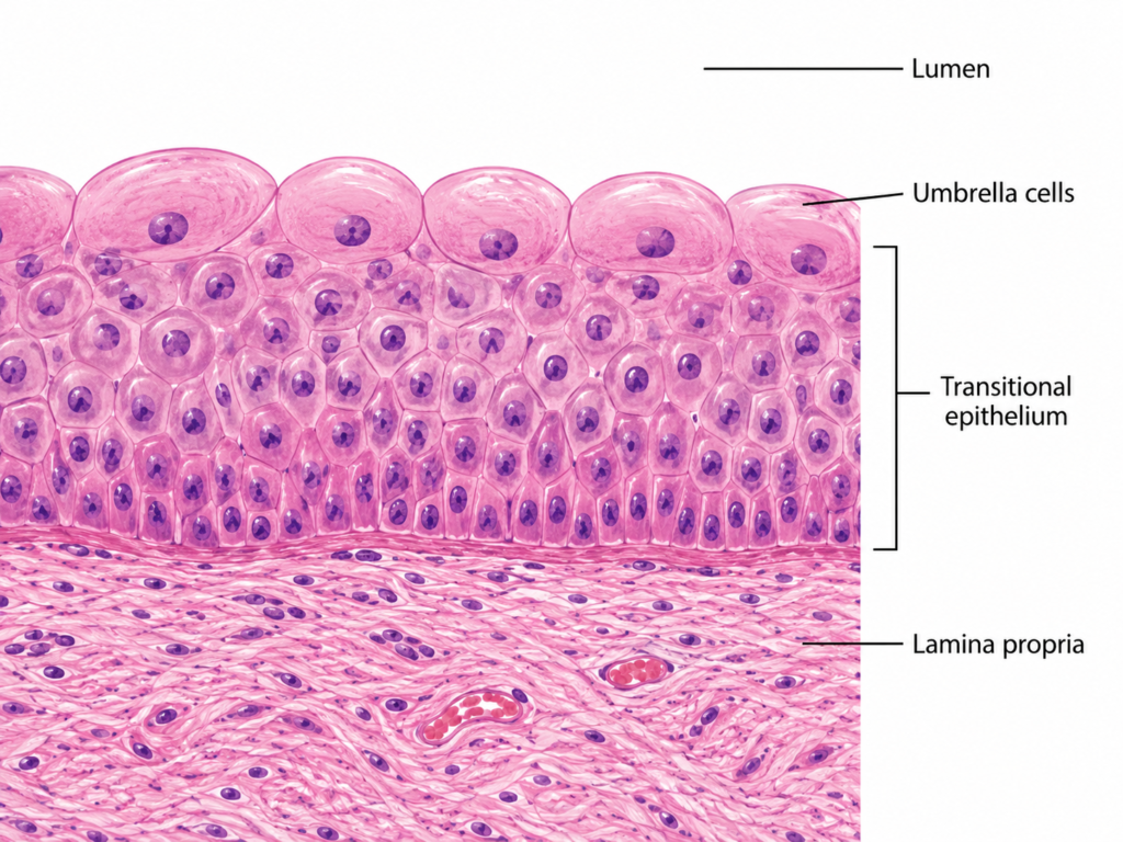
- Transitional epithelium appears as a multilayered epithelium under the microscope. The number of apparent layers is variable, and it also depends on whether the urinary organ is relaxed or distended.
- A thin basement membrane is present below the epithelial cells. It separates the urothelium from the underlying connective tissue or lamina propria.
- Basal cells are present close to the basement membrane. These are generally small cells and form the deepest region of the urothelium.
- Above these are the intermediate cells. They form the middle region and are larger than basal cells. Several intermediate cells can be seen between basal cells and the luminal surface.
- The most characteristic cells are present at the surface. These are large umbrella cells. They face directly toward the lumen and are much larger than cells present below them.
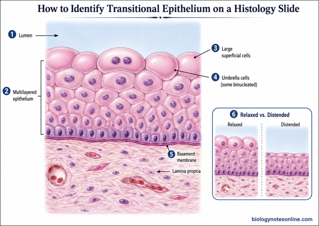
- In relaxed tissue, superficial umbrella cells are rounded or dome-shaped and project toward the lumen. This appearance is commonly used for identification of transitional epithelium on a histological slide.
- Their nuclei are generally rounded or oval. Some superficial cells contain two nuclei and binucleated umbrella cells can be seen.
- One large umbrella cell may extend over several of the smaller cells present below. During distension these superficial cells become broad and flattened, so their usual dome shape becomes much less prominent.
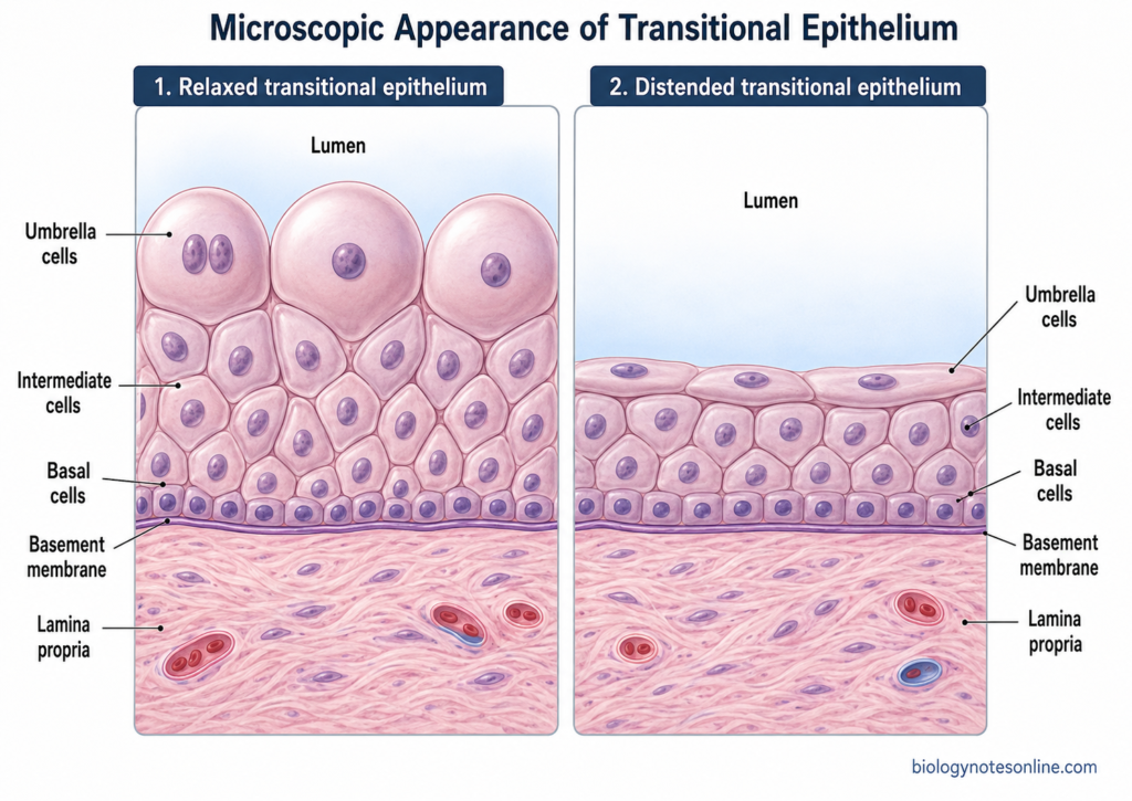
How to Identify It on a Histology Slide
- Find the lumen- First locate the free surface of the tissue. Transitional epithelium will be present directly along the urinary lumen.
- Look for a multilayered epithelium- Several layers of cells are present between the lumen and basement membrane. It should not appear as a single layer.
- Examine the superficial cells- Move toward the free luminal surface. The cells here are noticeably larger than the cells present toward the base.
- Look for umbrella cells- Large rounded or dome-shaped cells at the surface are an important identifying feature. Some may be binucleated.
- Identify the tissue below it- A basement membrane separates the epithelium from the underlying lamina propria, which is connective tissue.
- Check whether it is relaxed or distended- In relaxed tissue, the epithelium appears thicker and the umbrella cells are more rounded. A distended urothelium looks thinner. The superficial cells are flatter and spread over a greater surface.

Bladder vs Ureter Histology
| Features | Urinary Bladder | Ureter |
|---|---|---|
| Epithelium | The urinary bladder is lined by urothelium (transitional epithelium). It is well developed and changes in apparent thickness during filling and emptying. | The ureter is also lined by urothelium. The same basic epithelial type is present, with superficial umbrella cells facing urine. |
| Lumen shape | The lumen of bladder is broad and changes greatly with the amount of urine present. In an empty bladder, the mucosa is folded. | The lumen is usually narrow and commonly appears stellate or irregular in cross-section. This is due to prominent mucosal folds. |
| Mucosal folding | Mucosal folds are more obvious when the bladder is empty. They become reduced as the bladder is distended. | The mucosa forms distinct longitudinal folds. These give the ureter its characteristic irregular lumen on a histology slide. |
| Underlying connective tissue | Below the urothelium is the lamina propria. It contains connective tissue and blends toward the deeper wall. | A connective tissue lamina propria is also present below the urothelium. It forms the core of the mucosal folds. |
| Smooth muscle | The wall contains a thick smooth-muscle coat called detrusor muscle. Muscle bundles run in different directions and are not always separated into very clear layers. | Smooth muscle is more regularly arranged. In much of the ureter, inner longitudinal and outer circular layers are recognized, with an additional outer longitudinal layer becoming prominent toward the lower ureter. |
| Main slide appearance | Broad lumen, urothelium and a very thick irregular muscular wall. | Small irregular or star-shaped lumen, folded mucosa and a more organized muscular coat. |
Transitional Epithelium vs Stratified Squamous Epithelium
| Features | Transitional Epithelium | Stratified Squamous Epithelium |
|---|---|---|
| Type | It is a specialized stratified epithelium. It is also called urothelium. | It is a multilayered epithelium in which the superficial cells are squamous or flattened. |
| Major location | It is mainly found in the urinary tract, including renal pelvis, ureters and urinary bladder. | It is found mainly in areas subjected to friction, such as skin, oral cavity, esophagus and vagina, depending on keratinization. |
| Cell layers | Basal, intermediate and large superficial umbrella cells are present. | Basal cells are deeper. Cells become progressively flatter toward the surface. |
| Surface cells | The superficial cells are large. In relaxed condition they are rounded or dome-shaped. Some may be binucleated. | Superficial cells are flattened or squamous. In keratinized type, the surface cells lose nuclei and form a keratin layer. |
| Effect of stretching | The appearance changes with distension. Umbrella cells become flatter and the epithelium appears thinner. | It is not specially adapted for large repeated distension. Surface cells remain squamous in general. |
| Main function | It forms a strong urinary permeability barrier and also allows stretching of urinary organs. | Its major role is protection against mechanical abrasion and injury. |
| Special feature | Uroplakin-rich apical membrane and tight junctions are characteristic of the superficial urothelium. | Uroplakins are absent. Keratinization may be present in some stratified squamous epithelia. |
| Microscopic identification | Look for multilayered epithelium with large dome-shaped superficial cells, especially in relaxed tissue. | Look for several cell layers with distinctly flattened cells at the free surface. |
Clinical Significance
The urothelium is important in different urinary tract disorders. Damage to its superficial cells, barrier proteins or normal regenerative process can affect the underlying tissues.
The major clinical significance includes-
- Urinary tract infection (UTI)– The urothelium is commonly involved during urinary tract infection. Uropathogenic Escherichia coli can attach to the superficial urothelial cells. Some strains bind with uroplakins present on umbrella cells and can enter these cells. Infection may then cause inflammation and damage of the epithelial lining.
- Interstitial cystitis/bladder pain syndrome- In this condition, normal barrier function of urothelium may be altered. The epithelium becomes more permeable in some patients. Urinary substances can reach deeper bladder tissue and sensory nerves, which is associated with pain, urgency and frequent urination.
- Urothelial carcinoma- Cancer arising from urothelial cells is called urothelial carcinoma. It was previously commonly called transitional cell carcinoma. Most bladder cancers are of this type. It can also develop in urothelium of renal pelvis and ureter.
- Damage of urothelial barrier- Injury of superficial umbrella cells, tight junctions or apical membrane can reduce the normal permeability barrier. Urine components can then pass more easily into underlying tissue. Irritation and inflammatory changes may occur.
- Urothelial repair- Normal urothelium has a slow rate of cell turnover. But after injury, the cells rapidly start proliferation. Basal and intermediate cell populations take part in repair and lost superficial cells are formed again.
- Squamous metaplasia- During chronic irritation, the normal urothelium may be replaced by squamous epithelium in some regions. This is referred to as squamous metaplasia. Keratinizing type is clinically more important and persistent lesions need proper pathological evaluation.
Frequently Asked Questions (FAQs)
1. What is transitional epithelium?
Transitional epithelium is a specialized stratified epithelium found in the urinary tract. It can change its apparent shape and thickness when the tissue is stretched.
2. What is another name for transitional epithelium?
Another name for transitional epithelium is urothelium. This term is commonly used for the specialized epithelial lining of the urinary passages.
3. Where is transitional epithelium found?
It is found mainly in the renal calyces, renal pelvis, ureters, urinary bladder and proximal part of the urethra.
4. What is the main function of transitional epithelium?
The major function is to form a protective permeability barrier against urine. It also allows urinary organs, especially the bladder, to undergo considerable stretching.
5. Why is it called transitional epithelium?
It is called transitional epithelium because its microscopic appearance changes with distension. In relaxed condition it looks thicker, while during stretching the superficial cells become flatter.
6. Is transitional epithelium simple or stratified?
It is a stratified epithelium. Several layers of cells are present between the basement membrane and the luminal surface.
7. What are umbrella cells?
Umbrella cells are the large superficial cells of urothelium. They directly face the urine. In relaxed tissue, these cells are commonly rounded or dome-shaped and some may be binucleated.
8. What are the three cell layers of the urothelium?
The three main cell populations are basal cells, intermediate cells and superficial umbrella cells. Basal cells are found near the basement membrane, while umbrella cells form the luminal surface.
9. What happens to transitional epithelium when stretched?
During stretching, the epithelium becomes thinner in appearance. The large umbrella cells become broader and flatter, and their luminal surface area increases.
10. Why is the urinary bladder lined by transitional epithelium?
The bladder repeatedly fills with urine and then becomes empty. Transitional epithelium allows this change in volume and at the same time maintains a protective barrier between urine and underlying tissue.
11. Is transitional epithelium found in the ureter?
Yes. The ureters are lined by transitional epithelium. It forms the inner mucosal surface around the ureteral lumen.
12. Is transitional epithelium present in the kidney?
It is present in the urine-collecting regions associated with the kidney, especially the renal calyces and renal pelvis. It is not the epithelium of the renal tubules or glomeruli.
13. How can transitional epithelium be identified under a microscope?
First look for a multilayered epithelium facing a urinary lumen. The superficial cells are large and may appear rounded or dome-shaped. Smaller basal cells and intermediate cells are present below them.
14. What is the difference between transitional and stratified squamous epithelium?
Transitional epithelium has large superficial umbrella cells and is specially adapted for stretching. Stratified squamous epithelium has flattened superficial cells and is mainly adapted for protection against abrasion.
15. What is the difference between urothelium and transitional epithelium?
There is no major difference between these terms in normal histology. Urothelium is the more specific term used for transitional epithelium of the urinary tract.
References
- Birder, L. A. (2009). Urothelial signaling. Autonomic Neuroscience: Basic and Clinical, 153(1–2), 33–40. https://doi.org/10.1016/j.autneu.2009.07.005
- Birder, L. A., & Andersson, K.-E. (2013). Urothelial signaling. Physiological Reviews, 93(2), 653–680. https://doi.org/10.1152/physrev.00030.2012
- Birder, L. A., & de Groat, W. C. (2007). Mechanisms of disease: Involvement of the urothelium in bladder dysfunction. Nature Clinical Practice Urology, 4(1), 46–54. https://doi.org/10.1038/ncpuro0672
- Carattino, M. D., Prakasam, H. S., Ruiz, W. G., Clayton, D. R., McGuire, M., Gallo, L. I., & Apodaca, G. (2013). Bladder filling and voiding affect umbrella cell tight junction organization and function. American Journal of Physiology-Renal Physiology, 305(8), F1158–F1168. https://doi.org/10.1152/ajprenal.00282.2013
- Dixon, J. S., & Gosling, J. A. (1983). Histology and fine structure of the muscularis mucosae of the human urinary bladder. Journal of Anatomy, 136(Pt 2), 265–271. https://pubmed.ncbi.nlm.nih.gov/6682849/
- Eaton, A. F., Clayton, D. R., Ruiz, W. G., Griffiths, S. E., Rubio, M. E., & Apodaca, G. (2019). Expansion and contraction of the umbrella cell apical junctional ring in response to bladder filling and voiding. Molecular Biology of the Cell, 30(16), 2037–2052. https://doi.org/10.1091/mbc.E19-02-0115
- Hurst, R. E., Greenwood-Van Meerveld, B., Wisniewski, A. B., VanGordon, S., Lin, H., Kropp, B. P., & Towner, R. A. (2015). Increased bladder permeability in interstitial cystitis/painful bladder syndrome. Translational Andrology and Urology, 4(5), 563–571. https://doi.org/10.3978/j.issn.2223-4683.2015.10.03
- Iwasawa, T., Tsujino, T., Tsukahara, S., Fukushima, H., Ito, D., Tomiyama, E., Urabe, F., & Japanese Young Urologist Basic Research Collaboration. (2026). Translational advances in urothelial carcinoma: From bench to bedside. International Journal of Urology, 33(1), e70341. https://doi.org/10.1111/iju.70341
- Jackson, A. R., Narla, S. T., Bates, C. M., & Becknell, B. (2022). Urothelial progenitors in development and repair. Pediatric Nephrology, 37(8), 1721–1731. https://doi.org/10.1007/s00467-021-05239-w
- Le, N. B., Sona, S., Santo, B., Zhang, Y., Ou, R., Schweickart, R. A., Kochat, V., Padron, W. I., Rai, K., Lee, S.-H., Yi, J. M., Wessely, O., Lee, B. H., & Ting, A. H. (2026). Differentiation in the human urothelia is defined by distinct alternative polyadenylation. Cell Reports, 45(1), 116839. https://doi.org/10.1016/j.celrep.2025.116839
- Li, X., Hu, J., Yin, P., Liu, L., & Chen, Y. (2023). Mechanotransduction in the urothelium: ATP signalling and mechanoreceptors. Heliyon, 9(9), e19427. https://doi.org/10.1016/j.heliyon.2023.e19427
- Mathai, J. C., Zhou, E. H., Yu, W., Kim, J. H., Zhou, G., Liao, Y., Sun, T.-T., Fredberg, J. J., & Zeidel, M. L. (2014). Hypercompliant apical membranes of bladder umbrella cells. Biophysical Journal, 107(6), 1273–1279. https://doi.org/10.1016/j.bpj.2014.07.047
- Mescher, A. L. (2021). Junqueira’s basic histology: Text and atlas (16th ed.). McGraw Hill. https://accessmedicine.mhmedical.com/book.aspx?bookID=3047
- Montalbetti, N., Rued, A. C., Taiclet, S. N., Birder, L. A., Kullmann, F. A., & Carattino, M. D. (2017). Urothelial tight junction barrier dysfunction sensitizes bladder afferents. eNeuro, 4(3), ENEURO.0381-16.2017. https://doi.org/10.1523/ENEURO.0381-16.2017
- Pawlina, W., & Ross, M. H. (2020). Histology: A text and atlas: With correlated cell and molecular biology (8th ed.). Wolters Kluwer. https://medicine.lww.com/Book/Show/822948
- Qasrawi, F., Bergmann, F., Straube, P., Thiesler, H., Hildebrandt, H., Sinha, S., Flores, E. R., Trowe, M.-O., Rudat, C., & Kispert, A. (2026). ΔNP63 regulates epithelial stratification and differentiation in the murine ureter. American Journal of Physiology-Renal Physiology. Advance online publication. https://doi.org/10.1152/ajprenal.00468.2025
- Rausch, S., Gaisa, N., Youssef, R. F., Lotan, Y., Stenzl, A., & Kälble, T. (2014). Squamous cell lesions of the urinary bladder. Der Urologe, 53(3), 368, 370–374. https://doi.org/10.1007/s00120-013-3406-z
- Siegel, F., Torelli, A., Mattis, M., Debatin, J., Erben, P., & Gumbel, M. (2025). Proliferation and regeneration of the healthy human urothelium: A multi-scale simulation approach with 16 hypotheses of cell differentiation. PLOS ONE, 20(6), e0325132. https://doi.org/10.1371/journal.pone.0325132
- Thumbikat, P., Berry, R. E., Zhou, G., Billips, B. K., Yaggie, R. E., Zaichuk, T., Sun, T.-T., Schaeffer, A. J., & Klumpp, D. J. (2009). Bacteria-induced uroplakin signaling mediates bladder response to infection. PLOS Pathogens, 5(5), e1000415. https://doi.org/10.1371/journal.ppat.1000415
- Truschel, S. T., Wang, E., Ruiz, W. G., Leung, S.-M., Rojas, R., Lavelle, J., Zeidel, M., Stoffer, D., & Apodaca, G. (2002). Stretch-regulated exocytosis/endocytosis in bladder umbrella cells. Molecular Biology of the Cell, 13(3), 830–846. https://doi.org/10.1091/mbc.01-09-0435
- Veranič, P., Romih, R., & Jezernik, K. (2004). What determines differentiation of urothelial umbrella cells? European Journal of Cell Biology, 83(1), 27–34. https://doi.org/10.1078/0171-9335-00351
- Wang, J., Batourina, E., Schneider, K., Souza, S., Swayne, T., Liu, C., George, C. D., Tate, T., Dan, H., Wiessner, G., Zhuravlev, Y., Canman, J. C., Mysorekar, I. U., & Mendelsohn, C. L. (2018). Polyploid superficial cells that maintain the urothelial barrier are produced via incomplete cytokinesis and endoreplication. Cell Reports, 25(2), 464–477.e4. https://doi.org/10.1016/j.celrep.2018.09.042
- Wiessner, G. B., Plumber, S. A., Xiang, T., & Mendelsohn, C. L. (2022). Development, regeneration and tumorigenesis of the urothelium. Development, 149(9), dev198184. https://doi.org/10.1242/dev.198184
- Braun, M. (n.d.). Transitional epithelium (urothelium). Human Structure Virtual Microscopy, Indiana University School of Medicine. https://vmicro.iusm.iu.edu/hs_vm/docs/epi13.htm
- Braun, M. (n.d.). Urinary system, ureters and bladder. Human Structure Virtual Microscopy, Indiana University School of Medicine. https://vmicro.iusm.iu.edu/hs_vm/docs/lab11_5.htm
- Indiana University School of Medicine. (n.d.). Urinary bladder. Anatomy A215 Virtual Microscopy. https://medsci.indiana.edu/a215/virtualscope/docs/chap10_3.htm
- University of Minnesota, Department of Genetics, Cell Biology, and Development. (n.d.). Bladder: Transitional epithelia. Histology Guide. https://histologyguide.com/slideview/MH-018-transitional-epithelia/16-slide-1.html
- University of Minnesota, Department of Genetics, Cell Biology, and Development. (n.d.). Transitional epithelium. Histology Guide. https://histologyguide.com/slideview/MH-018-transitional-epithelia/02-slide-1.html
- University of Basel. (n.d.). Transitional epithelium (urinary bladder). Histology, Tales. https://tales.nmc.unibas.ch/en/histology-12/epithel-allgemeine-histologie-38/transitional-epithelium-urinary-bladder-221