

Endocytosis is a cellular process by which extracellular materials are taken inside the cell along with a portion of the plasma membrane. During this process, the plasma membrane bends inward (invagination) around the material and later pinches off, forming a membrane-bound vesicle inside the cell. This is referred to as an endocytic vesicle.
It is an energy-dependent vesicular trafficking process. The materials taken up by endocytosis can be extracellular fluid, macromolecules, particles and also proteins or lipids of the plasma membrane. Energy is required during membrane remodelling, formation of vesicle and its further trafficking inside the cell (ATP- or GTP-dependent cellular machinery are involved).
Endocytosis is therefore not simply a movement of molecules against the concentration gradient. Here, the material does not pass directly across the plasma membrane through a carrier or channel. It is taken inside by formation of vesicle from the plasma membrane.
Why Cells Use Endocytosis for Cargo That Cannot Simply Cross the Membrane
The lipid bilayer of plasma membrane acts as a barrier for most polar molecules, and large macromolecules or particles also cannot simply pass through this membrane. For these materials, endocytosis is used.
During this process, a portion of plasma membrane surrounds the extracellular material and bends inward, after which a vesicle is formed and the material is taken inside the cell without passing through the hydrophobic part of membrane as individual molecules. Eukaryotic cells can take up macromolecules, particulate substances and extracellular fluid by this process. Some substances are taken up selectively.
In receptor-mediated endocytosis, extracellular molecules first bind with specific receptors present on the cell surface and become concentrated before they are internalized within vesicles, which also makes uptake possible when the particular material is present in small amount in extracellular fluid.

Mechanism of Endocytosis
The mechanism of endocytosis differs according to the type of endocytic pathway. Different proteins, membrane lipids and cytoskeletal components may take part in it. The general process involves recognition or capture of material, remodeling of plasma membrane, formation of an endocytic carrier and its internalization into the cell.
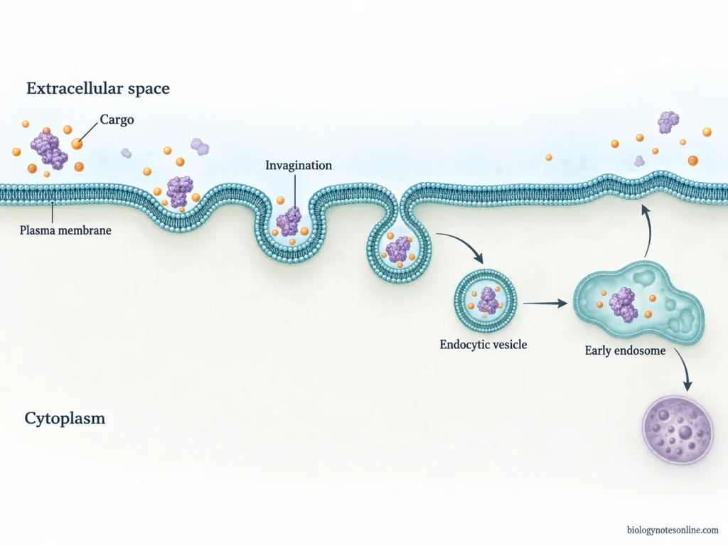
Cargo Recognition and Membrane Remodeling
- The material which has to be taken inside is first captured from the extracellular region. In some forms, extracellular fluid together with dissolved substances are taken up without any specific recognition. This is referred to as fluid-phase uptake.
- In selective endocytosis, specific extracellular cargo binds with receptors present on the plasma membrane. The receptor-cargo complexes can then accumulate at the region from where internalization takes place. Receptor-mediated endocytosis is based on this type of cargo selection.
- After capture of the material, the plasma membrane begins to change its shape. It may bend inward and form an invagination. In phagocytosis, however, the membrane extends around the large particle rather than simply forming a small inward pit.
- Different proteins and membrane lipids are involved in producing and maintaining membrane curvature. Clathrin is involved during clathrin-mediated endocytosis. It is not required in every endocytic pathway.
- Actin filaments also take part in many endocytic processes. Polymerization of actin can generate force required for movement and deformation of membrane, especially during phagocytosis, macropinocytosis and several clathrin-independent pathways. Its requirement varies depending upon the pathway and condition of the cell.
Budding, Scission and Internalization
- During this process, the remodeled plasma membrane develops into a bud, pit, membrane tubule or a larger membrane cup. The type of structure formed depends upon the endocytic pathway.
- The extracellular material becomes surrounded by the plasma membrane. However, all endocytic pathways do not produce the same type of coated vesicle. Some form clathrin-coated vesicles, while clathrin-independent pathways can produce other vesicular or tubular carriers. Phagocytosis and macropinocytosis form much larger membrane structures.
- The developing carrier is then separated from the plasma membrane, or the membrane surrounding the material is closed. This results in internalization of the material.
- In clathrin-mediated endocytosis, dynamin accumulates around the neck of the developing bud and helps in its membrane fission. Dynamin is a GTPase. But it is not involved in every pathway. Other endocytic mechanisms can carry out membrane separation with the help of different proteins, changes in membrane lipids and actin-based forces.
- After separation, the newly formed carrier is present inside the cell. Its cargo can then be transported to the endosomal system or other intracellular compartments depending upon the pathway.
Energy Requirements
- Endocytosis is an energy-dependent process because the plasma membrane has to be remodeled, moved and finally separated during formation of an intracellular carrier.
- Cytoskeletal rearrangement and different protein activities also require energy. ATP-dependent processes and GTP-binding proteins are involved at different stages of endocytosis.
- ATP is used during actin polymerization and remodeling. In pathways where dynamin is involved, GTP hydrolysis is required for its activity during membrane fission.
- Endocytosis is different from passive diffusion. In passive diffusion, molecules move directly through the membrane without formation of an intracellular vesicle and without direct use of cellular metabolic energy.
- It is also different from pump-mediated membrane transport. Pumps and membrane transporters move particular ions or molecules across the plasma membrane. During endocytosis, a portion of the plasma membrane itself is remodeled and taken inside together with the enclosed material.
Types and Pathways of Endocytosis
There are different types of endocytosis based on the nature and size of the material taken up and the membrane machinery used during the process. Broadly, endocytosis is divided into phagocytosis and pinocytosis. Pinocytosis further occurs by different pathways.
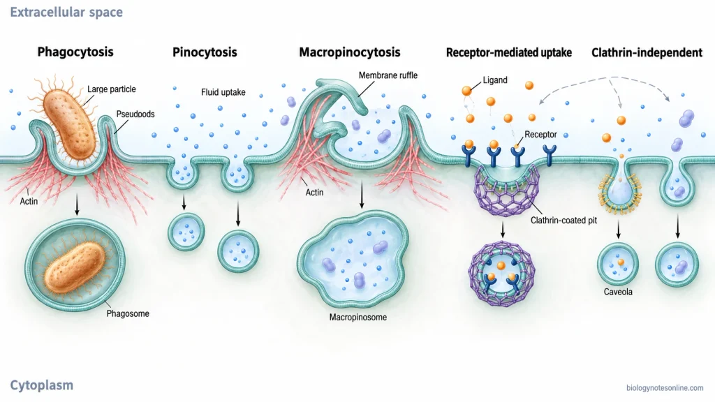
Endocytosis
- Phagocytosis – uptake of large particles by formation of a phagosome.
- Pinocytosis – uptake of fluid and smaller extracellular materials. It includes:
- Clathrin-mediated endocytosis (CME)
- Macropinocytosis
- Caveolae-associated endocytosis
- Other clathrin-independent endocytic pathways
- CLIC/GEEC pathway
- Arf6-associated pathways
- Flotillin-associated pathways
- Other pathways involving different small GTPases and membrane components
1. Phagocytosis
Phagocytosis is the uptake of large particles such as microorganisms, dead cells and cellular debris into the cell. The particle usually binds with receptors present on the cell surface. Following this, the plasma membrane extends around the particle with rearrangement of actin.
The membrane extensions surround the particle and finally close. A large membrane-bound compartment is formed, called a phagosome. This type of endocytosis is mainly carried out efficiently by specialized phagocytic cells in animals.
2. Pinocytosis
Pinocytosis is the uptake of extracellular fluid, dissolved substances and smaller extracellular materials. Here, a portion of plasma membrane is taken inside along with the material.
It is not a single endocytic pathway. Some pinocytic pathways use clathrin, while other pathways occur without formation of a clathrin coat. The membrane proteins, lipids and cytoskeletal components involved are also different.
a. Clathrin-Mediated Endocytosis (CME)
Clathrin-mediated endocytosis (CME) is one of the major pathways of pinocytosis. In this process, cargo molecules or membrane proteins are generally recognized by receptors and adaptor proteins. Clathrin is then assembled at the cytoplasmic surface of plasma membrane.
The membrane bends inward forming a clathrin-coated pit. With further bending, a membrane bud is produced which later gets separated from the plasma membrane. Dynamin and different accessory proteins are involved during formation and scission of this carrier.
After internalization, the clathrin coat is removed. The vesicle can now enter the endosomal pathway.
Receptor-mediated endocytosis commonly occurs through this pathway. A specific cargo first binds with its membrane receptor and is then taken inside the cell. However, receptor-mediated endocytosis itself is not a separate structural type of endocytosis. Some receptor-bound cargos can also enter by clathrin-independent pathways.
b. Macropinocytosis
Macropinocytosis is a form of pinocytosis in which extracellular fluid is taken up in relatively large amount. During this process, actin-dependent ruffling and extension of plasma membrane takes place.
These membrane folds later close and large intracellular vesicles are formed. They are called macropinosomes. A typical clathrin-coated pit is not formed during this process. Macropinosomes are much larger compared to many other endocytic carriers.
c. Caveolae-Associated Endocytosis
Caveolae-associated endocytosis is associated with small flask-shaped invaginations of plasma membrane called caveolae. These membrane regions contain high amounts of cholesterol and sphingolipids. Caveolin is an important structural protein present in caveolae.
Caveolae can take part in internalization and transcellular transport in particular cell types. Their function and activity varies among different cells.
d. Other Clathrin-Independent Endocytic Pathways
Apart from these, several pathways carry out endocytosis without formation of a clathrin coat. Their mechanism is not the same. Some require dynamin and others are dynamin-independent. Actin, membrane lipids and small GTPases can also be involved.
CLIC/GEEC pathway- It is a clathrin-independent pathway which produces clathrin-independent carriers (CLICs) from the plasma membrane. Clathrin and dynamin are not required in this pathway. GPI-anchored proteins (GPI-APs) and a portion of fluid-phase cargo can be taken up and delivered to GPI-AP-enriched early endosomal compartments (GEECs). Cdc42 and actin remodeling are involved during this process.
Arf6-associated pathways- These are also clathrin-independent forms of endocytosis. Arf6 and its associated membrane-remodeling machinery participate in uptake and trafficking of particular membrane cargos.
Flotillin-associated pathways- These pathways involve membrane regions containing flotillin proteins. Flotillins are associated with particular lipid-rich membrane domains and can participate in uptake of selected cargos.
Other clathrin-independent pathways also occur with the involvement of different small GTPases, membrane lipids and cytoskeletal components. The cargo and membrane carriers formed are different among these pathways.
How Does Clathrin-Mediated Endocytosis Work?
Clathrin-mediated endocytosis (CME) internalizes selected membrane proteins and receptor-bound cargo through clathrin-coated pits (CCPs) formed at the plasma membrane. The membrane bends during the process and finally produces a clathrin-coated vesicle (CCV). Several adaptor and accessory proteins take part in these events.
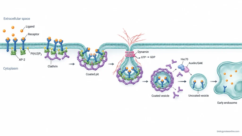
The following are the major steps of clathrin-mediated endocytosis-
Step 1- Cargo selection and initiation
In the first step, the cargo that has to be taken up becomes concentrated at a particular region of the plasma membrane. Membrane proteins may contain sorting signals, whereas extracellular ligands are first bound with their particular receptors. The AP-2 (adaptor protein-2) complex and other cargo-specific adaptor proteins recognize these membrane cargo and also interact with phosphatidylinositol-4,5-bisphosphate (PI(4,5)P₂) present in the membrane.
Step 2- Formation of clathrin-coated pit
The adaptor proteins now recruit clathrin from the cytoplasm. Clathrin molecules assemble into a lattice on the cytoplasmic side of plasma membrane, resulting in formation and growth of the clathrin-coated pit. The coat is connected with membrane through adaptor proteins rather than clathrin binding directly with membrane.
Step 3- Membrane bending and invagination
As the coat develops, the membrane bends further towards the cytoplasm. Different accessory proteins also help during this process. The pit becomes deeply invaginated and the selected cargo remains enclosed within it. At the later stage, only a narrow membrane neck connects the developing vesicle with the plasma membrane. Actin can also provide additional force during clathrin-mediated endocytosis, particularly when greater membrane force is required.
Step 4- Scission of the vesicle
The narrow neck is finally cut off from the plasma membrane. Dynamin, a large GTPase, assembles around this neck and takes part in membrane scission with the help of other accessory proteins. After scission, a free clathrin-coated vesicle is formed inside the cytoplasm.
Step 5- Removal of clathrin coat
The clathrin coat does not remain permanently around the newly formed vesicle. It is rapidly removed after vesicle formation. Auxilin/GAK and the ATPase chaperone Hsc70 take part in disassembly of the clathrin coat, and the coat components can again be used in another cycle of endocytosis.
Step 6- Transport towards endosomes
The uncoated vesicle then proceeds towards the endosomal system and its cargo is delivered to early endosomes. Here, sorting takes place. Some membrane proteins and receptors are returned back towards the plasma membrane, while other cargo can proceed further through the endosomal pathway for degradation or transport to another cellular location.
Endosomal Sorting and Cargo Fate
After endocytosis, the internalized material is transferred into the endosomal system. Early endosome mainly acts as the sorting compartment. From here, different cargo may be recycled, transported to another cellular compartment or moved toward degradation.
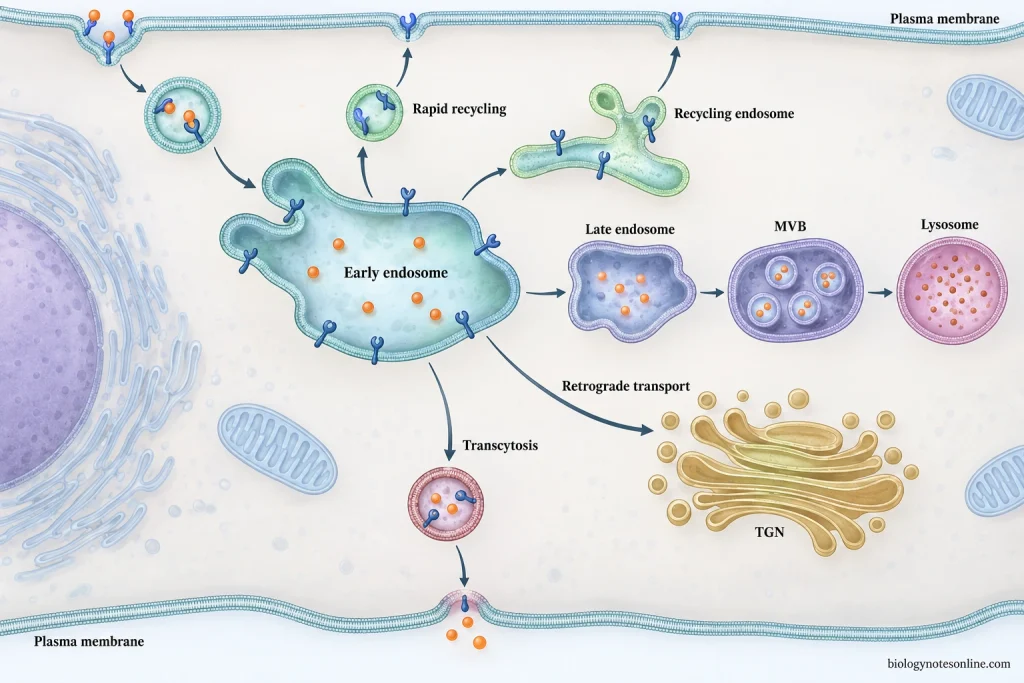
The process is as follows-
Step 1- Entry into Early Endosome
Newly formed endocytic carriers fuse with the early endosome and deliver their membrane and internal cargo. Early endosomes are mainly associated with Rab5 and phosphatidylinositol-3-phosphate (PI3P).
Step 2- Acidification and Cargo Separation
The internal region of early endosome becomes acidic by the activity of V-ATPase (vacuolar H+-ATPase). This lower pH causes separation of many receptor-ligand complexes. The separated receptor and its cargo can now move through different pathways.
Step 3- Sorting of Cargo
In this step, cargo is separated according to its further destination. Some of the important routes are-
- Recycling to plasma membrane- Some receptors and membrane components are returned back to the cell surface. Rab4 is associated with rapid recycling, while Rab11 mainly takes part through recycling endosomes.
- Retrograde transport- Selected cargo is transported from endosome toward the trans-Golgi network (TGN). Retromer and sorting nexins are involved in this route.
- Transcytosis- It occurs mainly in polarized cells. The internalized material is transported from one surface of the cell toward another region of plasma membrane.
- Degradative sorting- Cargo which has to be degraded remains within the maturing endosome. Ubiquitin can act as a sorting signal for many membrane proteins. ESCRT (Endosomal Sorting Complex Required for Transport) machinery takes part in their selection.
Step 4- Formation of Multivesicular Body
During degradative sorting, portions of endosomal membrane bud inward into the lumen. Small intraluminal vesicles (ILVs) are produced.
An endosome containing many ILVs is called a multivesicular body (MVB). ESCRT complexes are involved during formation and sorting of these vesicles.
Step 5- Maturation into Late Endosome
Early endosome gradually changes into a late endosome. During this process, Rab5 is replaced by Rab7 and the compartment becomes more acidic.
Most of the recycling cargo has already been removed at this stage.
Step 6- Delivery to Lysosome
The late endosome finally interacts and fuses with the lysosomal compartment. Cargo present in it is exposed to lysosomal hydrolytic enzymes and broken down into smaller components.
Functions of Endocytosis
Endocytosis has different functions in uptake of extracellular materials, regulation of plasma membrane and cellular signalling. Some of the important functions are-
- Endocytosis is used to take up nutrients and macromolecules from the extracellular region. In receptor-mediated endocytosis, particular substances are taken up selectively. LDL particles and transferrin-bound iron are some examples.
- It controls the amount of receptors, transporters and other proteins present on the plasma membrane. After internalization, these proteins may be returned back to the membrane or transferred for degradation. Thus, the surface composition of cell can be changed according to its requirement.
- Endocytosis also takes part in cell signalling. Activated receptors are internalized from the cell surface and can continue signalling from endosomes, get recycled or degraded. Degradation of these receptors decreases further cellular response. This is referred to as receptor down-regulation.
- Phagocytosis helps in uptake of microorganisms, dead cells and cellular debris. This function is mainly performed by specialized phagocytic cells. It is important for removal of unwanted materials and defense against invading organisms.
- In immune cells, extracellular antigens are taken up by endocytosis and later processed for antigen presentation. This process is important in dendritic cells and other antigen-presenting cells.
- Endocytosis is also used for recovery of plasma membrane added during exocytosis. At nerve terminals, membrane of synaptic vesicles is taken back after release of neurotransmitter. It can then be used again for formation of functional synaptic vesicles.
- In polarized cells, endocytosis helps in movement of materials from one side of the cell to another. This is referred to as transcytosis. Receptors together with their bound materials are internalized from one membrane surface and delivered toward the opposite surface.
- Endocytosis controls the surface level of adhesion molecules and proteins associated with cell polarity. Their internalization and recycling take part during cell migration and maintenance of cell polarity.
Examples of Endocytosis in Cells
Different cells use endocytosis for different materials and functions. Some of the representative examples are as follows-
| Example | Pathway | Cargo | Biological purpose |
|---|---|---|---|
| Macrophage or neutrophil engulfing microorganisms | Phagocytosis | Bacteria and other large particles | The microorganism is enclosed in a phagosome and later degraded after interaction with lysosomes. It is important in defence against infection. |
| LDL uptake by animal cells | Receptor-mediated endocytosis, mainly through clathrin-coated pits | LDL (low-density lipoprotein) carrying cholesterol | LDL first binds with the LDL receptor (LDLR) and is then taken inside the cell. Defects in LDL receptor-mediated uptake can result in familial hypercholesterolemia, with high LDL cholesterol in blood. |
| Extracellular-fluid sampling by dendritic cells | Macropinocytosis | Extracellular fluid, soluble molecules and antigens | Large amounts of surrounding fluid are taken up into macropinosomes. It is used by immature dendritic cells for continuous sampling of soluble antigens. |
Endocytosis and Exocytosis Compared
Endocytosis and exocytosis are vesicular transport processes associated with the plasma membrane. Endocytosis takes materials into the cell, whereas exocytosis carries materials towards the cell exterior. Both processes also take part in continuous recycling of plasma membrane.
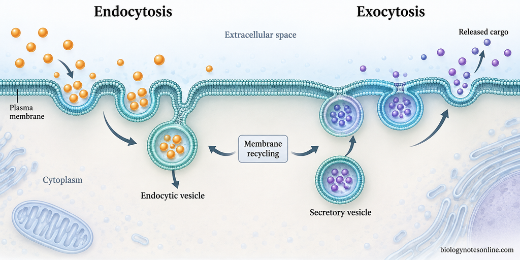
The major differences between endocytosis and exocytosis are as follows-
| Features | Endocytosis | Exocytosis |
|---|---|---|
| Definition | It is a process by which extracellular materials are taken inside the cell within membrane-bound vesicles. | It is a process by which intracellular vesicles fuse with plasma membrane and release their contents outside the cell. |
| Direction of transport | Material moves from outside towards inside of the cell. | Material is carried from inside towards the cell exterior. |
| Plasma membrane | A portion of plasma membrane bends inward and pinches off. An endocytic vesicle is formed. | Vesicle membrane fuses with the plasma membrane and becomes part of it during release. |
| Materials transported | Extracellular fluid, macromolecules, membrane proteins and large particles can be taken up. | Secretory proteins, neurotransmitters and other vesicular contents can be released. Membrane proteins and lipids are also delivered to cell surface. |
| Major forms | Phagocytosis, pinocytosis and receptor-mediated endocytosis are common forms. | Exocytosis mainly occurs by constitutive or regulated secretion. |
| Major function | It is used for nutrient uptake, receptor internalization, membrane turnover and uptake of particles. | It is used for secretion and delivery of newly synthesized proteins and lipids to plasma membrane. |
| Effect on membrane | Removes some membrane from the cell surface during vesicle formation. | Adds vesicle membrane to the cell surface during fusion. |
| Relationship | Endocytosed membrane components can be recycled back to plasma membrane. | Exocytosis returns membrane to cell surface, helping to balance membrane removed during endocytosis. |
Endocytosis, Active Transport, and Common Classification Confusions
Is Endocytosis Active Transport?
Endocytosis is an energy-dependent transport process and it is broadly considered as a type of active or bulk transport. Energy is required during membrane bending, vesicle formation and other steps of the process.
It is, however, different from the primary and secondary active transport carried out by membrane pumps and carriers. These transporters generally move ions or small solutes across the membrane, commonly against their concentration or electrochemical gradient. Endocytosis does not transport the complete cargo through such a membrane transporter. Instead, the plasma membrane surrounds the material and a vesicle is formed inside the cell. Movement against a concentration gradient, therefore, is not the characteristic feature that defines endocytosis.
Endocytosis vs. Phagocytosis and Pinocytosis
Endocytosis is the broader cellular uptake process. Phagocytosis and pinocytosis are particular forms of endocytosis.
Phagocytosis is used for uptake of large particles such as microorganisms, dead cells and cellular debris, resulting in formation of large phagosomes. Pinocytosis, on the other hand, involves the uptake of extracellular fluid and dissolved materials into endocytic carriers. The mechanisms involved in fluid uptake are not all same.
Receptor-Mediated Endocytosis Is Not Always Synonymous With Clathrin-Mediated Endocytosis
Receptor-mediated endocytosis is based on selective uptake, where a particular cargo first interacts with its receptor present on the cell surface. Clathrin-mediated endocytosis (CME) is based on the molecular machinery used during uptake. Thus, both terms are related but are not the same.
Clathrin-mediated endocytosis is the best-characterized major pathway of receptor-mediated uptake and many receptors are internalized through clathrin-coated pits. But receptor cargo can also be taken up by different clathrin-independent endocytic pathways (CIE). Therefore, receptor-mediated endocytosis cannot always be referred to as another name for clathrin-mediated endocytosis.
References
- Aguilar, R. C., & Wendland, B. (2005). Endocytosis of membrane receptors: Two pathways are better than one. Proceedings of the National Academy of Sciences of the United States of America, 102(8), 2679–2680. https://doi.org/10.1073/pnas.0500213102
- Alberts, B., Johnson, A., Lewis, J., Raff, M., Roberts, K., & Walter, P. (2002a). Glossary. In Molecular biology of the cell (4th ed.). Garland Science. https://www.ncbi.nlm.nih.gov/books/NBK21052/
- Alberts, B., Johnson, A., Lewis, J., Raff, M., Roberts, K., & Walter, P. (2002b). Transport from the trans Golgi network to the cell exterior: Exocytosis. In Molecular biology of the cell (4th ed.). Garland Science. https://www.ncbi.nlm.nih.gov/books/NBK26892/
- Alberts, B., Johnson, A., Lewis, J., Raff, M., Roberts, K., & Walter, P. (2002c). Transport into the cell from the plasma membrane: Endocytosis. In Molecular biology of the cell (4th ed.). Garland Science. https://www.ncbi.nlm.nih.gov/books/NBK26870/
- Blum, J. S., Wearsch, P. A., & Cresswell, P. (2013). Pathways of antigen processing. Annual Review of Immunology, 31, 443–473. https://doi.org/10.1146/annurev-immunol-032712-095910
- Carlsson, A. E. (2018). Membrane bending by actin polymerization. Current Opinion in Cell Biology, 50, 1–7. https://doi.org/10.1016/j.ceb.2017.11.007
- Charpentier, J. C., & King, P. D. (2021). Mechanisms and functions of endocytosis in T cells. Cell Communication and Signaling, 19, 92. https://doi.org/10.1186/s12964-021-00766-3
- Cooper, G. M. (2000). Endocytosis. In The cell: A molecular approach (2nd ed.). Sinauer Associates. https://www.ncbi.nlm.nih.gov/books/NBK9831/
- Day, K. J., & Stachowiak, J. C. (2020). Biophysical forces in membrane bending and traffic. Current Opinion in Cell Biology, 65, 72–77. https://doi.org/10.1016/j.ceb.2020.02.017
- Elkin, S. R., Lakoduk, A. M., & Schmid, S. L. (2016). Endocytic pathways and endosomal trafficking: A primer. Wiener Medizinische Wochenschrift, 166(7–8), 196–204. https://doi.org/10.1007/s10354-016-0432-7
- Groves, E., Dart, A. E., Covarelli, V., & Caron, E. (2008). Molecular mechanisms of phagocytic uptake in mammalian cells. Cellular and Molecular Life Sciences, 65(13), 1957–1976. https://doi.org/10.1007/s00018-008-7578-4
- Merrifield, C. J., & Kaksonen, M. (2014). Endocytic accessory factors and regulation of clathrin-mediated endocytosis. Cold Spring Harbor Perspectives in Biology, 6(11), a016733. https://doi.org/10.1101/cshperspect.a016733
- Naslavsky, N., & Caplan, S. (2018). The enigmatic endosome: Sorting the ins and outs of endocytic trafficking. Journal of Cell Science, 131(13), jcs216499. https://doi.org/10.1242/jcs.216499
- Norbury, C. C. (2006). Drinking a lot is good for dendritic cells. Immunology, 117(4), 443–451. https://doi.org/10.1111/j.1365-2567.2006.02335.x
- Palm, W. (2019). Metabolic functions of macropinocytosis. Philosophical Transactions of the Royal Society B: Biological Sciences, 374(1765), 20180285. https://doi.org/10.1098/rstb.2018.0285
- Royle, S. J. (2006). The cellular functions of clathrin. Cellular and Molecular Life Sciences, 63(16), 1823–1832. https://doi.org/10.1007/s00018-005-5587-0
- Rye, C., Wise, R., Jurukovski, V., DeSaix, J., Choi, J., & Avissar, Y. (2016a). 5.3 Active transport. In Biology. OpenStax. https://openstax.org/books/biology/pages/5-3-active-transport
- Rye, C., Wise, R., Jurukovski, V., DeSaix, J., Choi, J., & Avissar, Y. (2016b). 5.4 Bulk transport. In Biology. OpenStax. https://openstax.org/books/biology/pages/5-4-bulk-transport
- Saheki, Y., & De Camilli, P. (2012). Synaptic vesicle endocytosis. Cold Spring Harbor Perspectives in Biology, 4(9), a005645. https://doi.org/10.1101/cshperspect.a005645
- Sandvig, K., Kavaliauskiene, S., & Skotland, T. (2018). Clathrin-independent endocytosis: An increasing degree of complexity. Histochemistry and Cell Biology, 150(2), 107–118. https://doi.org/10.1007/s00418-018-1678-5
- Schmid, S. L. (2017). Reciprocal regulation of signaling and endocytosis: Implications for the evolving cancer cell. Journal of Cell Biology, 216(9), 2623–2632. https://doi.org/10.1083/jcb.201705017
- Šamaj, J., Baluška, F., Voigt, B., Schlicht, M., Volkmann, D., & Menzel, D. (2004). Endocytosis, actin cytoskeleton, and signaling. Plant Physiology, 135(3), 1150–1161. https://doi.org/10.1104/pp.104.040683
- Tokarev, A. A., Alfonso, A., & Segev, N. (2009). Overview of intracellular compartments and trafficking pathways. In N. Segev (Ed.), Trafficking inside cells: Pathways, mechanisms and regulation (pp. 3–14). Springer. https://doi.org/10.1007/978-0-387-93877-6_1
- Traub, L. M., & Bonifacino, J. S. (2013). Cargo recognition in clathrin-mediated endocytosis. Cold Spring Harbor Perspectives in Biology, 5(11), a016790. https://doi.org/10.1101/cshperspect.a016790
- von Zastrow, M., & Sorkin, A. (2021). Mechanisms for regulating and organizing receptor signaling by endocytosis. Annual Review of Biochemistry, 90, 709–737. https://doi.org/10.1146/annurev-biochem-081820-092427
спасибо автор большоеё