

Monocytes are a type of white blood cell or leukocyte found in the blood and they form an important cellular component of the innate immune system. They are large mononuclear phagocytic cells and are included in the mononuclear phagocyte system. These cells have a single non-segmented nucleus and can perform phagocytosis during immune defence.
Monocytes are also classified under agranulocytes. The term agranulocyte does not mean that their cytoplasm is completely without granules. It mainly indicates the absence of prominent specific granules that are characteristic of granulocytes. Fine azurophilic granules can still be present in monocytes, which mainly represent lysosomal granules within the cytoplasm.
Key Characteristics of Monocytes
The following are the key characteristics of monocytes:
- Monocytes are the largest leukocytes normally present in peripheral blood. Their size is generally about 12–20 µm in diameter. In normal adults, they form about 2–8% of the circulating leukocytes.
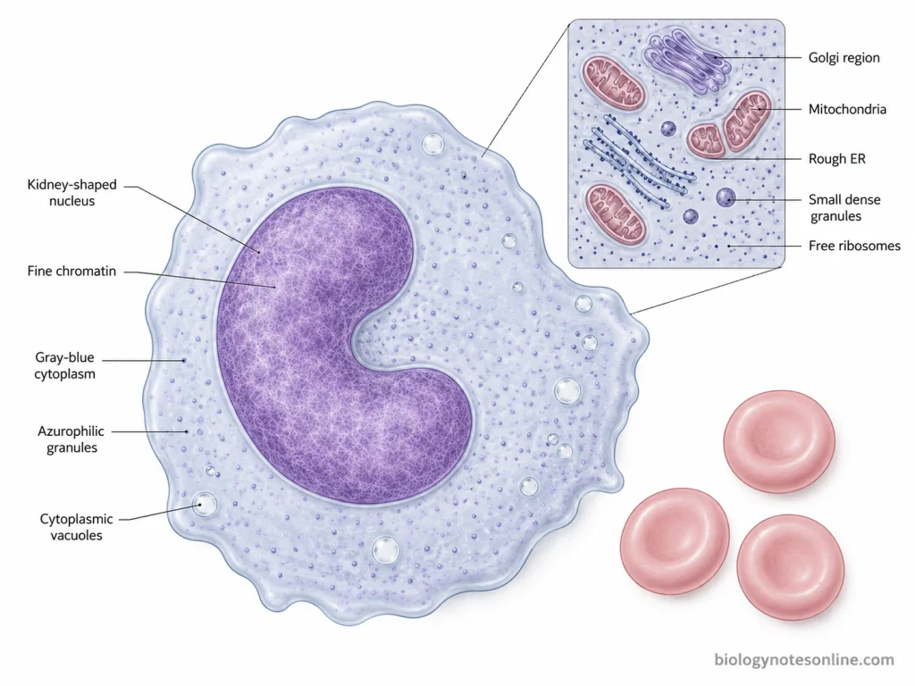
- The nucleus is large and usually irregular. Kidney-shaped and horseshoe-shaped nucleus is commonly seen, while sometimes it may appear bilobed. The chromatin is comparatively delicate.
- A large amount of pale gray to bluish cytoplasm is present. Fine azurophilic granules occur in the cytoplasm and cytoplasmic vacuoles may also be seen.
- Monocytes are produced from the myeloid lineage in bone marrow and after formation they are released into circulation. They remain in blood for a short period. During normal conditions or inflammation, these cells can later enter different tissues.
- These are important cells of innate immunity. Monocytes can recognize microorganisms and other danger signals with the help of different pattern-recognition receptors such as Toll-like receptors (TLRs).
- One of their major properties is phagocytosis. Microorganisms, damaged cells and cellular debris are engulfed by monocytes. Different cytokines are also produced during immune response.
- During infection or tissue injury, monocytes move from circulating blood into the affected tissues. In tissue, depending upon the signals present around the cells, they can give rise to monocyte-derived macrophages and dendritic cells.
- Monocytes can also process antigens and express MHC class II molecules. These molecules are involved in antigen presentation. Thus, the cells also have a role in connecting innate and adaptive immune responses.
- Human blood monocytes are not present as one uniform type. Based mainly on the expression of CD14 and CD16, three major populations are recognized. These are classical (CD14++CD16−), intermediate (CD14++CD16+) and non-classical (CD14+CD16++) monocytes.
- Classical monocytes form the major population in blood and show more phagocytic and inflammatory activity. Intermediate monocytes are more associated with processing and presentation of antigen.
- Non-classical monocytes move along the vascular endothelium. This type of movement is referred to as patrolling.
- Monocytes can perform different functions according to the signals present around them, and thus show considerable functional plasticity. They participate in microbial killing, inflammation and removal of damaged material. Tissue repair is also one of their functions.
Structure and Morphology of Monocytes
The following are the structural and morphological features of monocytes:
- Monocytes are large cells and are considered the largest leukocytes normally seen in the peripheral blood. They are generally about 12–20 µm in diameter, almost twice the size of a red blood cell.
- The cell outline is not always regular. Monocytes may have an irregular shape, and the cytoplasmic margin sometimes forms blunt projections or pseudopods.
- A large nucleus is present and its shape is quite variable. It is commonly indented, folded, kidney-shaped or horseshoe-shaped. In some cells the folding becomes deeper, giving a lobulated appearance.
- The nuclear chromatin has a characteristic fine and lacy appearance. It is less densely packed and appears lighter than the compact chromatin of lymphocytes. Mature monocytes generally do not show a distinct nucleolus.
- Cytoplasm is moderate to abundant and surrounds the irregular nucleus. It stains pale gray to blue with the usual blood stains. Due to the fine granules distributed throughout it, the cytoplasm frequently has a ground-glass appearance.
- Fine azurophilic granules are present in the cytoplasm. These granules are very small and lightly stained, usually reddish-blue or lilac in appearance. The azurophilic granules contain lysosomal enzymes and represent primary lysosomal granules of the monocyte.
- Cytoplasmic vacuoles may also be seen. They appear as clear or unstained spaces within the gray-blue cytoplasm and are more prominent in some monocytes than others.
- Under electron microscope, monocytes show an irregular nucleus with an extensive Golgi region. Numerous ribosomes, small parts of rough endoplasmic reticulum (RER), mitochondria and small dense granules are also present in the cytoplasm.
- Monocytes do not have one completely fixed appearance. Variation can be seen in their cell outline, degree of nuclear indentation, amount of cytoplasm, granules and the number of vacuoles present in the cell.
Origin and Development of Monocytes
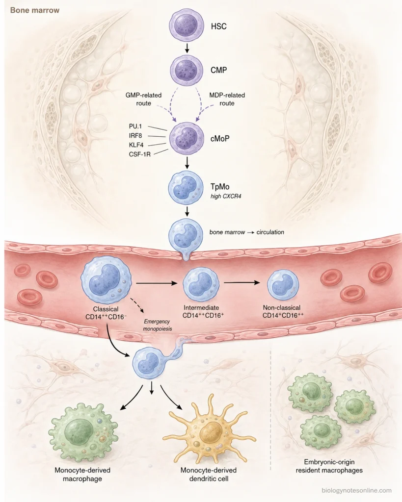
Monocytes are produced mainly in the bone marrow after birth from hematopoietic stem and progenitor cells. Their formation includes progressive restriction of the myeloid cells and finally the formation of mature circulating monocytes. The developmental process can be written in the following steps-
Step 1
The process begins with hematopoietic stem cells (HSCs) present in the bone marrow. These cells produce different hematopoietic progenitors, and the cells entering the myeloid pathway give rise to common myeloid progenitors (CMPs).
Step 2
CMPs further produce myeloid progenitors with more restricted capacity. Granulocyte-monocyte progenitors (GMPs) are formed during this process. Monocyte development can proceed through GMP-related and monocyte-dendritic cell progenitor (MDP) routes, so the pathway is not always a single fixed sequence.
Step 3
Further restriction results in formation of the common monocyte progenitor (cMoP). This progenitor is committed mainly to the monocyte lineage and has been identified in both mouse and human bone marrow. cMoPs can arise from different myeloid progenitor routes.
Step 4
During this stage the monocyte differentiation program becomes more prominent. Transcription factors such as PU.1, IRF8 and KLF4 take part in directing the progenitor toward monocyte development. CSF-1R (CD115) signalling is also important for development and survival of cells of this lineage.
Step 5
The cMoP then forms a transitional immature monocyte in the bone marrow. This cell is referred to as a transitional premonocyte (TpMo) and shows high expression of CXCR4. These cells can still proliferate and remain mainly retained within the bone marrow.
Step 6
TpMos gradually mature and CXCR4 expression becomes lower. Mature classical monocytes are now formed which have greater motility and functional maturity. After their last division, these cells can be released from the bone marrow into the circulation.
Step 7
The first major monocyte population appearing from the human bone marrow is the classical monocyte (CD14++CD16−). These cells remain in blood for a relatively short period. A part of the classical population then develops into intermediate monocytes (CD14++CD16+).
Step 8
Intermediate monocytes further give rise to non-classical monocytes (CD14+CD16++). Thus, in human blood the general maturation sequence is classical → intermediate → non-classical monocytes. This sequence has also been demonstrated by in vivo labeling studies.
Step 9
During infection or inflammatory condition, production and release of monocytes from the bone marrow can increase. This increased formation is referred to as emergency monopoiesis. Classical monocytes can be rapidly supplied to the blood during such conditions.
Step 10
After circulation, some monocytes leave the blood and enter tissues. Depending upon the local signals, these cells may develop into monocyte-derived macrophages or monocyte-derived dendritic cells. However, all tissue macrophages are not produced through this route, because many resident macrophage populations have an embryonic origin.
Types of Human Monocytes
Human monocytes present in the peripheral blood are not all similar. Based mainly on the expression of CD14 and CD16, they are classified into three major types. These are classical, intermediate and non-classical monocytes.
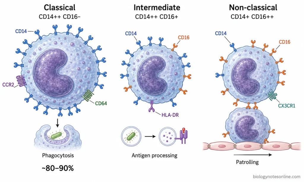
- Classical monocytes- These are represented as CD14++CD16− and form the major population of monocytes in normal human blood, generally about 80–90%. They express high amount of CCR2 and CD64. Classical monocytes show strong phagocytic activity and are rapidly recruited during inflammatory conditions.
- Intermediate monocytes- They are CD14++CD16+ monocytes and occur in much smaller number. Both CD14 and CD16 are expressed on their surface. A high expression of HLA-DR (MHC class II) is commonly present. These cells are more involved in antigen processing and presentation and show some features between classical and non-classical monocytes.
- Non-classical monocytes- These cells have comparatively low CD14 and high CD16 expression and are written as CD14+CD16++. They form a minor population in circulating blood. CX3CR1 is highly expressed in these cells. Non-classical monocytes move slowly along the vascular endothelium, and this movement is referred to as patrolling. They take part in vascular surveillance, inflammatory response and removal of cellular debris.
How Do Monocytes Reach Sites of Infection or Tissue Injury?
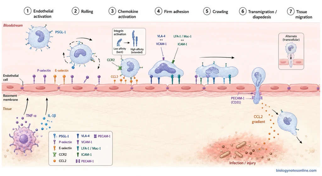
During infection or injury, monocytes present in circulating blood move towards the affected tissue. This process involves activation of endothelium, adhesion of monocytes and finally their movement across the blood vessel. The process takes place in the following steps-
Step 1- Activation of endothelium
At the infected or injured region, inflammatory mediators such as TNF-α and IL-1β activate the nearby vascular endothelial cells. Adhesion molecules are increased on their surface and chemokines are also presented there.
Step 2- Rolling of monocytes
The circulating monocytes are first captured on the activated endothelium and begin to roll slowly over it. P-selectin and E-selectin interact with ligands such as PSGL-1 present on the monocyte.
Step 3- Chemokine activation
In this step, chemokines present on the endothelial surface bind with their receptors on monocytes. CCL2 binds with CCR2, particularly during recruitment of classical monocytes. The monocyte integrins are now activated.
Step 4- Firm adhesion
After integrin activation, the rolling becomes stopped and monocyte attaches firmly to endothelial surface. VLA-4 binds with VCAM-1. LFA-1 and Mac-1 can bind with ICAM-1.
Step 5- Crawling
The attached monocyte changes its shape and starts moving over the inner surface of endothelium. It crawls towards a suitable region from where it can cross the vessel wall.
Step 6- Transmigration
Now the monocyte passes across the endothelial layer. This is referred to as transmigration or diapedesis. Usually, it moves between two adjacent endothelial cells, but movement through an endothelial cell can also occur. PECAM-1 is one of the molecules involved in this step.
Step 7- Migration into affected tissue
After crossing the endothelium and basement membrane, monocyte enters into the tissue. It moves along the increasing chemotactic signals towards the site of infection or tissue injury. Here, the recruited monocyte performs its immune functions.
Monocyte Count in a Blood Test
Monocyte count is commonly reported as a part of complete blood count (CBC) with differential. The result can be given as a percentage of total white blood cells or as an absolute monocyte count (AMC). Both values are useful, but their meaning is different.
Monocyte Percentage vs Absolute Monocyte Count
Monocyte percentage tells what proportion of the total white blood cells are monocytes. Thus, it is a relative value.
The absolute monocyte count gives the actual concentration of monocytes present in blood, usually expressed as cells/µL or ×10⁹/L. When it is not directly reported, the absolute value can be obtained from the total WBC count and monocyte percentage. Absolute differential counts are generally more useful for deciding whether the number of a particular white blood cell type is actually increased or decreased.
For example, the percentage of monocytes may appear increased when another white blood cell population becomes low, even if the actual number of monocytes has not increased. This is referred to as a relative increase.
The interpretation can be summarized as follows-
| Measurement | What it shows | Possible significance |
|---|---|---|
| Monocyte percentage | Percentage of total WBCs made up by monocytes. | A high or low percentage can result from changes in monocytes or changes in other white blood cells. |
| Absolute monocyte count (AMC) | Actual concentration of monocytes in blood. | Used to determine whether monocytes themselves are increased or decreased compared with the laboratory reference interval. |
| Percentage with AMC | Both relative and actual monocyte values are considered. | Gives better interpretation, especially when total WBC or other leukocyte counts are abnormal. |
What Is a Normal Monocyte Range?
There is no one monocyte range that is used by every laboratory. Reference values differ with laboratory method, population and age, and some laboratories also provide sex-specific values.
For example, Mayo Clinic Laboratories currently gives an adult AMC reference interval of 0.26–0.81 ×10⁹/L, whereas UChicago Medicine Medical Laboratories uses 0.16–0.92 ×10³/µL for persons 16 years and above. UChicago also reports an adult relative monocyte interval of 4–12%. These are laboratory reference values and not one universal normal range.
Therefore, the normal range printed along with the CBC result should be used. A monocyte value should be compared with the reference interval supplied by the laboratory which performed the test.
What Are High Monocytes (Monocytosis)?
An increase of the absolute monocyte count above the appropriate reference interval is referred to as monocytosis. It may be temporary or persistent.
Some of the conditions associated with monocytosis are-
- Infections, particularly some chronic infections. Monocytes may also rise during the recovery phase of an acute infection.
- Chronic inflammatory and autoimmune conditions can produce an increased monocyte count.
- Monocytosis may appear during recovery of the bone marrow after chemotherapy or other marrow suppression.
- Hematologic diseases are another cause. Persistent monocytosis can be present in chronic myelomonocytic leukemia (CMML) and some other myeloid neoplasms.
Monocytosis alone does not confirm any particular disease. The duration of the increase, other CBC findings, blood-cell morphology and the clinical condition are also considered before finding its cause.
What Are Low Monocytes (Monocytopenia)?
A monocyte count below the laboratory reference interval is called monocytopenia. Isolated monocytopenia is relatively uncommon and sometimes it occurs along with reduction of other blood cell types.
Low monocyte counts can be seen in-
- Bone marrow failure or suppression, including aplastic anemia and after chemotherapy.
- Severe infections such as sepsis.
- Treatment with some drugs, particularly glucocorticoids.
- Certain hematologic conditions, including hairy cell leukemia and myelodysplastic disorders.
A single low result does not always indicate an immune deficiency. Other white blood cell counts, medicines, recent illness and persistence of the low value are important for its interpretation.
Functions of Monocytes
Monocytes perform different functions during immune response and after entering into tissues. Some of the important functions of monocytes are as follows-
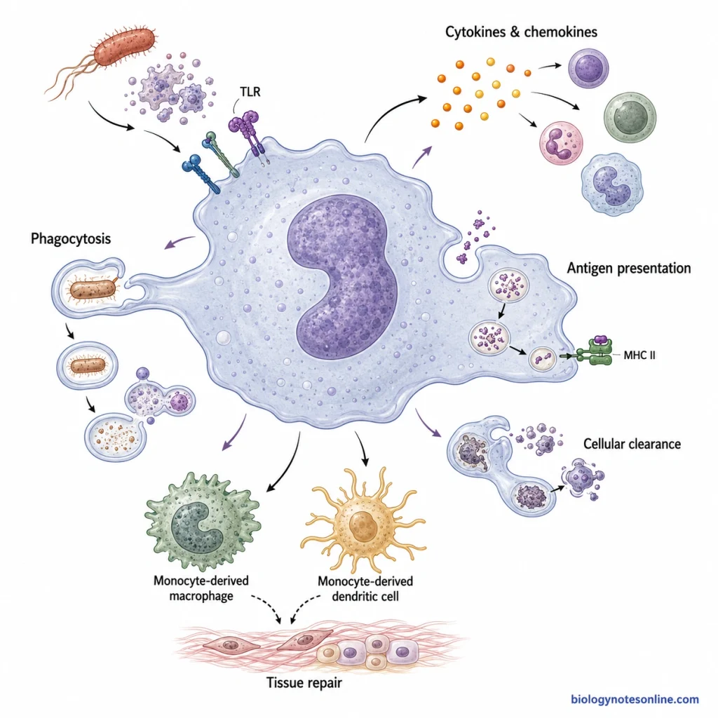
- One of the major functions of monocytes is phagocytosis. They engulf bacteria and other microorganisms and destroy them inside the cell.
- Monocytes recognize microorganisms and different danger signals through receptors present on their surface. This is important for activation of the innate immune response.
- Different cytokines and chemokines are produced by activated monocytes. Their production increases during infection and inflammatory conditions.
- Monocytes take part in antigen presentation. Antigens are taken up and processed by these cells and the antigenic peptides are presented through MHC molecules.
- During infection or tissue injury, circulating monocytes move from the blood into affected tissues. This migration allows the cells to reach the site where immune response is taking place.
- Some monocytes after entering tissues can develop into monocyte-derived macrophages or monocyte-derived dendritic cells. The type of cell formed depends upon the signals present in the tissue.
- Dead cells, damaged cells and cellular debris are also removed by monocytes. This is an important function during cellular clearance.
- Non-classical monocytes move along the surface of vascular endothelium. This characteristic movement is referred to as patrolling.
- Monocytes can regulate the inflammatory response. Depending upon the signals received by the cells, inflammatory or anti-inflammatory activities can be produced.
- Monocytes and monocyte-derived cells also take part in tissue repair. They participate in recovery of the damaged tissue after injury.
Biological and Clinical Importance of Monocytes
Monocytes are important during normal immune response as well as in different disease conditions. They take part in infection, inflammation, tissue repair and also have clinical importance.
- Host defence- Monocytes are recruited to infected tissues. They engulf and destroy different microorganisms.
- Inflammation- Monocytes produce different cytokines and inflammatory mediators. These substances take part in development and regulation of inflammation.
- Antigen presentation- Monocytes can process and present antigens through MHC molecules. Thus, they also participate in adaptive immune response.
- Vascular surveillance- Non-classical monocytes move along the vascular endothelium. This patrolling activity helps in detecting vascular damage.
- Tissue repair- Monocytes and monocyte-derived macrophages participate in repair of injured tissues. Abnormal or prolonged activity can also contribute to fibrosis.
- Cell differentiation- After entering tissues, some monocytes can form monocyte-derived macrophages or dendritic cells, especially during inflammatory conditions.
- Clinical evaluation- Changes in blood monocyte count are useful during clinical examination. Increased counts can occur in infections, inflammatory disorders and hematologic diseases.
- CMML- Persistent monocytosis is an important finding in chronic myelomonocytic leukemia (CMML). Other blood, bone marrow and molecular findings are also considered during diagnosis.
- Clinical biomarkers- Changes in classical, intermediate and non-classical monocyte populations are studied in different inflammatory diseases. These changes may have value as clinical biomarkers.
- Disease progression- Excessive or prolonged monocyte activity can take part in atherosclerosis, fibrosis and some cancers. Monocytes and their derived cells are therefore also studied as possible treatment targets.
References
- Caër, C., & Wick, M. J. (2020). Human intestinal mononuclear phagocytes in health and inflammatory bowel disease. Frontiers in Immunology, 11, 410. https://doi.org/10.3389/fimmu.2020.00410
- Canè, S., Ugel, S., Trovato, R., Marigo, I., De Sanctis, F., Sartoris, S., & Bronte, V. (2019). The endless saga of monocyte diversity. Frontiers in Immunology, 10, 1786. https://doi.org/10.3389/fimmu.2019.01786
- Chong, S. Z., Evrard, M., Devi, S., Chen, J., Lim, J. Y., See, P., Zhang, Y., Adrover, J. M., Lee, B., Tan, L., Li, J. L. Y., Liong, K. H., Phua, C., Balachander, A., Boey, A., Liebl, D., Tan, S. M., Chan, J. K. Y., Balabanian, K., . . . Ng, L. G. (2016). CXCR4 identifies transitional bone marrow premonocytes that replenish the mature monocyte pool for peripheral responses. Journal of Experimental Medicine, 213(11), 2293–2314. https://doi.org/10.1084/jem.20160800
- Cormican, S., & Griffin, M. D. (2020). Human monocyte subset distinctions and function: Insights from gene expression analysis. Frontiers in Immunology, 11, 1070. https://doi.org/10.3389/fimmu.2020.01070
- De Kleer, I., Willems, F., Lambrecht, B., & Goriely, S. (2014). Ontogeny of myeloid cells. Frontiers in Immunology, 5, 423. https://doi.org/10.3389/fimmu.2014.00423
- Doig, K., & Thompson, L. A. (2017). A methodical approach to interpreting the white blood cell parameters of the complete blood count. Clinical Laboratory Science, 30(3), 186–193. https://doi.org/10.29074/ascls.30.3.186
- Espinoza, V. E., & Emmady, P. D. (2023). Histology, monocytes. In StatPearls. StatPearls Publishing. https://www.ncbi.nlm.nih.gov/books/NBK557618/
- Gerhardt, T., & Ley, K. (2015). Monocyte trafficking across the vessel wall. Cardiovascular Research, 107(3), 321–330. https://doi.org/10.1093/cvr/cvv147
- Ginhoux, F., & Jung, S. (2014). Monocytes and macrophages: Developmental pathways and tissue homeostasis. Nature Reviews Immunology, 14(6), 392–404. https://doi.org/10.1038/nri3671
- Jakubzick, C. V., Randolph, G. J., & Henson, P. M. (2017). Monocyte differentiation and antigen-presenting functions. Nature Reviews Immunology, 17(6), 349–362. https://doi.org/10.1038/nri.2017.28
- Kapellos, T. S., Bonaguro, L., Gemünd, I., Reusch, N., Saglam, A., Hinkley, E. R., & Schultze, J. L. (2019). Human monocyte subsets and phenotypes in major chronic inflammatory diseases. Frontiers in Immunology, 10, 2035. https://doi.org/10.3389/fimmu.2019.02035
- Karlmark, K. R., Tacke, F., & Dunay, I. R. (2012). Monocytes in health and disease: Minireview. European Journal of Microbiology and Immunology, 2(2), 97–102. https://doi.org/10.1556/EuJMI.2.2012.2.1
- Kolypetri, P., & Weiner, H. L. (2023). Monocyte regulation by gut microbial signals. Trends in Microbiology, 31(10), 1044–1057. https://doi.org/10.1016/j.tim.2023.05.006
- Leick, M., Azcutia, V., Newton, G., & Luscinskas, F. W. (2014). Leukocyte recruitment in inflammation: Basic concepts and new mechanistic insights based on new models and microscopic imaging technologies. Cell and Tissue Research, 355(3), 647–656. https://doi.org/10.1007/s00441-014-1809-9
- Malcovati, L. (2024). Navigating the misty lands of monocytosis. Blood, 143(12), 1062–1064. https://doi.org/10.1182/blood.2023023332
- Mangaonkar, A. A., Tande, A. J., & Bekele, D. I. (2021). Differential diagnosis and workup of monocytosis: A systematic approach to a common hematologic finding. Current Hematologic Malignancy Reports, 16(3), 267–275. https://doi.org/10.1007/s11899-021-00618-4
- Marionneaux, S. (2020). Nonmalignant leukocyte disorders. In E. M. Keohane, C. N. Otto, & J. M. Walenga (Eds.), Rodak’s hematology: Clinical principles and applications (6th ed., pp. 445–465). Elsevier. https://doi.org/10.1016/B978-0-323-53045-3.00035-0
- Nichols, B. A., Bainton, D. F., & Farquhar, M. G. (1971). Differentiation of monocytes: Origin, nature, and fate of their azurophil granules. Journal of Cell Biology, 50(2), 498–515. https://doi.org/10.1083/jcb.50.2.498
- Olingy, C. E., Dinh, H. Q., & Hedrick, C. C. (2019). Monocyte heterogeneity and functions in cancer. Journal of Leukocyte Biology, 106(2), 309–322. https://doi.org/10.1002/JLB.4RI0818-311R
- Osa-Andrews, B., van Wijk, X. M. R., Herrera Rivera, N., Seifert, R. P., Harris, N. S., & Marin, M. J. (2025). An introduction to the complete blood count for clinical chemists: White blood cells. The Journal of Applied Laboratory Medicine, 10(2), 459–475. https://doi.org/10.1093/jalm/jfaf004
- Patel, A. A., Zhang, Y., Fullerton, J. N., Boelen, L., Rongvaux, A., Maini, A. A., Bigley, V., Flavell, R. A., Gilroy, D. W., Asquith, B., Macallan, D., & Yona, S. (2017). The fate and lifespan of human monocyte subsets in steady state and systemic inflammation. Journal of Experimental Medicine, 214(7), 1913–1923. https://doi.org/10.1084/jem.20170355
- Planchon, M. S., Fishman, J. A., & El Khoury, J. (2024). Modulation of monocyte effector functions and gene expression by human cytomegalovirus infection. Viruses, 16(12), 1809. https://doi.org/10.3390/v16121809
- Randolph, G. J., Jakubzick, C., & Qu, C. (2008). Antigen presentation by monocytes and monocyte-derived cells. Current Opinion in Immunology, 20(1), 52–60. https://doi.org/10.1016/j.coi.2007.10.010
- Riley, L. K., & Rupert, J. (2015). Evaluation of patients with leukocytosis. American Family Physician, 92(11), 1004–1011. https://www.aafp.org/pubs/afp/issues/2015/1201/p1004.html
- Robbins, C. S., & Swirski, F. K. (2010). The multiple roles of monocyte subsets in steady state and inflammation. Cellular and Molecular Life Sciences, 67(16), 2685–2693. https://doi.org/10.1007/s00018-010-0375-x
- Sampath, P., Moideen, K., Ranganathan, U. D., & Bethunaickan, R. (2018). Monocyte subsets: Phenotypes and function in tuberculosis infection. Frontiers in Immunology, 9, 1726. https://doi.org/10.3389/fimmu.2018.01726
- Shi, C., & Pamer, E. G. (2011). Monocyte recruitment during infection and inflammation. Nature Reviews Immunology, 11(11), 762–774. https://doi.org/10.1038/nri3070
- Skinner, B. M., & Johnson, E. E. P. (2017). Nuclear morphologies: Their diversity and functional relevance. Chromosoma, 126(2), 195–212. https://doi.org/10.1007/s00412-016-0614-5
- Sreejit, G., Fleetwood, A. J., Murphy, A. J., & Nagareddy, P. R. (2020). Origins and diversity of macrophages in health and disease. Clinical & Translational Immunology, 9(12), e1222. https://doi.org/10.1002/cti2.1222
- Tahir, S., & Steffens, S. (2021). Nonclassical monocytes in cardiovascular physiology and disease. American Journal of Physiology-Cell Physiology, 320(5), C761–C770. https://doi.org/10.1152/ajpcell.00326.2020
- Teh, Y. C., Chooi, M. Y., & Chong, S. Z. (2023). Behind the monocyte’s mystique: Uncovering their developmental trajectories and fates. Discovery Immunology, 2(1), kyad008. https://doi.org/10.1093/discim/kyad008
- Terry, R. L., & Miller, S. D. (2014). Molecular control of monocyte development. Cellular Immunology, 291(1–2), 16–21. https://doi.org/10.1016/j.cellimm.2014.02.008
- Uribe-Querol, E., & Rosales, C. (2020). Phagocytosis: Our current understanding of a universal biological process. Frontiers in Immunology, 11, 1066. https://doi.org/10.3389/fimmu.2020.01066
- Wacleche, V. S., Tremblay, C. L., Routy, J.-P., & Ancuta, P. (2018). The biology of monocytes and dendritic cells: Contribution to HIV pathogenesis. Viruses, 10(2), 65. https://doi.org/10.3390/v10020065
- Williams, H., Mack, C., Baraz, R., Marimuthu, R., Naralashetty, S., Li, S., & Medbury, H. (2023). Monocyte differentiation and heterogeneity: Inter-subset and interindividual differences. International Journal of Molecular Sciences, 24(10), 8757. https://doi.org/10.3390/ijms24108757
- Wolf, A. A., Yáñez, A., Barman, P. K., & Goodridge, H. S. (2019). The ontogeny of monocyte subsets. Frontiers in Immunology, 10, 1642. https://doi.org/10.3389/fimmu.2019.01642
- Wong, K. L., Tai, J. J.-Y., Wong, W.-C., Han, H., Sem, X., Yeap, W.-H., Kourilsky, P., & Wong, S.-C. (2011). Gene expression profiling reveals the defining features of the classical, intermediate, and nonclassical human monocyte subsets. Blood, 118(5), e16–e31. https://doi.org/10.1182/blood-2010-12-326355
- Wynn, T. A., & Vannella, K. M. (2016). Macrophages in tissue repair, regeneration, and fibrosis. Immunity, 44(3), 450–462. https://doi.org/10.1016/j.immuni.2016.02.015
- Zhu, Y. P., Thomas, G. D., & Hedrick, C. C. (2016). 2014 Jeffrey M. Hoeg Award lecture: Transcriptional control of monocyte development. Arteriosclerosis, Thrombosis, and Vascular Biology, 36(9), 1722–1733. https://doi.org/10.1161/ATVBAHA.116.304054
- Ziegler-Heitbrock, L., Ancuta, P., Crowe, S., Dalod, M., Grau, V., Hart, D. N., Leenen, P. J. M., Liu, Y.-J., MacPherson, G., Randolph, G. J., Scherberich, J., Schmitz, J., Shortman, K., Sozzani, S., Strobl, H., Zembala, M., Austyn, J. M., & Lutz, M. B. (2010). Nomenclature of monocytes and dendritic cells in blood. Blood, 116(16), e74–e80. https://doi.org/10.1182/blood-2010-02-258558
- Zini, G., Chang, Y. H., d’Onofrio, G., Frater, J., Germing, U., Merino, A., Pozdnyakova, O., Ross, D., Siqueira Filho, C. R., Takami, A., & Erber, W. (2026). ICSH recommendations for monocyte cell lineage morphologic identification, nomenclature harmonization, and utilization as a biomarker. International Journal of Laboratory Hematology, 48(1), 12–25. https://doi.org/10.1111/ijlh.70029