

Opsonization is an immune process by which microorganism, foreign particle or damaged cell is coated with molecules called opsonins. It makes the target more suitable for recognition by the phagocytic cells. The coating itself is not phagocytosis. After this coating, receptors present on macrophages and neutrophils can attach with the target more strongly, which helps in its engulfment and removal.
The major opsonins are antibodies and complement proteins. IgG is the important antibody opsonin. Another major one is C3b, which becomes deposited on the microbial surface during complement activation. C3b can be further converted into iC3b. It still acts as an important opsonic fragment although its activity in the complement amplification pathway is lost.
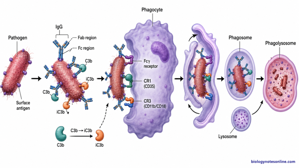
The phagocytic cells have receptors for these coated molecules. Fcγ receptors recognize the Fc portion of IgG. In case of complement opsonization, different complement receptors are involved, where C3b is recognized mainly by CR1 and iC3b has strong interaction with CR3 and CR4. Macrophages and neutrophils then bind with these opsonized particles. Phagocytosis becomes more efficient.
Quick facts about Opsonization
The following are some of the important facts about opsonization–
- Opsonin – It is a molecule which coats a target and increases its recognition by phagocytic cells.
- IgG – It is the classic antibody opsonin. The Fc portion remains available for binding with Fcγ receptors.
- C3b – One of the major complement opsonins deposited on microbial surfaces. It increases phagocytic recognition.
- iC3b – This is formed from C3b and remains an important opsonin. CR3 and CR4 are important receptors for this fragment.
- Macrophages and neutrophils – These are among the major phagocytic cells involved in removal of opsonized microorganisms.
- Opsonization and phagocytosis are related processes but not the same. Opsonization marks and promotes attachment of the target, while during phagocytosis the particle is actually taken inside the cell.
What Is Opsonization?
Opsonization is an immune process in which the surface of a microorganism, foreign particle or cell is coated with molecules called opsonins, making it more easily recognized by phagocytic cells. IgG antibodies and complement-derived C3 fragments, mainly C3b and iC3b, are important opsonins. It is a marking process.
An opsonin is a molecule that binds with a target and increases its recognition and uptake by a phagocyte. After coating, these molecules form recognizable sites on the target surface. The Fc portion of IgG is recognized by Fcγ receptors. Complement fragments are recognized by their respective complement receptors present on phagocytic cells, where iC3b is an important ligand for CR3. This makes attachment with the target stronger and phagocytosis becomes more efficient.
The target can be a microorganism, particle or a cell which has to be removed. Phagocytosis does not always require opsonization. Some particles can be taken up directly through other phagocytic receptors, but their uptake may be much lower than the opsonized particles. In some cases the difference is large.
For example, the encapsulated bacterium Streptococcus pneumoniae is efficiently opsonized by specific antibodies together with complement, which helps its uptake by neutrophils. Opsonization is therefore the coating or tagging of the target. Phagocytosis is the actual uptake of that target by the phagocytic cell.
How Opsonization Works
The process of opsonization takes place by coating of microorganism or other target with opsonins. After this, the coated target is recognized by receptors of phagocytic cells. The steps are as follows-
1. Binding of opsonin
In the first step, opsonins bind or become deposited on the surface of microorganism. IgG antibodies bind with their specific surface antigens. Complement activation also causes deposition of C3b on microbial surface, some of which can be converted to iC3b. These act as the coating molecules.
2. Recognition by phagocytic receptors
The coated opsonins are then recognized by specific receptors present on phagocytes. The Fc portion of IgG binds with Fcγ receptors. C3b and iC3b are recognized by complement receptors. For iC3b, CR3 and CR4 are important receptors.
3. Strong attachment of the target
Many opsonin molecules can be present over the same microbial surface. Due to this, several receptors of the phagocyte bind with the coated target at the same time. Receptors become clustered. This produces a much stronger attachment of the microorganism with the phagocytic cell.
4. Actin rearrangement
After binding of receptors, intracellular signals are produced inside the phagocyte. In Fcγ receptor-mediated phagocytosis, signaling involving ITAM, Syk, PI3K and small GTPases takes part. These signals cause rearrangement of the actin cytoskeleton around the attached particle.
5. Formation of phagosome
The cell membrane now begins to extend around the microorganism. It gradually surrounds the complete target and closes around it. The microorganism is therefore taken inside a membrane-bound vesicle called phagosome.
6. Maturation of phagosome
The newly formed phagosome undergoes further maturation inside the cell. During this process, it interacts with endosomal compartments and then lysosomal components. The compartment becomes acidic and develops into the degradative phagolysosome.
7. Killing and degradation
Inside the phagolysosome, the engulfed microorganism is exposed to lysosomal enzymes and antimicrobial substances. Phagocytes such as neutrophils can also generate reactive oxygen species during this process. The microorganism is killed and its different components are degraded.
Antibody-Mediated Opsonization
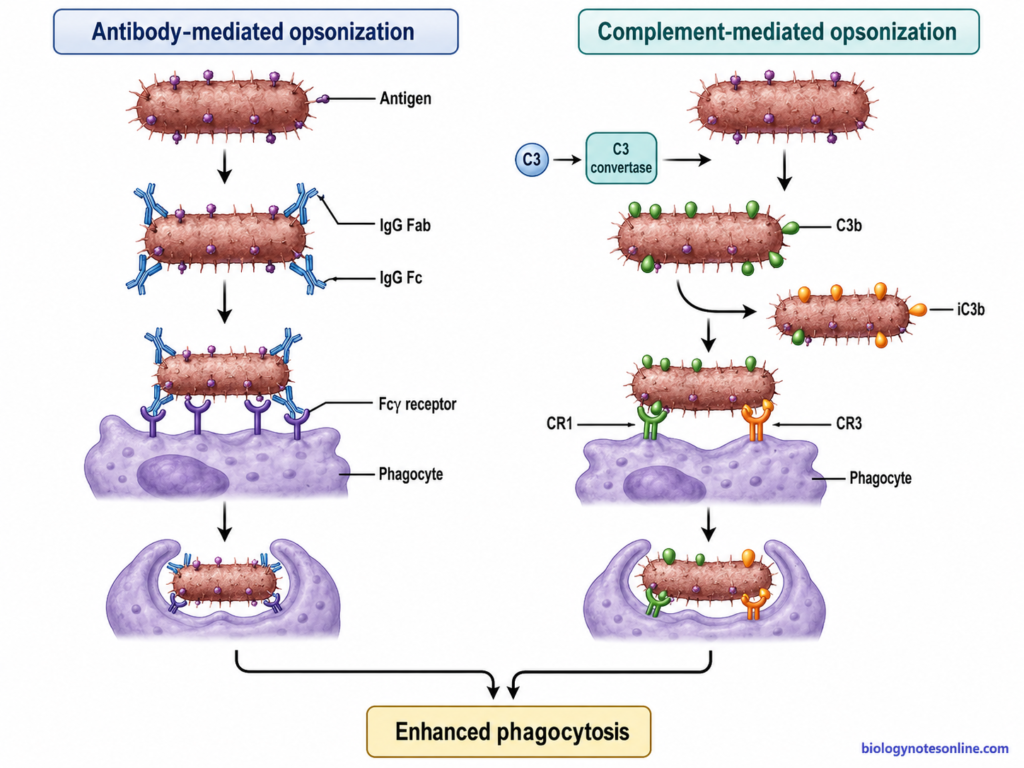
- Antibody-mediated opsonization is a process in which antibodies bind on the surface of microorganism and make it more readily recognized by phagocytic cells. IgG is the major antibody involved in this type of opsonization.
- The Fab regions of IgG bind with specific antigenic sites present on the microorganism. Its Fc portion remains exposed outside. This exposed part is used for recognition by the phagocyte.
- Neutrophils, macrophages and other phagocytic cells have Fc gamma receptors (FcγRs) on their surface. These receptors bind with Fc region of IgG coating the target. Different Fcγ receptors are present according to the type of phagocytic cell.
- Usually several IgG molecules become attached over the microbial surface. More than one Fcγ receptor can therefore bind with the antibody-coated target. The receptors become clustered at the site of contact.
- After Fcγ receptor binding, signals are produced inside the phagocytic cell. Activating Fcγ receptors use ITAM-dependent signaling, followed by activation of Syk kinase. It causes further signaling and rearrangement of actin cytoskeleton.
- The cell membrane then begins to extend around the attached microorganism. Pseudopodia are formed with the help of actin. These gradually surround the antibody-coated particle.
- The microorganism is taken inside the phagocyte and enclosed within a membrane-bound phagosome. The phagosome later matures and interacts with lysosomal compartments, forming the phagolysosome.
- Inside this compartment, the engulfed microorganism is exposed to lysosomal enzymes and other antimicrobial substances. In neutrophils and some other phagocytes, reactive oxygen species also take part in microbial killing.
- Antibody coating is especially useful for microorganisms which are poorly taken up directly by phagocytes. Encapsulated bacteria are an important example, where antibody binding makes their recognition by Fc receptors much more effective.
Complement-Mediated Opsonization
- Complement-mediated opsonization involves the coating of microorganisms with complement components, which are then recognized by complement receptors present on phagocytic cells.
- The process can begin through the classical, lectin or alternative pathway of complement activation. All three pathways result in the formation of C3 convertase.
- C3 convertase cleaves the complement protein C3 into C3a and C3b. It is C3b that mainly takes part in coating of the microbial surface.
- After cleavage of C3, a reactive thioester group of C3b becomes exposed. This allows the C3b molecule to form covalent attachment with suitable groups present on the surface of microorganism. Many molecules can be deposited in this way.
- Once the surface is coated with C3b, it can be recognized by complement receptors of phagocytic cells. CR1 (CD35) is an important receptor for C3b.
- Surface-bound C3b can also be cleaved by factor I, with the help of its regulatory cofactors, producing iC3b. It cannot form a C3 convertase. However, iC3b remains an important opsonin.
- iC3b is recognized mainly by CR3 (CD11b/CD18) and CR4 (CD11c/CD18). These receptors are found on different leukocytes including neutrophils, monocytes and macrophages.
- Binding of the complement-coated microorganism with these receptors increases its attachment to the phagocyte. Several receptor-opsonin interactions may occur over the same particle. The particle is therefore held more effectively on the cell surface.
- After receptor activation, changes in the actin cytoskeleton take place. The cell membrane moves around the bound microorganism and it is taken inside the phagocytic cell. A membrane-bound phagosome is formed.
- The phagosome then undergoes maturation and fuses with lysosomal compartments. This forms a degradative phagolysosome, where the microorganism is exposed to lysosomal enzymes and other antimicrobial substances.
- CRIg is another complement receptor which binds C3b and iC3b. It is mainly expressed by certain tissue macrophages and also helps in uptake of complement-opsonized microorganisms.
Major Opsonins and Their Receptors
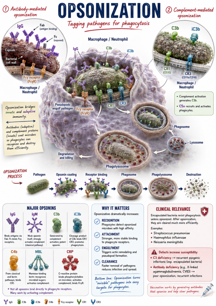
The major opsonins involved in phagocytosis are IgG antibodies and complement-derived C3 fragments. These molecules become attached to the target surface and are recognized by their specific receptors. Different opsonins have different receptors on phagocytic cells. The following are the important opsonins and their receptors-
1. IgG
IgG is the major antibody opsonin. It binds to antigen by its Fab regions while the Fc portion remains exposed outside. This Fc portion is recognized by Fc gamma receptors (FcγRs) present on neutrophils, macrophages and other immune cells.
The important activating receptors include FcγRI (CD64), FcγRIIA (CD32A) and FcγRIIIA (CD16A), although their distribution is different according to the cell type. FcγRI has high affinity for IgG. Binding and clustering of activating Fcγ receptors can initiate uptake of the IgG-coated particle.
2. C3b
C3b is one of the major complement opsonins. It is formed after cleavage of complement component C3 and becomes covalently deposited on the surface of microorganisms or other targets. Large amounts may be deposited during complement activation.
The important receptor for C3b is complement receptor 1 (CR1 or CD35). CR1 can bind C3b and also C4b. It is present on several blood cells including phagocytic cells, although its function also includes complement regulation and immune-complex clearance.
3. iC3b
iC3b is produced by proteolytic processing of surface-bound C3b. It cannot continue the complement convertase reactions like C3b, but it remains a very important opsonin.
Its major receptors are CR3 (CD11b/CD18, Mac-1) and CR4 (CD11c/CD18). Both receptors show strong recognition of iC3b-coated targets. CR3 is especially abundant on neutrophils, monocytes and macrophages and has an important role in opsonophagocytosis.
4. C4b
C4b is another complement fragment which can act as an opsonin. It is produced during activation of the classical and lectin complement pathways. Its opsonic role is less prominent compared with C3-derived fragments.
CR1 (CD35) is an important receptor for C4b. The same receptor also binds C3b.
5. C1q, MBL and Ficolins
C1q, mannose-binding lectin (MBL) and some ficolins can also show opsonic activity. They bind to microbial or altered surfaces and can help in their clearance. These molecules also have a major role in starting complement activation.
CR1 can interact with C1q, MBL and ficolin-2. Their removal mechanisms are more variable and other receptors can also be involved, so these are not as simple as the IgG-Fcγ receptor or iC3b-CR3 interaction.
6. CRIg
CRIg (complement receptor of the immunoglobulin superfamily) is a complement receptor mainly associated with tissue-resident macrophages. It binds C3b and iC3b deposited on target surfaces. This receptor can take part in phagocytosis of complement-opsonized microorganisms.
Major Opsonins and Their Phagocyte Receptors
| Opsonin | Immune source/pathway | How it binds the target | Main receptor(s) to learn | Main outcome |
|---|---|---|---|---|
| IgG | Adaptive humoral immunity | Fab region binds specific antigen on target surface | Fcγ receptors (FcγR) | Fc receptor-mediated attachment and phagocytosis. |
| C3b | Complement system | Becomes covalently deposited on target after C3 cleavage | CR1 (CD35) | Complement recognition and increased attachment to phagocytes. |
| iC3b | Complement system, formed from C3b | Remains attached on target after C3b degradation | CR3 (CD11b/CD18), CR4 (CD11c/CD18) | Strong opsonophagocytosis of complement-coated particles. |
| C4b | Classical and lectin complement pathways | Covalently attaches to target during complement activation | CR1 (CD35) | Helps complement recognition and clearance. |
| C1q | Classical complement pathway | Binds antibodies, microbial structures or altered cell surfaces | Calreticulin (cC1qR)–CD91/LRP1 in some phagocytic systems | Helps uptake of coated targets, especially apoptotic cells. |
| MBL | Lectin complement pathway | Carbohydrate-recognition domains bind mannose and related sugars on target | Calreticulin–CD91/LRP1 described in macrophages | Direct opsonic activity and activation of complement, leading to increased uptake. |
| CRP / pentraxins | Soluble innate pattern-recognition proteins | Bind selected microbial and damaged-cell surface ligands | FcγRI, FcγRIIA where supported | Can promote phagocytosis directly and also activate classical complement. |
For learning, the three major relationships are IgG → FcγR, C3b → CR1, and iC3b → CR3/CR4. The receptor relationships of C1q, MBL and pentraxins are more context-dependent and should not be treated as equally simple one-opsonin–one-receptor pairs.
Opsonization and Phagocytosis
- Opsonization and phagocytosis are related immune processes, but both are not the same. Opsonization refers to coating or marking of a microorganism with opsonins such as IgG, C3b or iC3b. It mainly helps in recognition.
- Phagocytosis is the process in which a phagocytic cell takes a particle inside the cell. The particle becomes enclosed in a membrane-bound vesicle called phagosome and later undergoes intracellular degradation.
- During opsonization, opsonin molecules first become attached on the target surface. These molecules now act as recognizable ligands for receptors present on macrophages, neutrophils and other phagocytic cells. Fcγ receptors recognize IgG-coated particles while complement receptors recognize complement-coated particles.
- After receptor binding, the target becomes firmly attached with the phagocyte. Many opsonin molecules can bind over the same particle, therefore several receptors may interact at the same time. This gives a stronger overall attachment or avidity and increases the efficiency of uptake.
- Opsonization commonly takes place before phagocytosis. It makes the particle more easy for recognition and ingestion. The actual taking of particle inside the cell is phagocytosis.
- Phagocytosis, however, does not always require opsonization. Some microorganisms can be recognized directly by receptors present on phagocytes without previous coating by antibody or complement. This is called non-opsonic phagocytosis.
- In non-opsonic phagocytosis, microbial structures can directly interact with phagocytic receptors. Pattern-recognition receptors, lectin-type receptors and some integrins may take part depending on the microorganism.
- An encapsulated bacterium gives a common example. The capsule of Streptococcus pneumoniae interferes with normal phagocytic uptake. When the bacterium becomes properly coated with antibody and complement products, recognition by phagocytes is increased and its removal becomes more effective.
- Opsonization = marking and attachment of target.
- Phagocytosis = engulfment of the target by phagocytic cell.
Opsonization vs Phagocytosis
| Feature | Opsonization | Phagocytosis |
|---|---|---|
| Definition | Opsonization is the coating or marking of a microorganism or particle with opsonins. | Phagocytosis is the process by which a phagocytic cell engulfs a microorganism or particle. |
| Main purpose | It helps in better recognition and attachment of the target. | It takes the target inside the phagocytic cell. |
| Main molecules involved | IgG, C3b and iC3b are the major opsonins. | Actin, phagocytic receptors and membrane components take part in the process. |
| Receptors involved | Opsonins are recognized by Fcγ receptors, CR1, CR3 and CR4. | Receptor binding starts the engulfment process. Both opsonic and non-opsonic receptors may be involved. |
| Sequence | It generally takes place before opsonic phagocytosis. | It occurs after recognition and attachment of the target. |
| What happens | Opsonins become attached on the target surface. | Cell membrane surrounds the target and forms a phagosome. |
| Need for opsonin | Opsonins are required for this process. | Opsonins are not always required. Some particles can undergo non-opsonic phagocytosis. |
| Effect on uptake | It makes attachment with phagocytes more efficient. | It results in actual uptake of the particle. |
| Final outcome | Target becomes more suitable for recognition and clearance. | The engulfed target is taken into a phagosome and later degraded after phagosome maturation. |
| Example | A bacterium coated with IgG or C3 fragments. | A macrophage or neutrophil engulfing the coated bacterium. |
Opsonization of Encapsulated Bacteria
- Encapsulated bacteria contain an outer polysaccharide capsule, which can interfere with their direct recognition and attachment to phagocytic cells. The effect is not exactly same in all encapsulated bacteria. In Streptococcus pneumoniae, the capsule can reduce complement deposition and also strongly inhibits neutrophil phagocytosis.
- Antibodies bind with capsular or other surface antigens of the bacteria. Complement components can also become deposited over the bacterial surface. C3b and its product iC3b are important in this coating. These now act as recognizable molecules for receptors of phagocytes.
- The antibody-coated part is recognized through Fc receptors, whereas C3-coated bacteria are recognized through complement receptors. Thus, the capsule is no longer the only surface presented to the phagocytic cell. Attachment becomes more effective.
- Neutrophils and macrophages can then bind and take up the opsonized bacteria. This is especially important for bacteria present in blood, where efficient removal is required before extensive multiplication takes place.
- The spleen has an important role in clearance of encapsulated bacteria from circulation. Splenic macrophages and the filtering action of spleen help in removal of these organisms, and loss of splenic function greatly increases the risk from encapsulated bacterial infection.
- Antibody and complement can work together during this process. Antibody may directly promote Fc receptor-mediated uptake and can also activate complement, producing more complement coating over the bacterial surface. The relative importance of these mechanisms varies between bacterial species and antibodies.
Examples of Opsonization
- Streptococcus pneumoniae – It contains a polysaccharide capsule which is a major factor for resistance to phagocytosis. The capsule reduces effective C3b/iC3b deposition and interferes with Fc receptor, complement receptor and even non-opsonic phagocytosis. When specific antibody and complement are present, opsonophagocytic uptake is greatly increased.
- Neisseria meningitidis – The meningococcal capsule has an important role in resistance against complement and phagocytosis. Antibodies together with complement can promote opsonophagocytosis by polymorphonuclear cells. In serogroup B, the polysialic acid capsule is particularly important in resistance to complement-mediated killing and opsonophagocytosis.
- Haemophilus influenzae type b – It has a polyribosyl ribitol phosphate (PRP) capsule. This capsule makes the bacterium resistant to phagocytosis in the absence of specific anticapsular antibody. Deposition of C3 helps binding with macrophages, and antibody further increases ingestion of the bacteria.
Biological Importance of Opsonization
The following are some of the important biological roles of opsonization–
- Opsonization increases the recognition of microorganisms by phagocytic cells. IgG, C3b or iC3b present over the microbial surface provide binding sites for their respective receptors. The uptake by macrophages and neutrophils is therefore increased.
- It is especially important in removal of encapsulated bacteria. The capsule can interfere with effective attachment and phagocytosis, while coating by antibody and complement makes these organisms more suitable for phagocytic uptake. Streptococcus pneumoniae is an important example.
- Antibody-mediated opsonization provides a connection between antibody response and phagocytic cells. The antibody first recognizes a specific antigen and its Fc portion can then bind with Fcγ receptors of phagocytes. In this way antigen-specific antibody helps in actual removal of the target.
- Complement also provides opsonic activity before a specific antibody response is formed. The alternative and lectin pathways can activate complement without antigen-specific antibody, producing deposition of C3 fragments on microbial surfaces. This is an important part of early innate defense.
- Opsonization is also involved in removal of apoptotic and damaged cells. Complement components and other opsonic molecules can become associated with apoptotic cell surface and promote their recognition by phagocytes. Rapid removal of these cells helps to prevent persistence of cellular contents and unwanted inflammatory reactions.
- Complement coating also helps in clearance of immune complexes from blood. Complement-coated immune complexes can bind with CR1 (CD35) present on erythrocytes. These are carried towards liver and spleen, where the complexes are removed by tissue macrophages.
- Efficient opsonization has major importance in protection against infection. Defects involving complement, particularly C3, can cause increased susceptibility to severe and recurrent infections, including infections by encapsulated organisms.
Clinical and Laboratory Relevance of Opsonization
- Reduced antibody or complement activity can decrease opsonization. Microbial clearance may therefore become poor.
- C3 deficiency affects formation of important complement opsonins. It is associated with severe and recurrent bacterial infections.
- Opsonophagocytic assay (OPA) is used to measure the functional activity of antibodies.
- Opsonophagocytic killing assay (OPKA) measures whether antibodies can help phagocytic cells in killing the target bacteria.
- In these assays, serum antibodies, complement and phagocytic cells are generally allowed to act against a selected bacterium. Bacterial survival or killing is then measured.
- OPKA is commonly used in vaccine studies.
- Vaccines against encapsulated bacteria can produce antibodies which support opsonization and phagocytic killing.
- Pneumococcal vaccines are an important example. The functional activity of vaccine-induced antibodies can be studied by opsonophagocytic assays.
- Reduced opsonization may occur in antibody or complement defects. However, every recurrent infection is not caused by an opsonization defect.
FAQ
- Crowley, M. T., Costello, P. S., Fitzer-Attas, C. J., Turner, M., Meng, F., Lowell, C., Tybulewicz, V. L. J., & DeFranco, A. L. (1997). A critical role for Syk in signal transduction and phagocytosis mediated by Fcγ receptors on macrophages. The Journal of Experimental Medicine, 186(7), 1027–1039.
- Advanced molecular physiology, biophysics, and clinical immunology of opsonization. (n.d.).
- CRIg: A macrophage complement receptor required for phagocytosis of circulating pathogens. (n.d.). ResearchGate.
- Cdc42 regulates Fcγ receptor-mediated phagocytosis through the activation and phosphorylation of Wiskott-Aldrich syndrome protein (WASP) and neural-WASP. (n.d.). PMC.
- Complement mediated opsonization. (n.d.). Sino Biological.
- Complement receptor of the immunoglobulin superfamily/CRIg. (n.d.). Creative Biolabs.
- Vandendriessche, S., Cambier, S., Proost, P., & Marques, P. E. (2021). Complement receptors and their role in leukocyte recruitment and phagocytosis. Frontiers in Cell and Developmental Biology, 9, Article 624025.
- Complement system part I – Molecular mechanisms of activation and regulation. (n.d.). PMC.
- Complement control protein factor H: The good, the bad, and the inadequate. (n.d.). PMC – NIH.
- Complement deficiencies and infections. (n.d.). PubMed.
- Complement receptor 3 mediates both sinking phagocytosis and … (n.d.).
- Complement receptor of the immunoglobulin family. (2022, March 27). In Wikipedia. https://en.wikipedia.org/w/index.php?title=Complement_receptor_of_the_immunoglobulin_family&oldid=1079568190.
- Development of a multi-specificity opsonophagocytic killing assay. (n.d.). PubMed – NIH.
- Efficiency of a pneumococcal opsonophagocytic killing assay improved by multiplexing and by coloring colonies. (n.d.). PMC.
- Encapsulated bacteria. (n.d.). Picmonic.
- Evaluate vaccine immunogenicity with OPKA. (n.d.). Pel-Freez Biologicals.
- Garapati, P. V. (2012). FCGR activation. Reactome Pathway Database.
- Józsi, M. (2017). Factor H family proteins in complement evasion of microorganisms. Frontiers in Immunology, 8, Article 571.
- Fc gamma R-mediated phagocytosis. (n.d.). CUSABIO.
- Fc receptor-mediated phagocytosis requires CDC42 and Rac1. (n.d.). PMC – NIH.
- Fcγ receptor–mediated phagocytosis in macrophages lacking the Src family tyrosine kinases Hck, Fgr, and Lyn. (n.d.). PMC.
- Moore, S. R., Menon, S. S., Cortes, C., & Ferreira, V. P. (2021). Hijacking factor H for complement immune evasion. Frontiers in Immunology, 12, Article 602277.
- Identification of two distinct mechanisms of phagocytosis controlled by different Rho GTPases. (n.d.). PubMed.
- Infections of people with complement deficiencies and patients … (n.d.).
- Measuring opsonic phagocytosis via Fcγ receptors and complement receptors on macrophages. (n.d.). PMC.
- InformationBoxTicket Lifestyles. (n.d.). Opsonization explained | Mechanism, antibodies, complement & examples | Immunology made simple [Video]. YouTube.
- Sapkota, A. (2025, August 8). Opsonization: Definition, mechanism, opsonins, examples. Microbe Notes.
- Cleveland Clinic. (2024, April 26). Opsonization: Process, purpose, effects & examples.
- Opsonophagocytic killing assay (OPKA). (n.d.). Synoptics.
- Opsonophagocytic killing assay to assess immunological responses against bacterial pathogens. (n.d.). PMC.
- Stoakes, S. F. (2023, July 21). Overview of opsonization. News-Medical.Net.
- Asuka, E., & Launico, M. V. (2023, May 1). Physiology, opsonization. In StatPearls. StatPearls Publishing.
- Pre-existing systemic inflammation impairs bacterial clearance in the spleen. (n.d.). PMC – NIH.
- Sadarangani, M. (2018). Protection against invasive infections in children caused by encapsulated bacteria. Frontiers in Immunology, 9, Article 2674.
- Regulation of phagocytosis by Rho GTPases. (n.d.). PMC – NIH.
- Role of complement system in phagocytosis. (n.d.). Creative Biolabs.
- Splenic red pulp macrophages eliminate the liver-resistant Streptococcus pneumoniae from the blood circulation of mice. (n.d.). PMC.
- Sun, X.-Z., & Liu, Y. (2026). The key role of complement receptor CRIg in Kupffer cell-mediated liver disease progression. Viruses, 18(4), Article 473.
- Janeway, C. A., Jr., Travers, P., Walport, M., & Shlomchik, M. J. (2001). The complement system and innate immunity. In Immunobiology: The immune system in health and disease (5th ed.). Garland Science.
- Biointron. (2024, November 22). What is opsonization?.