

Cytoskeleton is a network of protein filaments that are present throughout the cytoplasm of a cell. It forms the internal framework of the cell. It is involved in maintaining the cell shape and gives mechanical support to the cell.
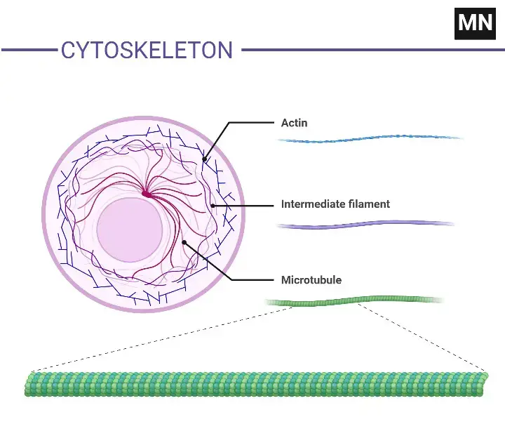
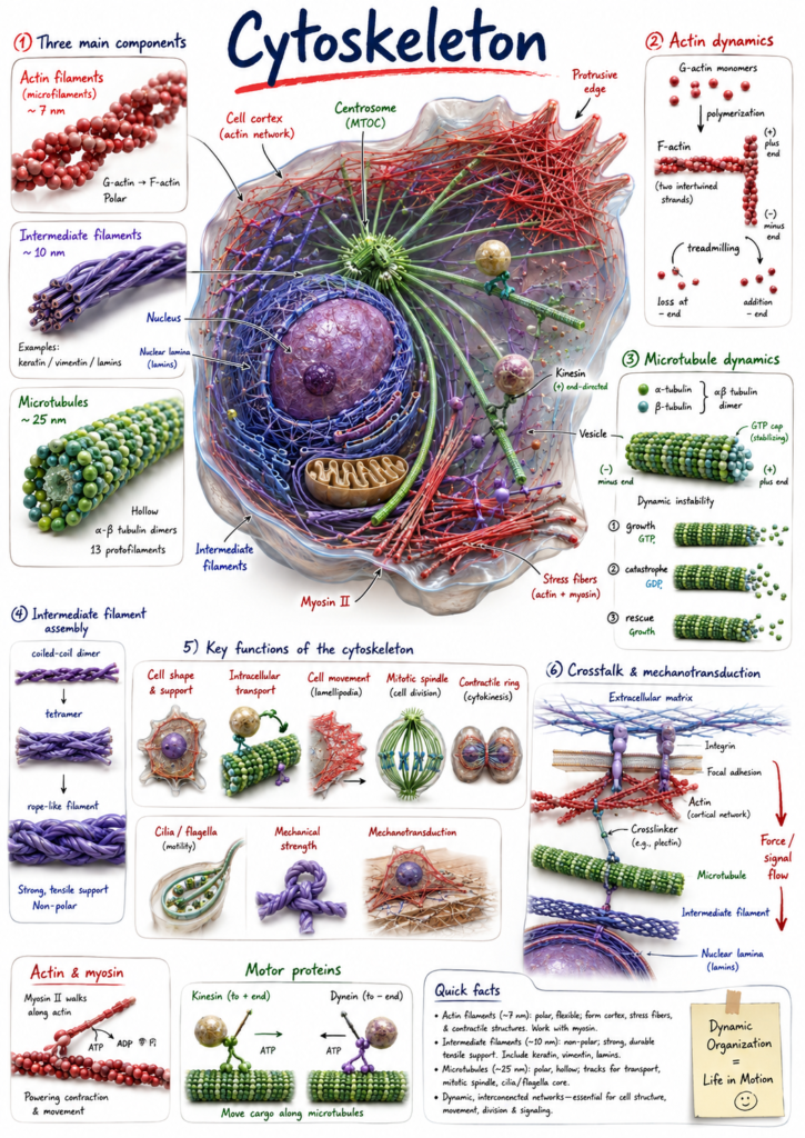
The cytoskeleton is not a fixed structure. Its filaments can assemble and disassemble according to different activities of the cell. In eukaryotic cells, the cytoskeleton is mainly made up of three types of filaments, microfilaments (actin filaments), intermediate filaments, and microtubules.
These filaments are different in their protein composition and structure. They are arranged in different regions of the cytoplasm. Some form a network below the cell membrane while others extend through the cytoplasm and help in the arrangement of different cellular components.
The cytoskeleton also has a role in movement of cell and its components. It is used in movement of organelles and vesicles, intracellular transport and cell division. Thus, it provides structural support to the cell and also participates in different cellular movements.
Why Cells Need a Cytoskeleton
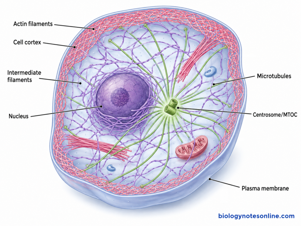
Cells need cytoskeleton for maintaining their internal organization and normal cell shape. The cell is not simply a membrane enclosing different organelles. Its internal components have to remain arranged in particular regions. Cytoskeletal filaments provide this internal support, and also help in maintaining the polarity of many cells.
The cytoplasm also contains vesicles, organelles and many other cellular materials which have to move from one region to another. Microtubules provide tracks for much of this movement. Motor proteins such as kinesin and dynein carry different cargo along them. Thus, the cytoskeleton has an important role in intracellular transport and also in positioning of organelles within the cell.
Cell movement requires continuous changes in cell shape. Actin filaments can be rapidly formed and removed at different regions of the cell. Actin together with myosin produces contractile force. These activities are used during crawling movement, changes of cell surface and several other forms of cellular movement.
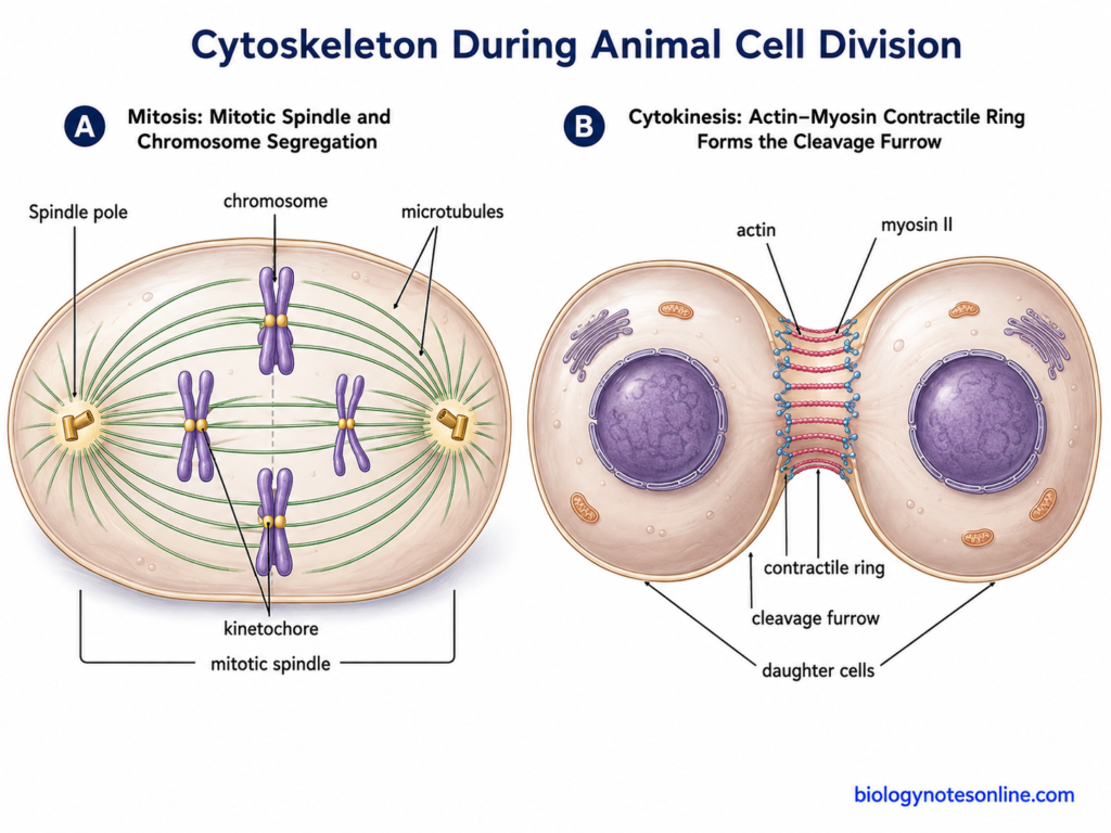
The cytoskeleton is also required when a cell divides. Microtubules form the mitotic spindle responsible for separation of chromosomes. Division of the cytoplasm involves another cytoskeletal system. In many animal and fungal cells, actin with myosin forms the contractile ring, which constricts during cytokinesis.
Mechanical stress is continuously produced on many cells. Cytoskeletal networks help the cell to resist such forces and maintain its structure. At the same time, the arrangement is not fixed. Filaments can reorganize when the cell grows, moves, divides or changes its shape. Due to these different activities, cytoskeleton is required for cell support, organization, movement and proper distribution of cellular components.
Key Characteristics of Cytoskeleton
Some of the important characteristics of cytoskeleton are-
- Cytoskeleton is a complex network of protein filaments present within the cytoplasm of a cell. It provides an internal framework, and is also associated with different movements occurring inside the cell.
- It is not enclosed by a membrane.
- The major cytoskeletal filaments include actin filaments (microfilaments), intermediate filaments and microtubules. Each is made of different protein components and they are also different in their structural organization.
- These filaments have different thickness. Actin filaments are about 6–8 nm in diameter, intermediate filaments are nearly 10 nm, while microtubules are much thicker, about 24–25 nm.
- Actin filaments and microtubules have two structurally different ends and hence they are polar. Intermediate filaments are non-polar.
- The cytoskeleton is not a fixed framework of cell. Actin and microtubules can show rapid formation and breakdown. Their organization changes during movement, division and other cellular activities.
- Intermediate filaments are relatively more stable and mainly provide mechanical strength. They can also undergo changes and reorganization, therefore these filaments are not completely permanent structures.
- Cytoskeletal proteins are arranged in different forms within the cell. Actin may form bundles or three-dimensional networks. A dense actin network is also present below the plasma membrane, called the cell cortex.
- Different accessory proteins remain associated with these filaments. Some proteins help in their formation, breakdown, branching and attachment with other structures of the cell.
- Motor proteins work along particular cytoskeletal filaments. Myosin is associated with actin. Kinesin and dynein are mainly associated with microtubules and are used for movement of different cellular materials.
- The distribution of cytoskeleton is not same in every type of cell. Intermediate filament proteins also differ according to cells, such as keratins, vimentin, desmin and neurofilament proteins.
- Cytoskeletal systems do not always function separately. Interaction of actin with microtubules occurs during cell migration, cell polarity and division, where the organization of one system can influence the other.
- Besides maintaining cell shape, the cytoskeleton has role in intracellular transport, movement of cell and its components, mechanical support and chromosome separation during cell division.
Cytoskeleton Across Different Cell Types and Organisms
The cytoskeleton is present in different types of cells, but its arrangement is not same in all. Animal and plant cells contain well-developed cytoskeletal systems. Prokaryotic cells also possess filament-forming proteins, although their system is different from the typical eukaryotic cytoskeleton.
Animal and Plant Cytoskeletons
Both animal and plant cells contain actin filaments and microtubules. Their distribution and organization differ because the structure of these cells is different.
- Animal cells– The cytoskeleton of animal cells commonly consists of actin filaments, microtubules, and intermediate filaments. Actin occurs in large amount below the plasma membrane and also through different regions of cytoplasm. Microtubules are generally extended outward from a microtubule-organizing region. In many animal cells, the centrosome acts as the major microtubule-organizing centre.
- Plant cells– Plant cells also contain a cytoskeleton. The presence of a rigid cell wall does not mean that cytoskeleton is absent. Actin filaments and microtubules are the major well-studied systems in plant cells and perform different functions in growth, intracellular movement and cell division.
- Cortical microtubules are commonly present just below the plasma membrane of plant cells. Their orientation is closely associated with deposition of cellulose in the cell wall. They guide the movement of cellulose synthase complexes, which affects the direction in which the cell expands.
- The plant cell wall and cytoskeleton are different structures. Cell wall forms a strong outer support. Cytoskeleton remains inside the cell and is involved in internal organization, transport and different changes occurring during growth.
- Most higher plant cells do not contain a typical centrosome like that of animal cells. Their microtubules can be organized from several regions of the cell.
- During plant cell division, microtubules form some special arrangements. The spindle is involved in chromosome separation. Later, the phragmoplast is formed between the daughter nuclei, which has an important role in formation of the new cell plate. Actin filaments are also associated with this region.
Prokaryotic Cytoskeletal Systems
The cytoskeleton was earlier considered mainly as a characteristic of eukaryotic cells. It is now known that bacteria also contain several filament-forming proteins. These, however, are not simply smaller forms of the three eukaryotic cytoskeletal filaments.
- FtsZ– It is a tubulin-related protein involved in bacterial cell division. FtsZ molecules assemble at the future division site and form a ring-like structure called the Z-ring. Other proteins required for division are then associated with this region.
- MreB– MreB is an actin-like protein present in many rod-shaped bacteria. It is associated with the inner surface of plasma membrane and works with proteins involved in cell-wall synthesis. MreB has a major role in maintaining the rod shape and normal growth of these bacterial cells.
- Bacterial cytoskeleton is not limited only to FtsZ and MreB. Some bacteria contain other filament-forming proteins such as crescentin and bactofilins, which are involved in cell shape and cellular organization.
- Thus, prokaryotic cells have their own cytoskeletal systems. Their proteins, arrangement and functions can be quite different from the classical cytoskeleton of animal and plant cells.
Components of the Cytoskeleton
The major components of cytoskeleton are of three types-
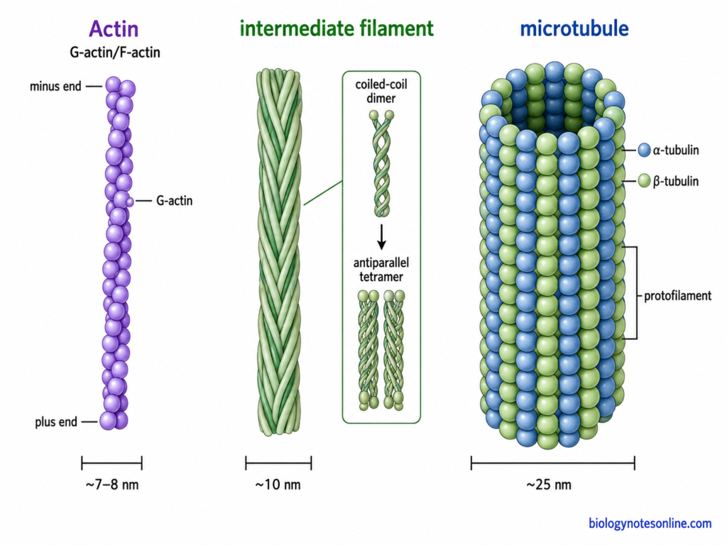
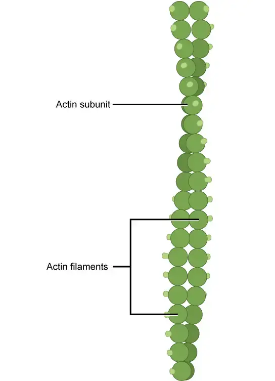
- Microfilaments (Actin filaments)– These are the thinnest cytoskeletal filaments, about 7 nm in diameter. They are made of actin protein. The globular form of actin (G-actin) joins one after another and forms filamentous actin (F-actin), which is arranged as two strands twisted around each other. A large number of these filaments occur below the plasma membrane. This region forms the cell cortex. Microfilaments take part in changes of cell shape, movement of cells and contraction of muscles. During cytokinesis, actin filaments also form the contractile ring for separation of the two cells.
- Intermediate filaments– The diameter is nearly 10 nm. These are strong, rope-like filaments and are more stable compared with microfilaments and microtubules. Different cells can contain different intermediate filament proteins. Keratins are present in epithelial cells, desmin in muscle cells, neurofilament proteins in neurons and lamins form a supporting network inside the nuclear envelope. Vimentin is another important type. Their major function is mechanical support. They resist stretching and mechanical stress on the cell.
- Microtubules– These are hollow tubes formed from α-tubulin and β-tubulin. Both proteins occur together as tubulin heterodimers. Usually 13 protofilaments make the wall of one microtubule, giving a diameter of about 25 nm. Microtubules are not permanent structures. They can grow or become shorter by addition and removal of tubulin units. Within the cell they provide tracks for movement of vesicles and different organelles. They also form the mitotic spindle during cell division. Cilia and flagella contain microtubules as their major structural component.
Actin Filaments vs Intermediate Filaments vs Microtubules
| Characteristics | Actin Filaments (Microfilaments) | Intermediate Filaments | Microtubules |
|---|---|---|---|
| Basic structure | Thin, solid protein filaments. | Strong rope-like filaments. | Hollow tubular structures. |
| Diameter | About 7 nm. | About 10 nm. | About 25 nm. |
| Main protein | Made up of actin. Globular actin (G-actin) polymerizes to form filamentous actin (F-actin). | Made from different intermediate filament proteins. Keratins, vimentin, desmin, neurofilament proteins and lamins are important examples. | Formed by α-tubulin and β-tubulin heterodimers. |
| Arrangement | Two actin strands are arranged around each other. They can form bundles and networks. | Protein dimers associate and form a rope-like filament. | Usually 13 protofilaments are arranged to form the wall of a microtubule. |
| Polarity | Polar. It has a plus and a minus end. | Non-polar. | Polar, having plus and minus ends. |
| Nature | Highly dynamic. Rapid assembly and disassembly occurs. | Comparatively more stable. Their arrangement can also be changed during different cellular activities. | Highly dynamic and show alternate periods of growth and shortening. |
| Nucleotide involved | ATP is bound to actin subunits and hydrolyzed after polymerization. | Polymerization does not depend on ATP or GTP hydrolysis. | GTP is associated with tubulin and β-tubulin-bound GTP is hydrolyzed after incorporation. |
| Common location | Large amount occurs below the plasma membrane, forming the cell cortex. | Extend through cytoplasm and provide mechanical connections. Nuclear lamins occur below the inner nuclear membrane. | Extend through the cytoplasm from microtubule-organizing regions. They are also present in spindle, cilia and flagella. |
| Motor protein | Mainly myosin. | No conventional motor protein moves along intermediate filaments. | Kinesin and dynein are the major microtubule motor proteins. |
| Major function | Cell movement, changes in cell shape, cytokinesis and muscle contraction. | Mainly provides mechanical strength and resistance against stretching. | Intracellular transport, organelle positioning, chromosome separation and maintenance of cell organization. |
| Role in cell division | Forms the contractile ring during cytokinesis in many animal cells. | Some intermediate filament systems undergo rearrangement during cell division. | Forms the mitotic spindle for chromosome separation. |
| Special structures formed | Microvilli, lamellipodia, filopodia and contractile structures. | Nuclear lamina and mechanically strong filament networks. | Mitotic spindle, cilia and flagella. |
| Main property | More associated with cell surface movement and force generation. | Gives tensile strength to cells and tissues. | Provides long intracellular tracks and organizes different regions of the cell. |
How Cytoskeletal Filaments Assemble and Remodel
The filaments of cytoskeleton are continuously organized according to different activities of the cell. New protein subunits can be added and the already formed filaments can also be disassembled. This assembly is especially rapid in actin filaments and microtubules. Intermediate filaments are generally more stable, but their arrangement can also change.
Actin Polymerization and Treadmilling
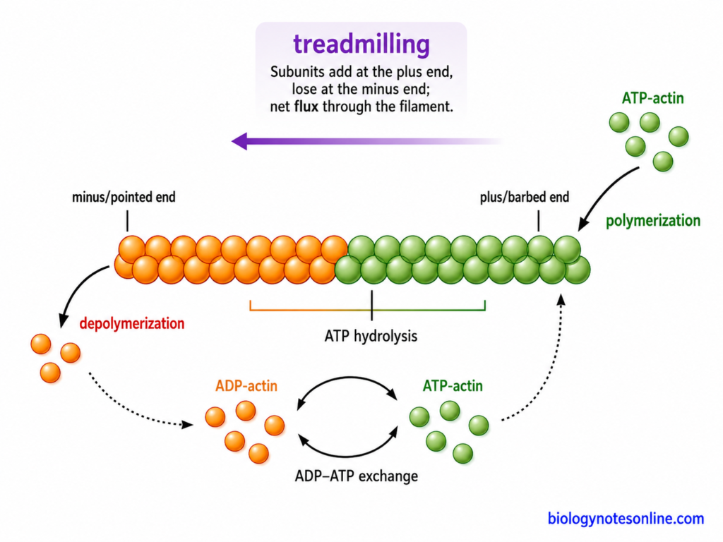
Actin filaments are formed by polymerization of globular actin (G-actin) molecules. The process involves nucleation followed by elongation of the filament. Actin filaments have two different ends, a plus or barbed end and a minus or pointed end.
The major events are as follows-
- Nucleation– In the initial step, a small group of actin molecules joins to form a stable nucleus. Formation of this nucleus is slow. Inside the cell, proteins such as Arp2/3 complex and formins help in nucleation of new actin filaments.
- Elongation– ATP-bound actin molecules are then added to the growing filament. Addition takes place at both ends but is usually faster at the plus end. After actin enters the filament, its bound ATP is hydrolyzed. Older portions of the filament therefore contain more ADP-actin.
- Treadmilling– Under suitable conditions, actin molecules are continuously added at the plus end while subunits are lost from the minus end. The filament can remain almost the same in length during this condition. This process is called treadmilling. It allows rapid renewal of actin without removing the complete filament.
Actin remodeling is also controlled by several actin-binding proteins. Cofilin promotes breakdown and turnover of older actin filaments, whereas profilin helps in supplying polymerization-competent actin. Other proteins can cap, cut, bundle or form branches in the filaments. Thus, actin networks can change rapidly in regions where cell movement or change of cell shape is taking place.
Microtubule Assembly and Dynamic Instability
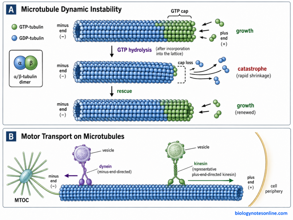
Assembly of microtubules takes place from α/β-tubulin heterodimers. In many animal cells, their nucleation occurs at the microtubule-organizing centre (MTOC), especially the centrosome, with the help of γ-tubulin ring complexes (γ-TuRCs). The minus end is commonly associated with the organizing centre. Plus end remains more dynamic.
Microtubule remodeling includes the following events-
- Growth– GTP-bound tubulin dimers are added mainly to the plus end. A region rich in GTP-tubulin is maintained at the growing end and is generally referred to as the GTP cap. It favors continued growth of the microtubule.
- GTP hydrolysis– After incorporation of the tubulin dimer into the microtubule, GTP associated with β-tubulin is hydrolyzed to GDP. The older microtubule lattice therefore contains mainly GDP-tubulin.
- Catastrophe– Sometimes the growing end loses its stabilizing GTP-rich cap. The protofilaments become unstable and rapid shortening begins. The change from growth to rapid shrinkage is called catastrophe.
- Rescue– A shrinking microtubule does not always disappear completely. It can again change to a growing state. This change from shortening to growth is known as rescue.
The repeated switching between growth and shortening is called dynamic instability. It enables microtubules to be rapidly rearranged, particularly during changes in cell organization and formation of the mitotic spindle.
Intermediate Filament Assembly and Remodeling
The assembly of intermediate filaments is different from actin filaments and microtubules. ATP or GTP hydrolysis is not required for their basic polymerization. They also do not possess distinct plus and minus ends.
The assembly takes place in a series of associations-
- Two intermediate filament protein molecules first coil around each other and form a coiled-coil dimer.
- Two dimers then associate in an antiparallel arrangement. A tetramer is formed. Because of this antiparallel arrangement, the filament has no overall polarity.
- Several tetramers associate laterally to produce short unit-length filaments (ULFs).
- These units join end-to-end. Further rearrangement and radial compaction produces the mature rope-like intermediate filament.
Intermediate filaments are relatively stable structures, but they are not permanently fixed. Their networks are reorganized during cell division, migration and other changes in cell structure. Phosphorylation is one important mechanism controlling this process. For example, phosphorylation of vimentin promotes filament disassembly, and phosphorylation of nuclear lamins contributes to breakdown of the nuclear lamina during mitosis. The proteins can later assemble again after changes in their phosphorylation state.
Coordination Between Cytoskeletal Networks
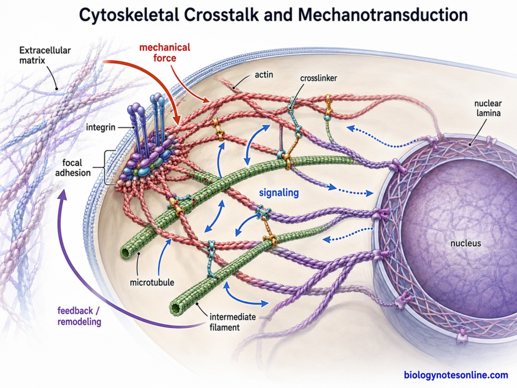
The different cytoskeletal filaments do not always work separately. Actin filaments, microtubules, and intermediate filaments can interact with one another and form a coordinated cellular system. Such interactions are generally referred to as cytoskeletal crosstalk.
The major types of coordination are as follows-
- Actin and microtubule coordination– Interaction between actin filaments and microtubules is common during cell movement, cell polarity and division. Microtubules can grow toward actin-rich regions of the cell. Actin organization can also influence where the microtubules are captured or stabilized. Different linking and regulatory proteins are involved in this interaction.
- Coordination during cell migration– A migrating cell needs continuous rearrangement of both filament systems. Actin polymerization produces protrusion at the leading edge. Microtubules extend toward this region and help in maintaining cell polarity, transport of materials and organization of adhesion sites. Their activities therefore occur together rather than as two independent processes.
- Linking proteins– Some proteins can interact with more than one cytoskeletal system. Spectraplakins, for example, can associate with actin and microtubules, and some members are also connected with intermediate filaments and cell junctions. These proteins provide physical links between different filament networks.
- Intermediate filament interactions– Intermediate filaments are also connected with actin and microtubule systems. Their main role is not only mechanical support. Proteins such as vimentin can influence organization of the other cytoskeletal networks, and intermediate filaments participate in transmission of mechanical forces from the cell surface toward internal regions and the nucleus.
- Coordination in intracellular transport– Some cellular materials may use both microtubule and actin tracks during their movement. Microtubules are generally used for longer-distance transport through the cytoplasm, whereas actin can be important near the cell cortex and at particular destination sites. Switching of cargo between these networks is controlled by motor and adaptor proteins.
- Mechanical coordination– The three filament systems have different mechanical properties. Actin forms flexible networks and contractile structures, microtubules resist compression and provide intracellular organization, while intermediate filaments are highly resistant to stretching. Their association gives the cell a mechanical system which can respond to different forces.
- Changes in one cytoskeletal system can also affect another. Disruption or rearrangement of microtubules, for example, may alter actin organization and cell polarity. Similar interactions occur with intermediate filament networks. This coordination is important during cell migration, division, adhesion and response to mechanical signals.
Functions of the Cytoskeleton
The cytoskeleton performs several structural and movement-related functions within the cell. Some of the important functions are-
- Maintaining cell shape and support – The cytoskeleton forms an internal supporting framework of the cell. Actin filaments support the cell cortex, while intermediate filaments provide mechanical strength. Microtubules also take part in maintaining the organization and polarity of many cells.
- Movement of organelles and vesicles – Cytoskeletal filaments provide tracks for transportation within the cytoplasm. Kinesin and dynein move different cellular materials along microtubules. Actin with myosin is also used for short-range movement and transport in many cells. In this way, vesicles and organelles can be transported to different regions of the cell.
- Cell movement – Actin filaments have a major role in movement of cells. Their rapid assembly at the cell surface forms structures such as lamellipodia, filopodia and pseudopodia. Actin together with myosin also produces force. These activities are used during crawling movement of many cells.
- Muscle contraction – In muscle cells, thin actin filaments interact with myosin filaments. Sliding of these filaments causes shortening of the contractile structure and produces muscle contraction. ATP is required for the activity of myosin motor proteins.
- Cell division – The cytoskeleton has important functions during both nuclear and cytoplasmic division. Microtubules form the mitotic spindle, which is involved in segregation and movement of chromosomes. After this, actin and myosin form the contractile ring during cytokinesis in many animal cells. This divides the cytoplasm between the daughter cells.
- Movement of cilia and flagella – Microtubules form the main internal framework of eukaryotic cilia and flagella. Their movement is produced by the activity of axonemal dynein, which acts between adjacent microtubules. These structures are used for movement of the cell or for movement of materials over the cell surface.
- Cell adhesion and mechanical resistance – Cytoskeletal filaments are connected with different cell junctions and adhesion sites. Actin filaments are linked with several cell-cell and cell-matrix adhesions. Intermediate filaments form strong mechanical connections at structures such as desmosomes and help cells to tolerate stretching and other mechanical stress.
- Cell signaling and response to mechanical force – The cytoskeleton is also involved in mechanotransduction. Forces received at the cell surface can be transmitted through cytoskeletal networks toward internal regions and the nucleus. Changes in these networks can therefore influence signaling, adhesion and cellular responses to the surrounding environment.
- Support of the nucleus – Lamins, a type of intermediate filament protein, form the nuclear lamina below the inner nuclear membrane. This network gives structural and mechanical support to the nucleus and helps in maintaining its shape.
Scientific and Medical Importance of the Cytoskeleton
The cytoskeleton has important role in different areas of cell biology and medical science. Abnormality of cytoskeletal proteins can affect cell strength, movement, transport and division. Some cytoskeletal proteins are also important targets of drugs.
The following are some of the important scientific and medical importance of cytoskeleton-
- Study of cell structure and movement– Cytoskeleton is studied for understanding cell shape, movement and internal organization. Actin filaments can be directly visualized by fluorescent phalloidin. This method is widely useful for studying the distribution and changes of F-actin within cells.
- Cancer cell migration and invasion– Movement of cancer cells depends greatly on rearrangement of actin cytoskeleton. The Arp2/3 complex produces branched actin networks and helps in formation of cellular protrusions. Experimental studies have shown its role in migration and invasion of cancer cells through three-dimensional extracellular matrix.
- Target of anticancer drugs– Microtubules are one of the important targets used in cancer chemotherapy. Drugs such as paclitaxel and vinblastine bind with tubulin and change normal microtubule dynamics. The mitotic spindle is affected and normal cell division cannot proceed properly. These microtubule-targeting agents are therefore important both in cancer treatment and experimental studies of microtubule function.
- Skin diseases– Mutations in intermediate filament proteins can make cells mechanically weak. Keratin 5 (K5) and keratin 14 (K14) are present in basal cells of epidermis. Mutations affecting these proteins are a major cause of epidermolysis bullosa simplex (EBS), where minor mechanical stress can produce cell damage and skin blistering.
- Neurological disorders– Neurons depend on microtubules and their motor proteins for transport over long axons. KIF5A is a neuronal kinesin involved in transport of mitochondria, neurofilaments and other materials. Defective KIF5A reduces axonal transport and is associated with neurological disorders such as hereditary spastic paraplegia.
- Ciliary diseases– Motile cilia contain a highly organized microtubule cytoskeleton with axonemal dynein proteins. Defects in these proteins affect normal beating of cilia. Mutations in DNAH5, which encodes an axonemal dynein heavy chain, are associated with primary ciliary dyskinesia (PCD). Recurrent respiratory problems and defects of body laterality may occur in affected patients.
- Lamin-related disorders– Lamins are intermediate filament proteins forming the nuclear lamina. Mutation in the LMNA gene can disturb the normal nuclear structure. Hutchinson-Gilford progeria syndrome (HGPS) is a well-known example, in which a mutant lamin A protein is produced and abnormal nuclear shape is commonly observed.
- Chromosome segregation studies– Microtubules form the mitotic spindle and attach with chromosomes during cell division. Errors in this system can produce improper chromosome segregation and aneuploidy. Altered spindle microtubule behavior is also studied in cancer cells because chromosome instability is common in many tumors.
- Development of experimental tools– Cytoskeletal proteins provide useful systems for studying polymerization, molecular motors and intracellular force. Fluorescent probes against actin and different microscopic methods allow changes of these filaments to be followed inside cells. The cytoskeleton is therefore widely used for studying cell mechanics, movement and changes occurring during normal or diseased conditions.
References
- Alberts, B., Johnson, A., Lewis, J., Raff, M., Roberts, K., & Walter, P. (2002). Molecular biology of the cell (4th ed.). Garland Science. https://www.ncbi.nlm.nih.gov/books/NBK21054/
- Atherton, J., Stouffer, M., Francis, F., & Moores, C. A. (2022). Visualising the cytoskeletal machinery in neuronal growth cones using cryo-electron tomography. Journal of Cell Science, 135(7), jcs259234. https://doi.org/10.1242/jcs.259234
- Bhattacharyya, K., & Klumpp, S. (2025). The unexpected structure and dynamics of vimentin networks. Journal of Cell Biology, 224(4), e202503012. https://doi.org/10.1083/jcb.202503012
- Binarová, P., Cenklová, V., Procházková, J., Doskočilová, A., Volc, J., Vrlík, M., & Bögre, L. (2006). γ-Tubulin is essential for acentrosomal microtubule nucleation and coordination of late mitotic events in Arabidopsis. The Plant Cell, 18(5), 1199–1212. https://doi.org/10.1105/tpc.105.038364
- Büchau, F., Vielmuth, F., Waschke, J., & Magin, T. M. (2022). Bidirectional regulation of desmosome hyperadhesion by keratin isotypes and desmosomal components. Cellular and Molecular Life Sciences, 79(5), 223. https://doi.org/10.1007/s00018-022-04244-y
- Burnette, D. T., Shao, L., Ott, C., Pasapera, A. M., Fischer, R. S., Baird, M. A., Der Loughian, C., Delanoe-Ayari, H., Paszek, M. J., Davidson, M. W., Betzig, E., & Lippincott-Schwartz, J. (2014). A contractile and counterbalancing adhesion system controls the 3D shape of crawling cells. Journal of Cell Biology, 205(1), 83–96. https://doi.org/10.1083/jcb.201311104
- Castle, B. T., McCubbin, S., Prahl, L. S., Bernens, J. N., Sept, D., & Odde, D. J. (2017). Mechanisms of kinetic stabilization by the drugs paclitaxel and vinblastine. Molecular Biology of the Cell, 28(9), 1238–1257. https://doi.org/10.1091/mbc.E16-08-0567
- Celler, K., Koning, R. I., Koster, A. J., & van Wezel, G. P. (2013). Multidimensional view of the bacterial cytoskeleton. Journal of Bacteriology, 195(8), 1627–1636. https://doi.org/10.1128/JB.02194-12
- Cooper, G. M. (2000). The cell: A molecular approach (2nd ed.). Sinauer Associates. https://www.ncbi.nlm.nih.gov/books/NBK9839/
- de Ceuninck van Capelle, C., Luo, L., Leitner, A., Tschanz, S. A., Latzin, P., Ott, S., Herren, T., Müller, L., & Ishikawa, T. (2025). Proteomic and structural comparison between cilia from primary ciliary dyskinesia patients with a DNAH5 defect. Frontiers in Molecular Biosciences, 12, 1593810. https://doi.org/10.3389/fmolb.2025.1593810
- Dogterom, M., & Koenderink, G. H. (2019). Actin–microtubule crosstalk in cell biology. Nature Reviews Molecular Cell Biology, 20, 38–54. https://doi.org/10.1038/s41580-018-0067-1
- Eriksson, M., Brown, W. T., Gordon, L. B., Glynn, M. W., Singer, J., Scott, L., Erdos, M. R., Robbins, C. M., Moses, T. Y., Berglund, P., Dutra, A., Pak, E., Durkin, S., Csoka, A. B., Boehnke, M., Glover, T. W., & Collins, F. S. (2003). Recurrent de novo point mutations in lamin A cause Hutchinson–Gilford progeria syndrome. Nature, 423(6937), 293–298. https://doi.org/10.1038/nature01629
- Fenton, A. K., & Gerdes, K. (2013). Direct interaction of FtsZ and MreB is required for septum synthesis and cell division in Escherichia coli. The EMBO Journal, 32(13), 1953–1965. https://doi.org/10.1038/emboj.2013.129
- Giri, A., Bajpai, S., Trenton, N., Jayatilaka, H., Longmore, G. D., & Wirtz, D. (2013). The Arp2/3 complex mediates multigeneration dendritic protrusions for efficient 3-dimensional cancer cell migration. The FASEB Journal, 27(10), 4089–4099. https://doi.org/10.1096/fj.12-224352
- Gunasekara, H., Perera, T., & Hu, Y. S. (2024). Phalloidin-PAINT: Enhanced quantitative nanoscale imaging of F-actin. Biophysical Journal, 123(18), 3051–3064. https://doi.org/10.1016/j.bpj.2024.07.003
- Heuser, T., Raytchev, M., Krell, J., Porter, M. E., & Nicastro, D. (2009). The dynein regulatory complex is the nexin link and a major regulatory node in cilia and flagella. Journal of Cell Biology, 187(6), 921–933. https://doi.org/10.1083/jcb.200908067
- Hohmann, T., & Dehghani, F. (2019). The cytoskeleton—A complex interacting meshwork. Cells, 8(4), 362. https://doi.org/10.3390/cells8040362
- Hotta, T., Kong, Z., Ho, C.-M. K., Zeng, C. J. T., Horio, T., Fong, S., Vuong, T., Lee, Y.-R. J., & Liu, B. (2012). Characterization of the Arabidopsis augmin complex uncovers its critical function in the assembly of the acentrosomal spindle and phragmoplast microtubule arrays. The Plant Cell, 24(4), 1494–1509. https://doi.org/10.1105/tpc.112.096610
- Karle, K. N., Möckel, D., Reid, E., & Schöls, L. (2012). Axonal transport deficit in a KIF5A−/− mouse model. Neurogenetics, 13(2), 169–179. https://doi.org/10.1007/s10048-012-0324-y
- Kocher, T., Peking, P., Klausegger, A., Murauer, E. M., Piñón Hofbauer, J., Wally, V., Lettner, T., Hainzl, S., Ablinger, M., Bauer, J. W., Reichelt, J., & Koller, U. (2017). Cut and paste: Efficient homology-directed repair of a dominant negative KRT14 mutation via CRISPR/Cas9 nickases. Molecular Therapy, 25(11), 2585–2598. https://doi.org/10.1016/j.ymthe.2017.08.015
- Li, S., Sun, T., & Ren, H. (2015). The functions of the cytoskeleton and associated proteins during mitosis and cytokinesis in plant cells. Frontiers in Plant Science, 6, 282. https://doi.org/10.3389/fpls.2015.00282
- Margolin, G., Gregoretti, I. V., Cickovski, T. M., Li, C., Shi, W., Alber, M. S., & Goodson, H. V. (2012). The mechanisms of microtubule catastrophe and rescue: Implications from analysis of a dimer-scale computational model. Molecular Biology of the Cell, 23(4), 642–656. https://doi.org/10.1091/mbc.E11-08-0688
- McFarlane, H. E., Döring, A., & Persson, S. (2014). The cell biology of cellulose synthesis. Annual Review of Plant Biology, 65, 69–94. https://doi.org/10.1146/annurev-arplant-050213-040240
- McQuillen, R., & Xiao, J. (2020). Insights into the structure, function, and dynamics of the bacterial cytokinetic FtsZ-ring. Annual Review of Biophysics, 49, 309–341. https://doi.org/10.1146/annurev-biophys-121219-081703
- Palmisano, M. G., Bremner, S. N., Hornberger, T. A., Meyer, G. A., Domenighetti, A. A., Shah, S. B., Kiss, B., Kellermayer, M., Ryan, A. F., & Lieber, R. L. (2015). Skeletal muscle intermediate filaments form a stress-transmitting and stress-signaling network. Journal of Cell Science, 128(2), 219–224. https://doi.org/10.1242/jcs.142463
- Pittenger, J. T., Hess, J. F., Budamagunta, M. S., Voss, J. C., & Fitzgerald, P. G. (2008). Identification of phosphorylation-induced changes in vimentin intermediate filaments by site-directed spin labeling and electron paramagnetic resonance. Biochemistry, 47(41), 10863–10870. https://doi.org/10.1021/bi801137m
- Risteski, P., Martinčić, J., Jagrić, M., Tintor, E., Petelinec, A., & Tolić, I. M. (2024). Microtubule poleward flux as a target for modifying chromosome segregation errors. Proceedings of the National Academy of Sciences of the United States of America, 121(47), e2405015121. https://doi.org/10.1073/pnas.2405015121
- Sanghvi-Shah, R., & Weber, G. F. (2017). Intermediate filaments at the junction of mechanotransduction, migration, and development. Frontiers in Cell and Developmental Biology, 5, 81. https://doi.org/10.3389/fcell.2017.00081
- Sapra, K. T., & Medalia, O. (2021). Bend, push, stretch: Remarkable structure and mechanics of single intermediate filaments and meshworks. Cells, 10(8), 1960. https://doi.org/10.3390/cells10081960
- Schneider, R., Ehrhardt, D. W., Meyerowitz, E. M., & Sampathkumar, A. (2022). Tethering of cellulose synthase to microtubules dampens mechano-induced cytoskeletal organization in Arabidopsis pavement cells. Nature Plants, 8(9), 1064–1073. https://doi.org/10.1038/s41477-022-01218-7
- van Teeffelen, S., & Gitai, Z. (2011). Rotate into shape: MreB and bacterial morphogenesis. The EMBO Journal, 30(24), 4856–4857. https://doi.org/10.1038/emboj.2011.430
- Verderame, M., Alcorta, D., Egnor, M., Smith, K., & Pollack, R. (1980). Cytoskeletal F-actin patterns quantitated with fluorescein isothiocyanate-phalloidin in normal and transformed cells. Proceedings of the National Academy of Sciences of the United States of America, 77(11), 6624–6628. https://doi.org/10.1073/pnas.77.11.6624
- Wilson, C. A., Tsuchida, M. A., Allen, G. M., Barnhart, E. L., Applegate, K. T., Yam, P. T., Ji, L., Keren, K., Danuser, G., & Theriot, J. A. (2010). Myosin II contributes to cell-scale actin network treadmilling through network disassembly. Nature, 465(7296), 373–377. https://doi.org/10.1038/nature08994